


Article Figures & Data
Figures
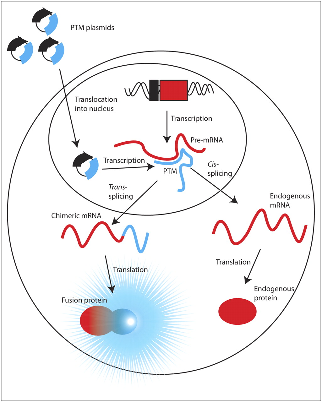
- FIGURE 1.
Schematic of SMaRT imaging strategy. SMaRT PTMs encoding a reporter gene and cloned into plasmid DNA are delivered to nucleus of cell, where they are transcribed (blue lines). PTMs can intercept portion of pre-mRNA (red lines) of arbitrary gene via complementary nucleic acid binding and trans-splice coding domain into target. This hybrid mRNA is translated into chimeric protein composed of portion of target gene and reporter gene, which can elicit signal. Remaining pool of endogenous pre-mRNA can cis-splice and be translated into endogenous protein.
- FIGURE 2.
(A) Various target genes and PTMs used in study. Target gene consisted of HSV1-sr39tk coding sequence fused to intron from HPV16-E6 (TK). As a control, orientation of intron was reversed (TKas). PTMs were generated with binding domains of lengths 80 bp (H1), 160 bp (H2), and 240 bp (H3) complementary or homologous (H1s, H2s, H3s) to intron of target gene. These PTMs contained branch point (black circle), polypyrimidine tract (black rectangle), and dinucleotide 3′ splice site (AG), as well as Renilla luciferase gene without its start codon ((-AUG)luc). Two safety PTMs were constructed with complementary sequences to branch point–polypyrimidine tract–AG signals (white circle, white rectangle, UC) either upstream (S1) or downstream (S2) of 80-bp binding domain. (B) Target gene can undergo either cis-splicing, in which single-amino-acid codon plus stop signal are spliced onto HSV1-sr39tk gene, or trans-splicing, in which luciferase gene is spliced onto HSV1-sr39tk gene.
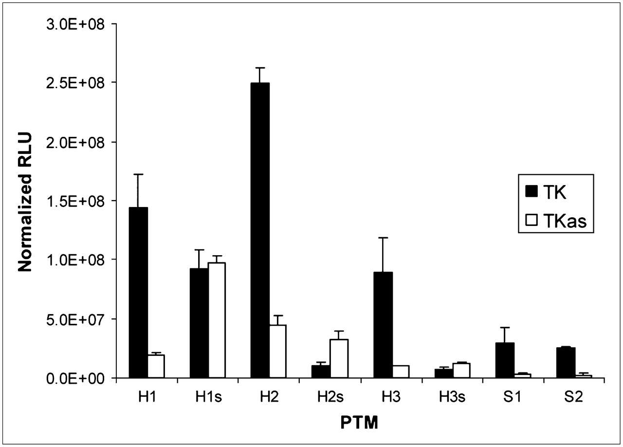
- FIGURE 3.
Cotransfection results. Units are reported in relative light units (RLUs) normalized for total protein content (mean ± SD). Each PTM was cotransfected with either target gene or control gene. Cells were lysed and assayed 24 h after transfection.
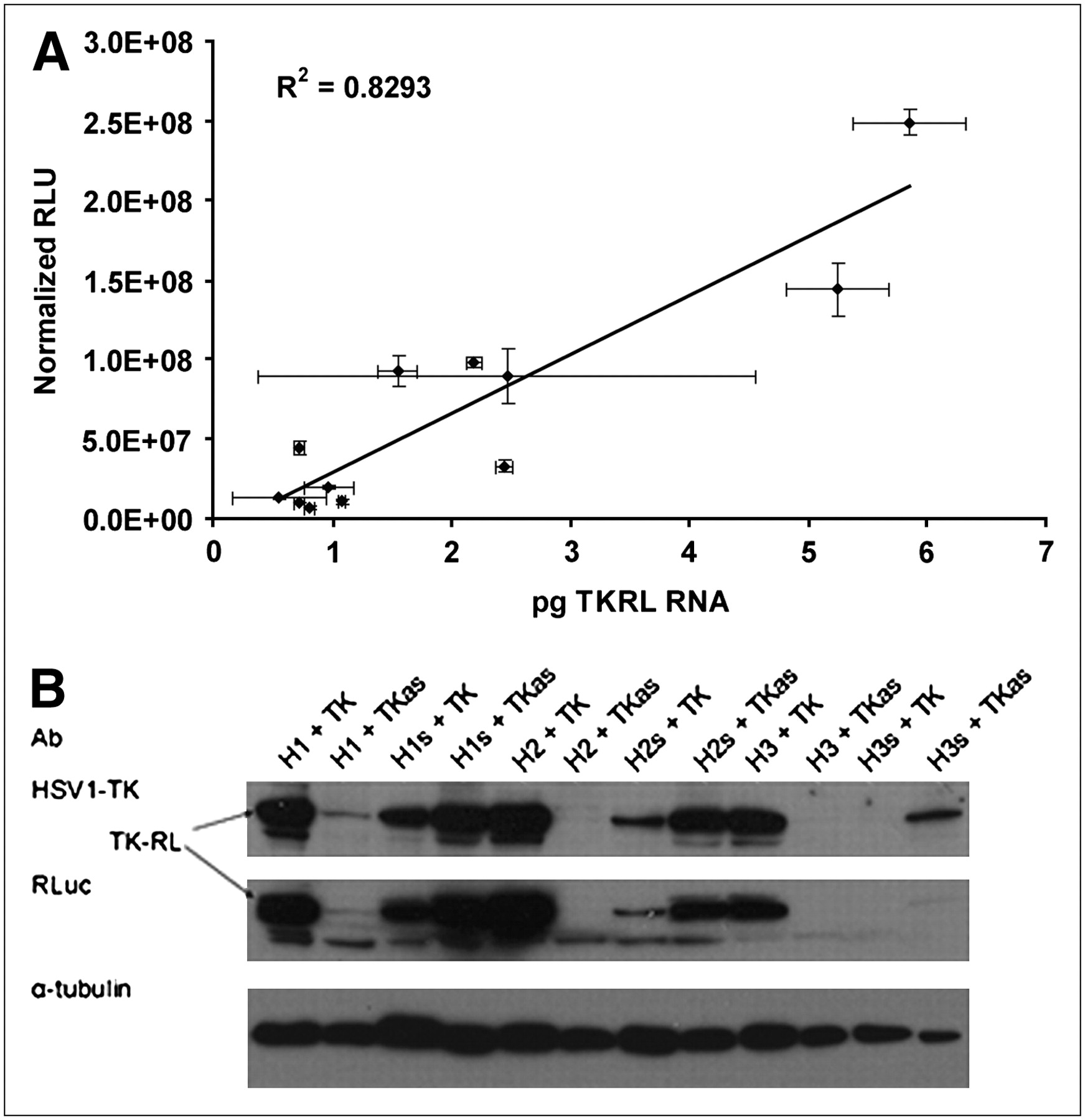
- FIGURE 4.
Cells were transfected with various combinations of PTM and target. Cells were lysed 24 h later, and both total RNA and total protein were collected for analysis. (A) Mass levels of trans-spliced mRNA quantified by real-time RT-PCR and correlated with absolute signals shown in Figure 3. (B) Western blot demonstrating qualitative agreement of trans-spliced protein levels with absolute signal and mRNA levels. RLU = relative light units.
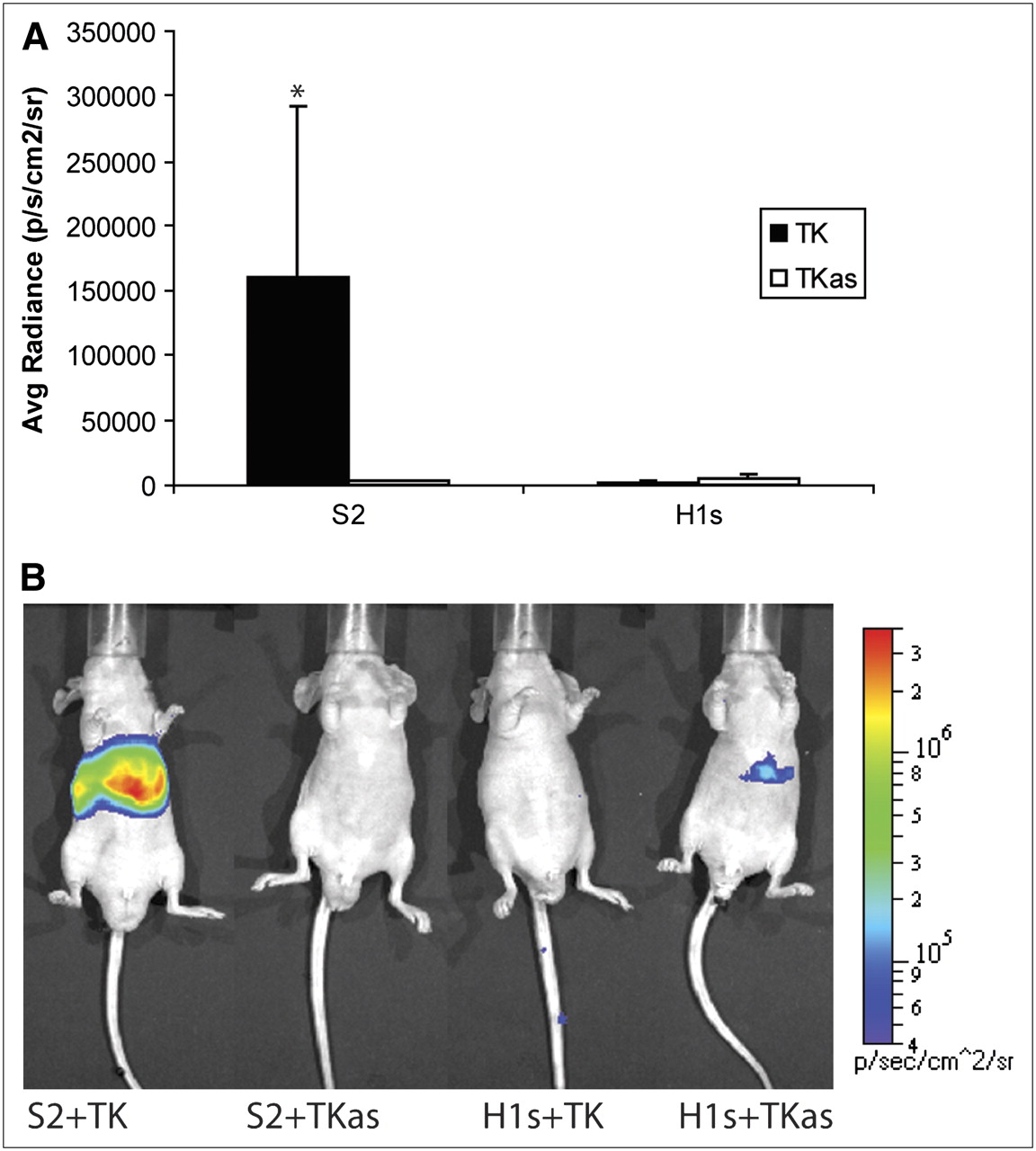
- FIGURE 5.
Summary of in vivo experiments. Nude mice (n = 12) were injected hydrodynamically with combination of PTM and target plasmids. Labels on abscissa refer to constructs shown in Figure 2. Twenty-four hours later, they were injected with coelenterazine and imaged for 1 min. ROIs of equal size for each mouse were drawn over entire liver, and average radiance was calculated. (A) Average radiance for each mouse (mean ± SEM). Values are reported in photons/s/cm2/steradian. *P = 0.0862, Kruskal–Wallis test. (B) Representative images of mice injected with PTM and target gene plus negative controls. Scale covers 2 orders of magnitude; units are reported in photons/cm2/s/steradian.


Tables
PTM Ranks Logs Normalizations H1 1.461 0.096 0.110 H2 1.168 0.106 0.077 H3 2.922 0.115 0.092 S1 1.422 0.097 0.113 S1 3.711 0.119 0.149 Indices were calculated to predict imaging potential of each PTM from available data using weighted average formula and numeric transformations including ranks, logs, and normalization. Values correspond to giving RACE data one third the weight of luciferase assay data.
PTM % aberrant trans-splicing % cis-spliced % correct trans-splicing % unspliced H1 (n = 48) 37 0 29 35 H2 (n = 18) 50 11 39 0 H3 (n = 15) 27 27 40 7 S1 (n = 35) 23 0 17 60 S2 (n = 36) 28 0 39 33 Total RNA was collected from transiently cotransfected 293T cells and subjected to RACE using Rluc-specific primer.


