


Article Figures & Data
Figures
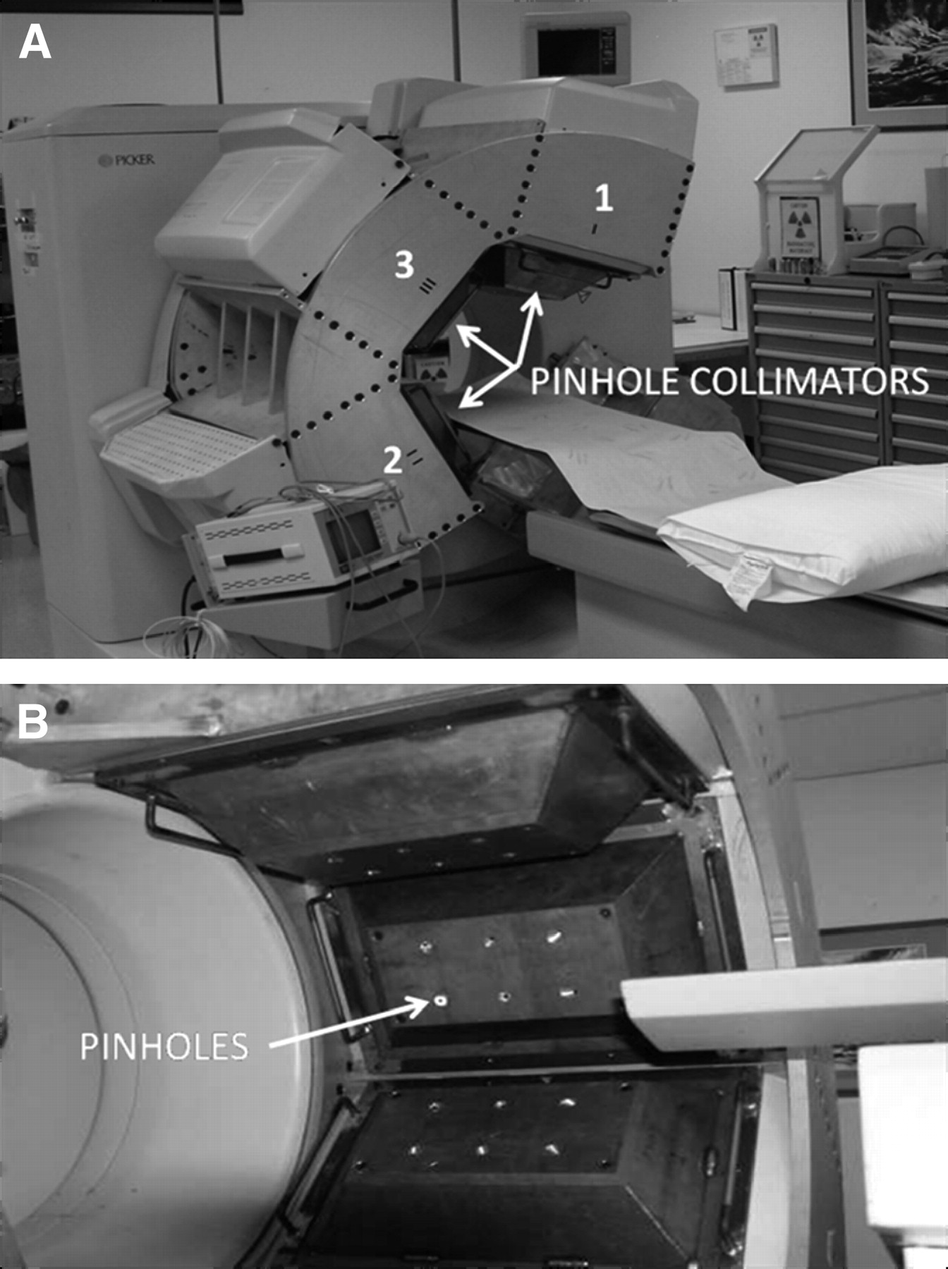
- FIGURE 1.
(A) Picker Prism 3000XP is shown as modified for multipinhole cardiac SPECT perfusion imaging. System is shown set up for patient to be positioned feet-in so that 3 detectors will surround left anterior oblique position of heart. Detector 3 has been remounted between detectors 1 and 2, and all 3 detectors were remounted 90° from the original mounting configuration. (B) Close-up of all 3 detectors with pinhole collimators in place.
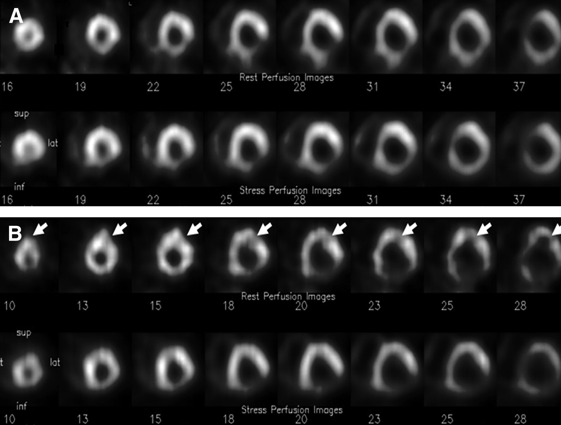
- FIGURE 2.
Short-axis reconstructions for multipinhole (A) and rotational (B) SPECT acquisitions performed on the same patient showing inferior-lateral defect. Resting images are shown above stress images. Arrows point to artifact caused by patient motion, which occurred during acquisition of resting rotational SPECT images. This type of motion artifact is eliminated by simultaneous multipinhole SPECT technique.
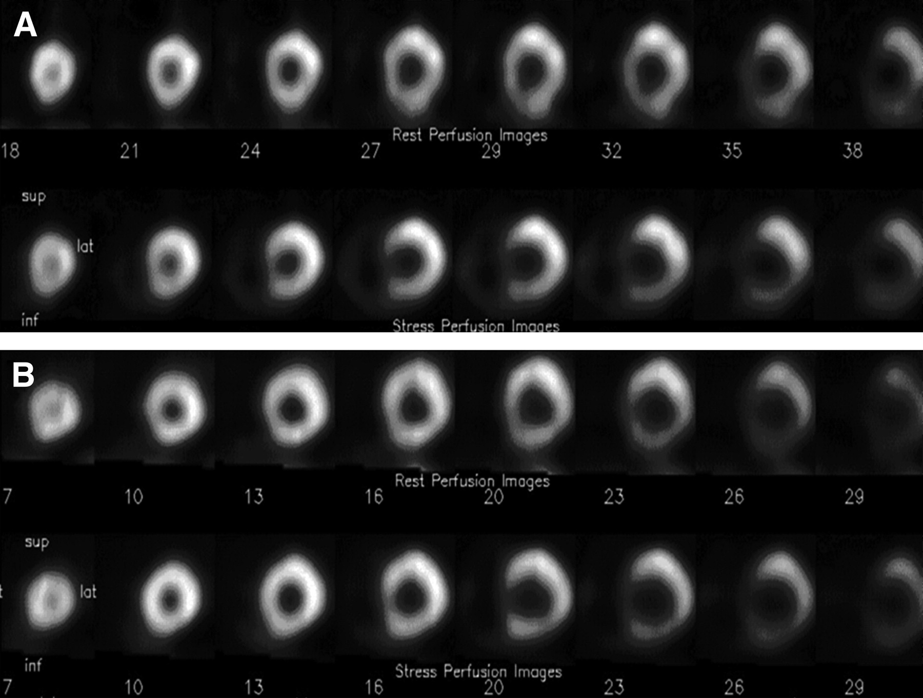
- FIGURE 3.
Short-axis reconstructions for multipinhole (A) and rotational (B) SPECT acquisitions performed on patient with anterior flow difference. Resting images are shown above stress images. Images from 2 modalities are visually and quantatively similar. (See curves in Fig. 4.) Same areas of anterior flow difference are identified in both sets of images, but images in A show larger stress defect than rotational SPECT results in B.
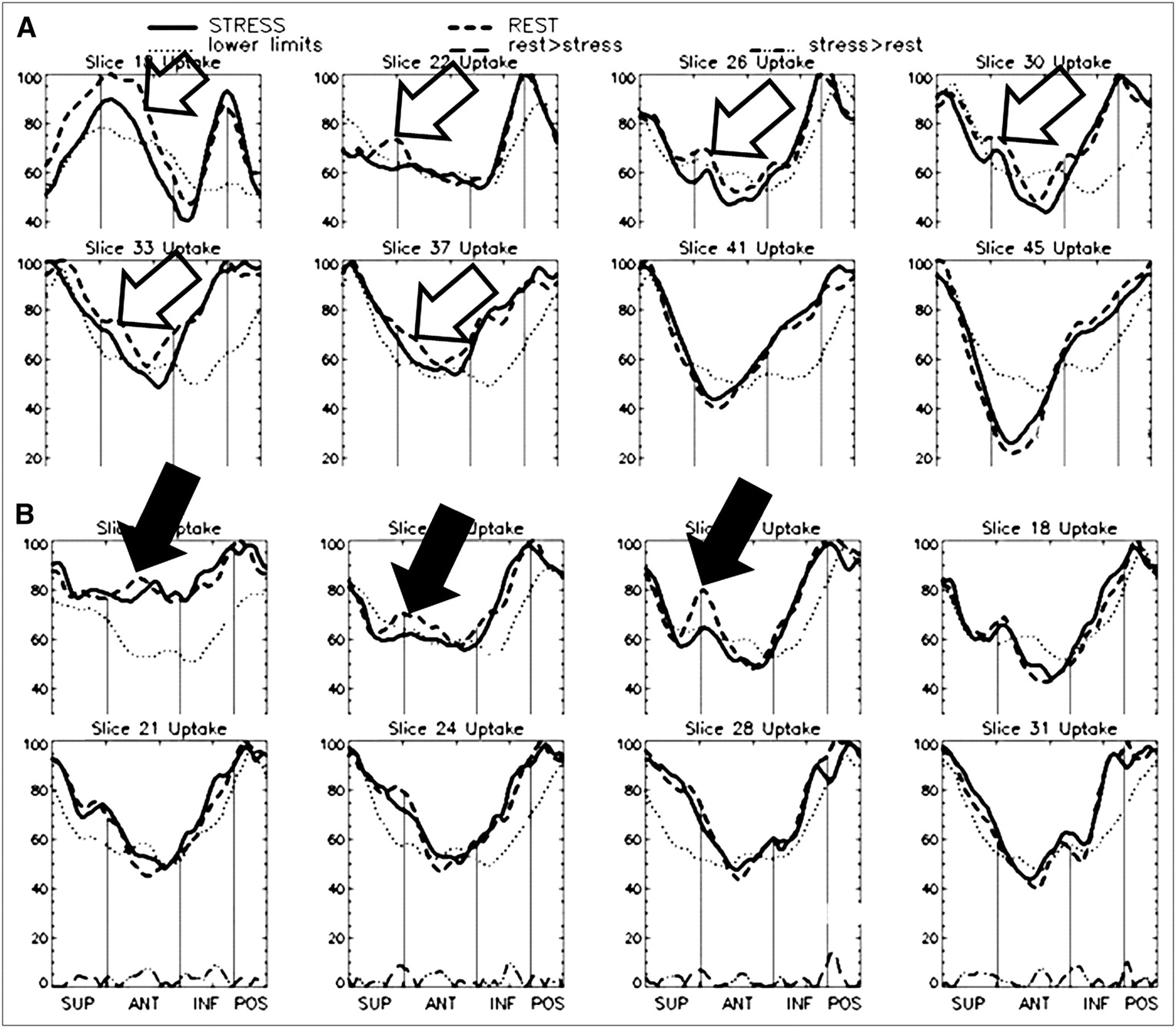
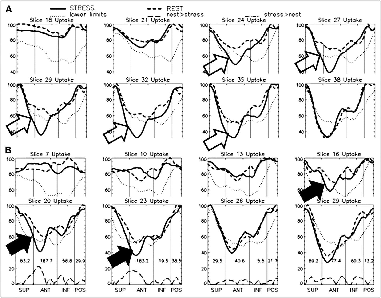
- FIGURE 4.
CPC of short-axis images shown in Figure 3 for multipinhole (A) and rotational (B) SPECT reconstructions. Same anterior regions of flow difference are identified in both sets of curves, but multipinhole analysis shows greater flow differentiation (open arrows) than rotational analysis (closed arrows). SUP = superior; ANT = anterior; INF = inferior; POS = posterior.
- FIGURE 5.
CPC of short-axis slices from multipinhole (A) and rotational (B) SPECT acquisitions on patient with anterior infarct. Low-level flow differences as seen in region of infarct in multipinhole study (open arrows) are also demonstrated, but to lesser extent, in rotational study (closed arrows). SUP = superior; ANT = anterior; INF = inferior; POS = posterior.
- FIGURE 6.
CPC of short-axis slices from multipinhole (A) and rotational (B) SPECT acquisitions on patient with inferior defect. Curves for both A and B analyses show inferior flow differences (open arrows), which are consistent with patient's history. Rotational study also shows flow differences in superior apical region (solid arrows), which are inconsistent with location of any known obstructions. SUP = superior; ANT = anterior; INF = inferior; POS = posterior.
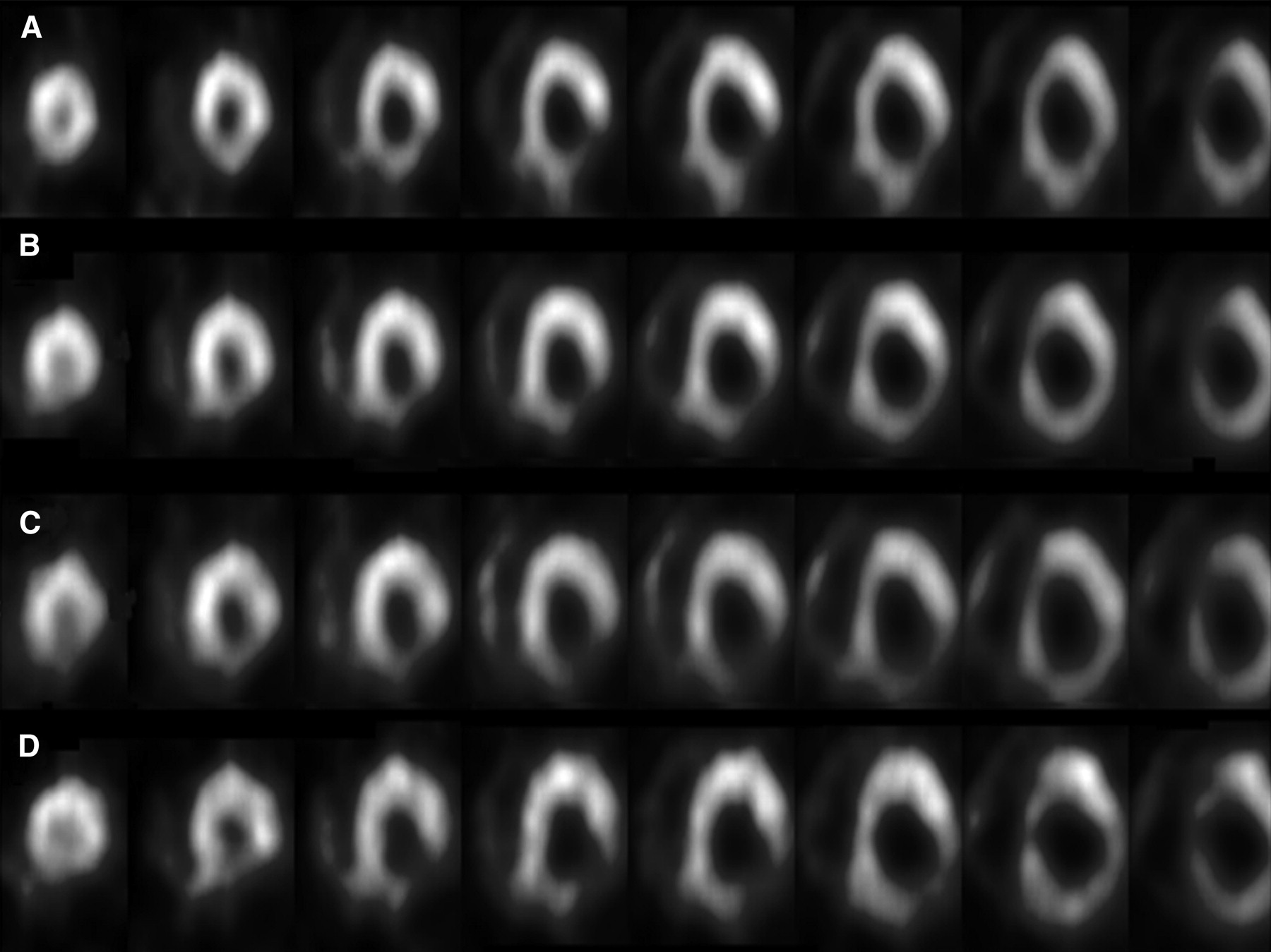
- FIGURE 7.
Short-axis reconstructions from multipinhole SPECT MPI demonstrating DPAC. (A) Resting images using 99mTc tetrofosmin. (B) Simultaneously acquired 201Tl stress reconstructions combining both peaks of 201Tl. (C and D) Separate reconstructions for lower peak (C) and upper peak (D) of 201Tl. Attenuation-compensated stress images in B visually compare better to resting images in A than stress images from either of 2 individual thallium peaks shown in C and D.


Additional Files
Supplemental Data
Files in this Data Supplement:


