


Article Figures & Data
Figures
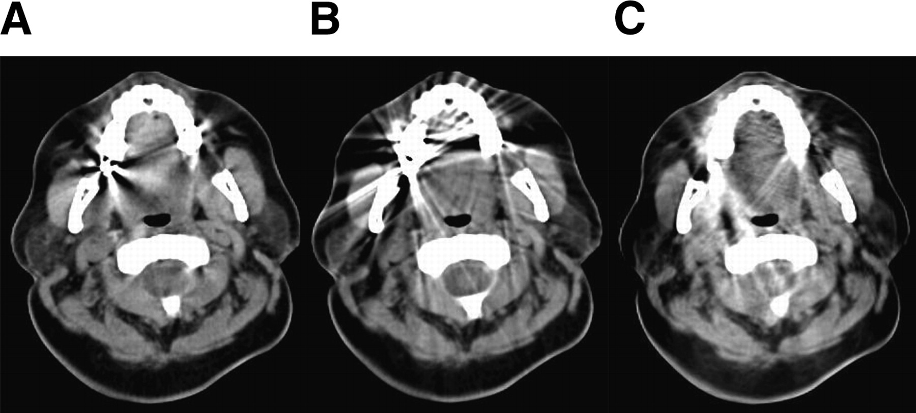
- FIGURE 1.
Original CT image (A); analytic CT metallic artifact reduction algorithm (B); and hybrid, iterative CT metallic artifact reduction algorithm (C). Window was 300 HU wide, and level was 30 HU for the 3 images.
- FIGURE 2.
Placement of ROIs used in the analysis.
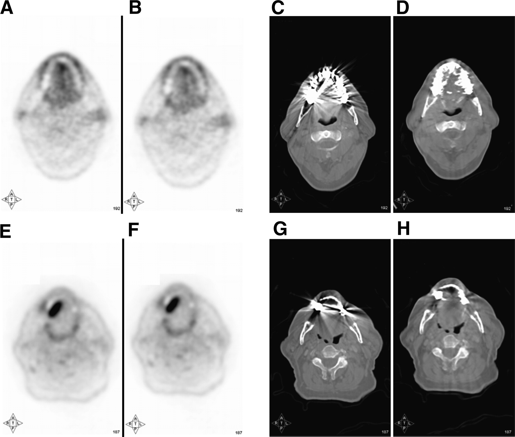
- FIGURE 3.
Two clinical examples showing PET images reconstructed using standard CT algorithm to calculate attenuation factors (A and E), PET images reconstructed using hybrid algorithm to obtain attenuation factors (B and F), CT images reconstructed using standard CT algorithm (C and G), and CT images reconstructed using hybrid algorithm (D and H).
- FIGURE 4.
Mean HU from standard CT reconstruction plotted against mean HU from metallic artifact reduction reconstruction. Blue circles correspond to overestimated areas in standard CT reconstruction, green circles correspond to areas unaffected, and red circles correspond to areas of underestimation. In insert, green circles correspond to areas unaffected in standard CT reconstruction in patients with dental implants, and magenta stars correspond to data from patients without dental implants. In each case, solid line represents trend line with slope equal to 0.3.
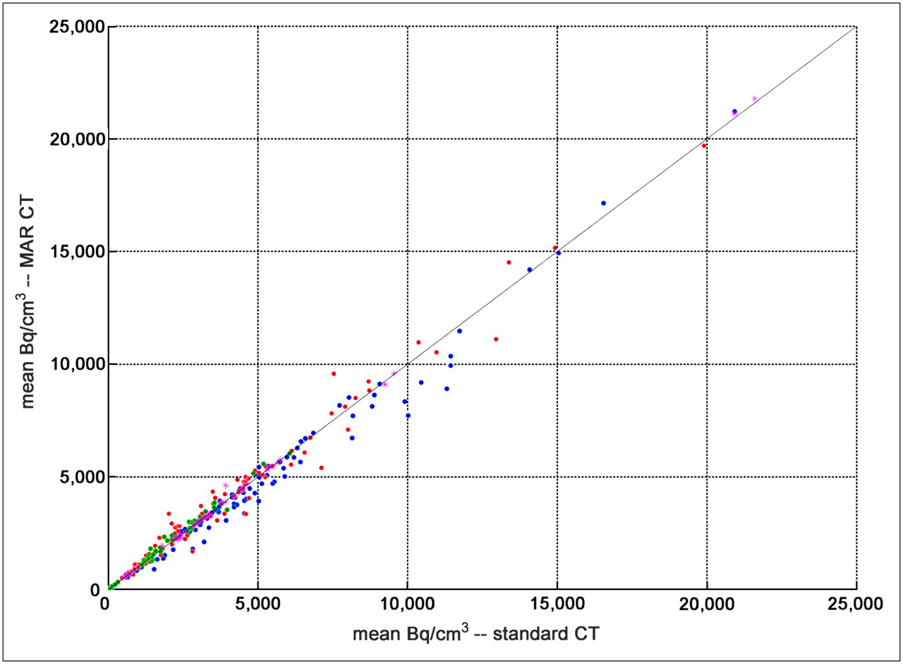
- FIGURE 5.
Mean Bq/cm3 using standard CT reconstruction to calculate attenuation correction factors plotted against mean Bq/cm3 from metallic artifact reduction reconstruction. Blue circles correspond to underestimated areas in metallic artifact reduction CT reconstruction, green circles correspond to areas unaffected, and red circles correspond to areas of overestimation. Magenta stars correspond to data from patients without dental implants. Solid line represents line of identity.
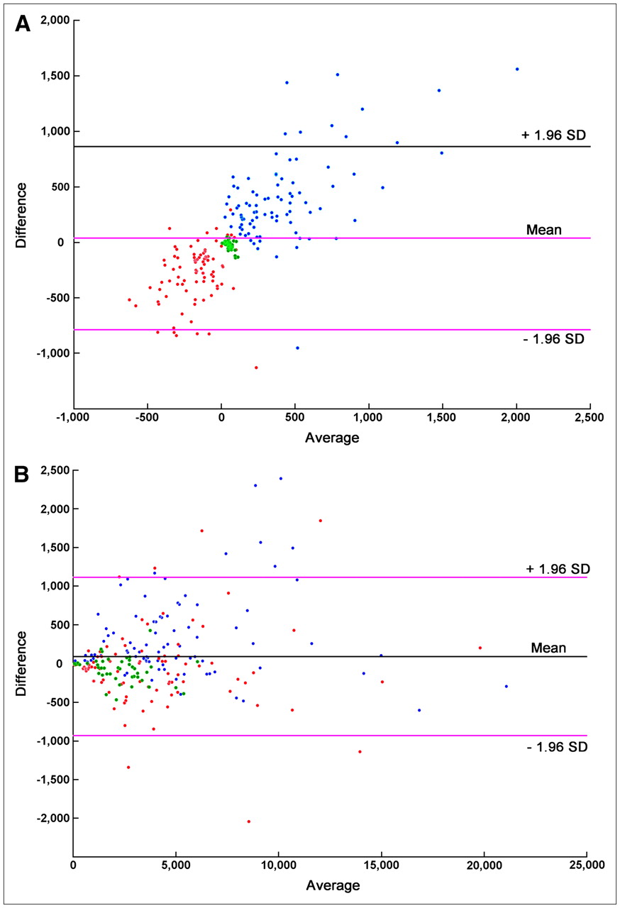
- FIGURE 6.
Bland–Altman plots for HUs resulting from the 2 CT reconstructions (A) and Bq/cm3 from the 2 PET reconstructions (B). In this plot, difference between results from the 2 reconstructions is plotted against average of the 2 reconstructions. Blue circles correspond to underestimated areas in metallic artifact reduction CT reconstruction, green circles correspond to areas unaffected, and red circles correspond to areas of overestimation.


