
Main menu

  • Home
  • Content
    • Current
    • Ahead of print
    • Past Issues
    • JNM Supplement
    • SNMMI Annual Meeting Abstracts
    • Continuing Education
    • JNM Podcasts
  • Subscriptions
    • Subscribers
    • Institutional and Non-member
    • Rates
    • Journal Claims
    • Corporate & Special Sales
  • Authors
    • Submit to JNM
    • Information for Authors
    • Assignment of Copyright
    • AQARA requirements
  • Info
    • Reviewers
    • Permissions
    • Advertisers
  • About
    • About Us
    • Editorial Board
    • Contact Information
  • More
    • Alerts
    • Feedback
    • Help
    • SNMMI Journals
  • SNMMI
    • JNM
    • JNMT
    • SNMMI Journals
    • SNMMI

User menu

  • Subscribe
  • My alerts
  • Log in
  • My Cart

Search

  • Advanced search
Journal of Nuclear Medicine
  • SNMMI
    • JNM
    • JNMT
    • SNMMI Journals
    • SNMMI
  • Subscribe
  • My alerts
  • Log in
  • My Cart
Journal of Nuclear Medicine

Advanced Search

  • Home
  • Content
    • Current
    • Ahead of print
    • Past Issues
    • JNM Supplement
    • SNMMI Annual Meeting Abstracts
    • Continuing Education
    • JNM Podcasts
  • Subscriptions
    • Subscribers
    • Institutional and Non-member
    • Rates
    • Journal Claims
    • Corporate & Special Sales
  • Authors
    • Submit to JNM
    • Information for Authors
    • Assignment of Copyright
    • AQARA requirements
  • Info
    • Reviewers
    • Permissions
    • Advertisers
  • About
    • About Us
    • Editorial Board
    • Contact Information
  • More
    • Alerts
    • Feedback
    • Help
    • SNMMI Journals
  • View or Listen to JNM Podcast
  • Visit JNM on Facebook
  • Join JNM on LinkedIn
  • Follow JNM on Twitter
  • Subscribe to our RSS feeds
OtherBASIC SCIENCE INVESTIGATIONS

Detection of Secondary Thalamic Degeneration After Cortical Infarction Using cis-4-18F-Fluoro- d-Proline

Karl-Josef Langen, Dagmar Salber, Kurt Hamacher, Gabriele Stoffels, Guido Reifenberger, Dirk Pauleit, Heinz H. Coenen and Karl Zilles
Journal of Nuclear Medicine September 2007, 48 (9) 1482-1491; DOI: https://doi.org/10.2967/jnumed.107.041699
Karl-Josef Langen
  • Find this author on Google Scholar
  • Find this author on PubMed
  • Search for this author on this site
Dagmar Salber
  • Find this author on Google Scholar
  • Find this author on PubMed
  • Search for this author on this site
Kurt Hamacher
  • Find this author on Google Scholar
  • Find this author on PubMed
  • Search for this author on this site
Gabriele Stoffels
  • Find this author on Google Scholar
  • Find this author on PubMed
  • Search for this author on this site
Guido Reifenberger
  • Find this author on Google Scholar
  • Find this author on PubMed
  • Search for this author on this site
Dirk Pauleit
  • Find this author on Google Scholar
  • Find this author on PubMed
  • Search for this author on this site
Heinz H. Coenen
  • Find this author on Google Scholar
  • Find this author on PubMed
  • Search for this author on this site
Karl Zilles
  • Find this author on Google Scholar
  • Find this author on PubMed
  • Search for this author on this site
  • Article
  • Figures & Data
  • Info & Metrics
  • PDF
Loading

Article Figures & Data

Figures

  • Tables
  • FIGURE 1. 
    • Download figure
    • Open in new tab
    • Download powerpoint
    FIGURE 1. 

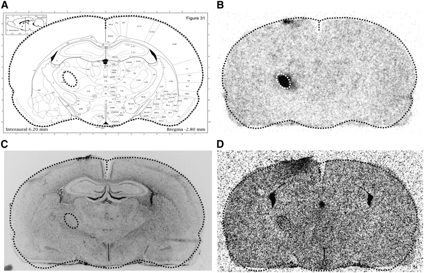
    Localization of TN (rat 10): Coronal brain slice in the anatomic map (A) was identified using the histologic slice (C). The corresponding autoradiogram of d-cis-18F-FPro (B) was adapted to the circumference of this map. Reprojection of focal tracer uptake in the midbrain to the anatomic map identifies the thalamic nucleus. The ventroposteromedial thalamic nucleus (VPM) is indicated by an ellipse. The corresponding autoradiogram using 3H-PK11195 (D) shows only minor tracer uptake in the VPM. (Image A modified and reprinted with permission of (15).)

  • FIGURE 2. 
    • Download figure
    • Open in new tab
    • Download powerpoint
    FIGURE 2. 

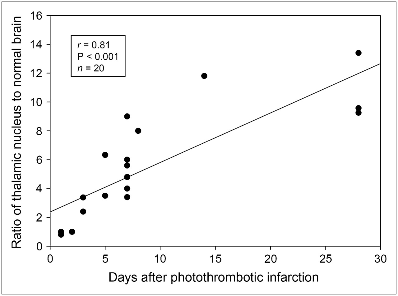
    d-cis-18F-FPro uptake in TN (ratio of tracer uptake in thalamic tissue divided by normal brain tissue) vs. time after cortical infarction. There is a significant correlation.

  • FIGURE 3. 
    • Download figure
    • Open in new tab
    • Download powerpoint
    FIGURE 3. 

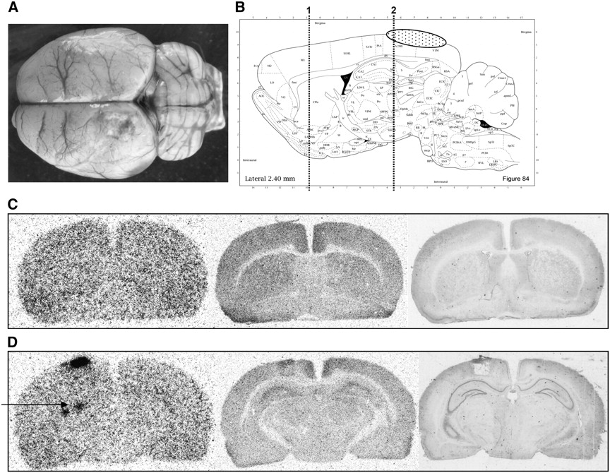
    (A) Dorsal view of rat brain 7 d after PT (rat 6). Cortical infarction involves parts of motor and somatosensory cortex. (B) Sagittal slice from anatomic rat brain atlas demonstrates position of infarction (stippled area) and position of coronal slices: 1, level of basal ganglia (C); 2, level of hippocampus/thalamus (D). Autoradiogram with d-cis-18F-FPro (C, left image) exhibits tracer uptake in area of cortical infarction and in caudate nucleus (arrow). Furthermore, there is focal d-cis-18F-FPro uptake in ventral posteromedial thalamic nucleus and posterior thalamic nuclear group (D, left image, arrows). Corresponding autoradiograms using 3H-DG (C and D, middle images) and histologic staining using toluidine blue (C and D, right images) show no abnormalities in caudate nucleus and in thalamus. (Image B modified and reprinted with permission of (15).)

  • FIGURE 4. 
    • Download figure
    • Open in new tab
    • Download powerpoint
    FIGURE 4. 

    (A) Dorsal view of rat brain 7 d after PT (rat 5) with infarction in visual cortex. (B) Sagittal slice from anatomic rat brain atlas demonstrates position of infarction (stippled area) and position of coronal slices: 1, level of the basal ganglia (C); 2, level of the hippocampus/thalamus (D). Autoradiograms with d-cis-18F-FPro (C and D, left images) exhibit tracer uptake in area of cortical infarction and in lateral posterior thalamic nucleus (LP) and dorsolateral geniculate nucleus (DLG) (D, arrow). Corresponding autoradiograms using 3H-DG (C and D, middle images) and histologic staining using toluidine blue (C and D, right images) show no abnormalities in thalamus. (Image B modified and reprinted with permission of (15).)
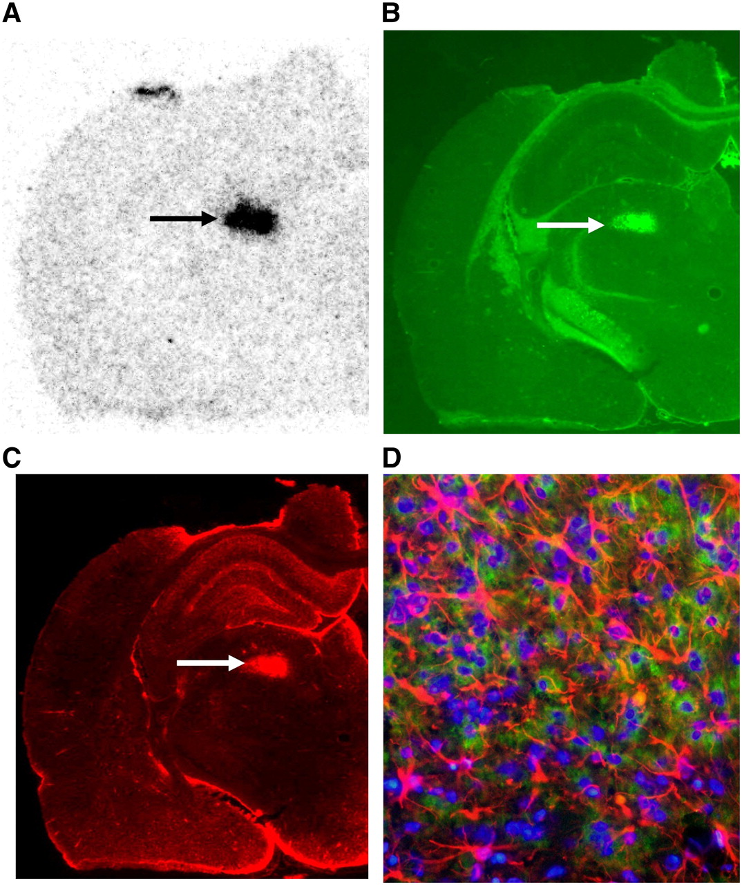
  • FIGURE 5. 
    • Download figure
    • Open in new tab
    • Download powerpoint
    FIGURE 5. 

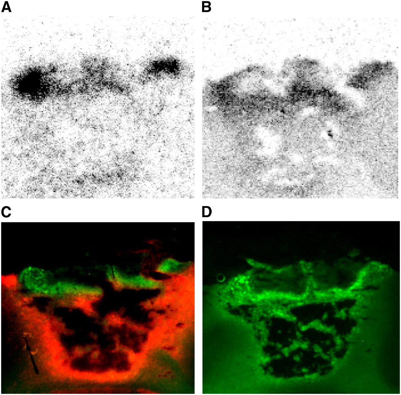
    (A–C) Coronal slices of left hemisphere at level of thalamus 28 d after PT (rat 11). (A) Autoradiogram using d-cis-18F-FPro shows high uptake in posterior thalamic nuclear group (arrow). (B) Immunofluorescent staining using CD11b shows activated microglia (arrow). (C) Immunofluorescent staining using GFAP shows reactive astrogliosis in same area (arrow) as in B. (D) Thalamic nucleus at higher magnification: blue = cell nuclei; red = astrocytes; green = activated microglia.

  • FIGURE 6. 
    • Download figure
    • Open in new tab
    • Download powerpoint
    FIGURE 6. 

    Cortical infarction 7 d after PT. (A and B) Autoradiographic distribution of d-cis-18F-FPro (A) and 3H-DG (B). (C) Double immunofluorescence labeling using CD11b shows activated microglia (green) and GFAP shows reactive astrogliosis (red). (D) Immunofluorescence labeling using CD68 shows macrophages. d-cis-18F-FPro and 3H-DG accumulation is similar to high density of activated microglia and macrophages but different from that of reactive astrocytosis.

Tables

  • Figures
    • View popup
    TABLE 1

    Summary of Data on Animals with Cortical Infarctions

    d-cis-FPro uptake3H-DG3H-PK111953H-MET
    Rat no.Time after PT (d)Cortical areaThalamic nucleus, striatumPT/brainTN/brainPT/brainTN/brainPT/brainTN/brainPT/brainTN/brain
    17Mot, SensCPu, Po, VPM, VPL12.64.0
    25Mot, SensCPu, Po, VL11.76.34.21.4
    38VisDLG, LP16.68.02.41.0
    47Sens, VisPo, VPM, DLG12.46.05.51.0
    57VisDLG, LP17.63.42.21.1
    67Mot, SensCPu, Po, VPM, VPL, VL9.44,03.01.2
    77MotCPu, VL16.84.83.01.0
    87SensPo, VPM, VPL10.79.02.70.9
    97SensPo, VPM, VPL6.65.62.30.8
    1014SensPo, VPM, VPL31.611.85.71.4
    1128SensPo, VPM4.19.6
    1228SensPo, VPM24.813.4
    1328SensPo, VPM7.09.38.01.0
    145Mot, SensCPu, Po, VPM11.83.52.71.1
    153Mot, SensCPu, Po, VPM13.43.41.00.8
    163Mot, SensCPu, Po, VPM20.82.40.60.9
    172Sens—3.3—0.9—
    182Sens—3.8—0.7—
    191Sens—3.4—1.0—
    201Sens—3.8—0.6—
    Mean12.16.72.51.06.81.21.41.0
    SD8.13.51.50.21.70.31.10.2
    • PT = cortical infarction induced by photothrombosis; ; 3H-DG = 3H-deoxyglucose; 3H-Pk11195 = ligand for peripheral benzodiazepine receptors; 3H-MET = 3H-methyl-l-methionine; PT/brain = ratio of maximal tracer uptake PT divided by mean uptake in normal brain tissue; TN/brain = ratio of maximal tracer uptake in thalamic nucleus divided by mean uptake in normal brain; Mot = motor cortex; Sens = somatosensory cortex; CPu = caudate putamen; Po = posterior thalamic nuclear group; VPM = ventral posteromedial thalamic nucleus; VPL = ventroposterolateral thalamic nucleus; VL = ventrolateral thalamic nucleus; Vis = visual cortex; DLG = dorsal lateral geniculate nucleus; LP = lateral posterior thalamic nucleus.

PreviousNext
Back to top

In this issue

Journal of Nuclear Medicine: 48 (9)
Journal of Nuclear Medicine
Vol. 48, Issue 9
September 2007
  • Table of Contents
  • Table of Contents (PDF)
  • About the Cover
  • Index by author
Print
Download PDF
Article Alerts
Sign In to Email Alerts with your Email Address
Email Article

Thank you for your interest in spreading the word on Journal of Nuclear Medicine.

NOTE: We only request your email address so that the person you are recommending the page to knows that you wanted them to see it, and that it is not junk mail. We do not capture any email address.

Enter multiple addresses on separate lines or separate them with commas.
Detection of Secondary Thalamic Degeneration After Cortical Infarction Using cis-4-18F-Fluoro- d-Proline
(Your Name) has sent you a message from Journal of Nuclear Medicine
(Your Name) thought you would like to see the Journal of Nuclear Medicine web site.
Citation Tools
Detection of Secondary Thalamic Degeneration After Cortical Infarction Using cis-4-18F-Fluoro- d-Proline
Karl-Josef Langen, Dagmar Salber, Kurt Hamacher, Gabriele Stoffels, Guido Reifenberger, Dirk Pauleit, Heinz H. Coenen, Karl Zilles
Journal of Nuclear Medicine Sep 2007, 48 (9) 1482-1491; DOI: 10.2967/jnumed.107.041699

Citation Manager Formats

  • BibTeX
  • Bookends
  • EasyBib
  • EndNote (tagged)
  • EndNote 8 (xml)
  • Medlars
  • Mendeley
  • Papers
  • RefWorks Tagged
  • Ref Manager
  • RIS
  • Zotero
Share
Detection of Secondary Thalamic Degeneration After Cortical Infarction Using cis-4-18F-Fluoro- d-Proline
Karl-Josef Langen, Dagmar Salber, Kurt Hamacher, Gabriele Stoffels, Guido Reifenberger, Dirk Pauleit, Heinz H. Coenen, Karl Zilles
Journal of Nuclear Medicine Sep 2007, 48 (9) 1482-1491; DOI: 10.2967/jnumed.107.041699
Twitter logo Facebook logo LinkedIn logo Mendeley logo
  • Tweet Widget
  • Facebook Like
  • Google Plus One
Bookmark this article

Jump to section

  • Article
    • Abstract
    • MATERIALS AND METHODS
    • RESULTS
    • DISCUSSION
    • CONCLUSION
    • Acknowledgments
    • Footnotes
    • References
  • Figures & Data
  • Info & Metrics
  • PDF

Related Articles

  • This Month in JNM
  • PubMed
  • Google Scholar

Cited By...

  • Intracortical transplantation of human induced pluripotent stem cell-derived progenitors ameliorates delayed thalamic degeneration following cortical stroke
  • Stem Cell-Derived Neurons Grafted in the Striatum Are Expelled Out of the Brain After Chronic Cortical Stroke
  • Google Scholar

More in this TOC Section

  • PET/MRI of Hypoxic Atherosclerosis Using 64Cu-ATSM in a Rabbit Model
  • Tumor Uptake of Anti-CD20 Fabs Depends on Tumor Perfusion
  • How Sensitive Is the Upper Gastrointestinal Tract to 90Y Radioembolization? A Histologic and Dosimetric Analysis in a Porcine Model
Show more Basic Science Investigations

Similar Articles

SNMMI

© 2025 SNMMI

Powered by HighWire