


Article Figures & Data
Figures
- FIGURE 1.
Bone marrow biopsy specimens from iliac crest. (A) Five-section composite image of single histologic section of biopsy sample. (B) Representative distance measurements of hematopoietic CD34+ cells to nearest bone trabecula surface (yellow lines), blood vessel fragment to nearest bone trabecula surface (blue lines), and separation distances between hematopoietic CD34+ cells and nearest blood vessel fragment (violet lines).
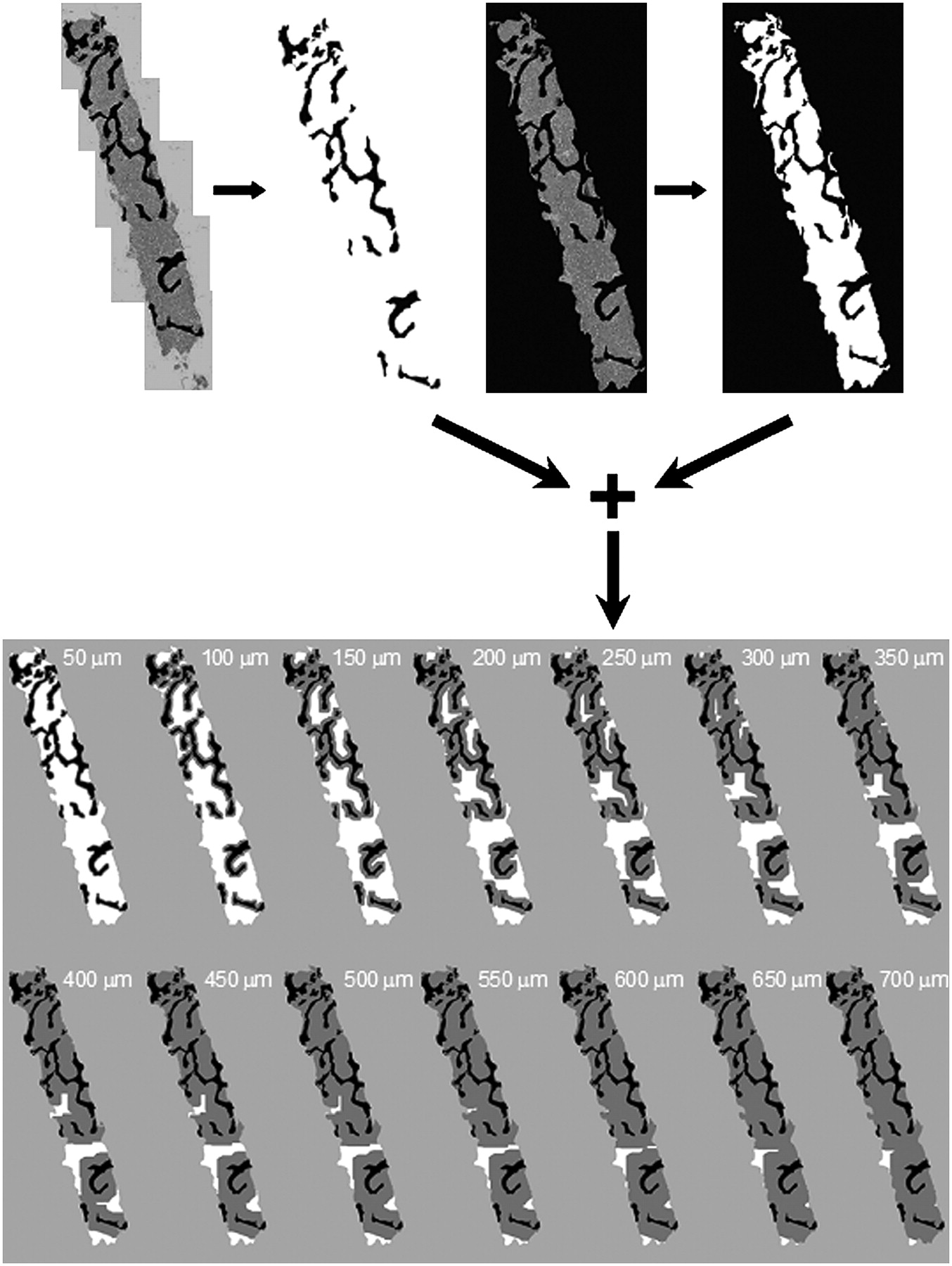
- FIGURE 2.
Image-processing steps to determine 50-μm areal contours from all surfaces of bone trabeculae.
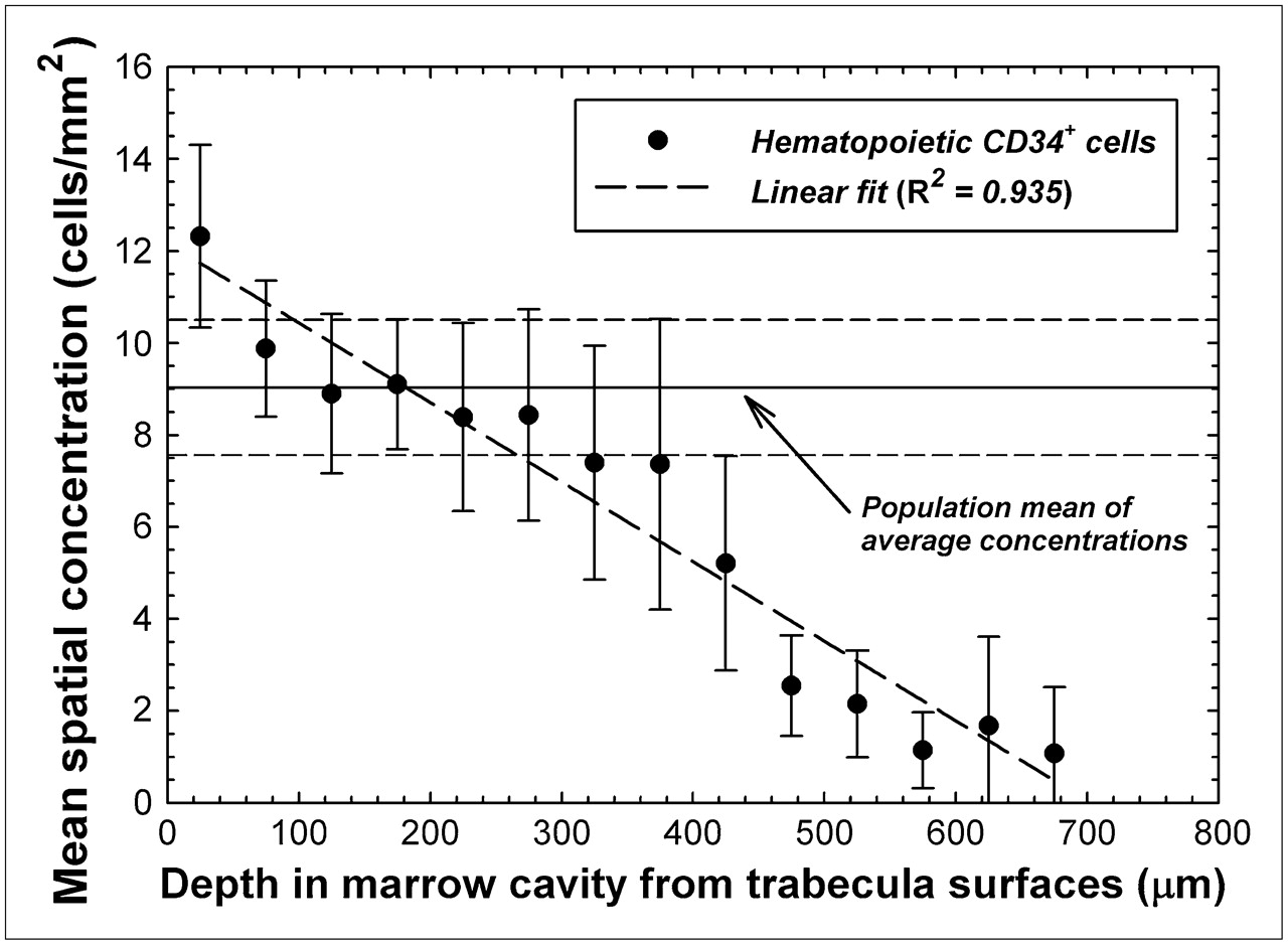
- FIGURE 3.
Mean spatial concentration of hematopoietic CD34+ cells as function of distance into bone marrow cavities of iliac crest. Data points indicate specimen-averaged mean values binned every 50 μm, and dashed line represents linear fit to data. Horizontal lines indicate mean and SD of average concentrations seen across study population.
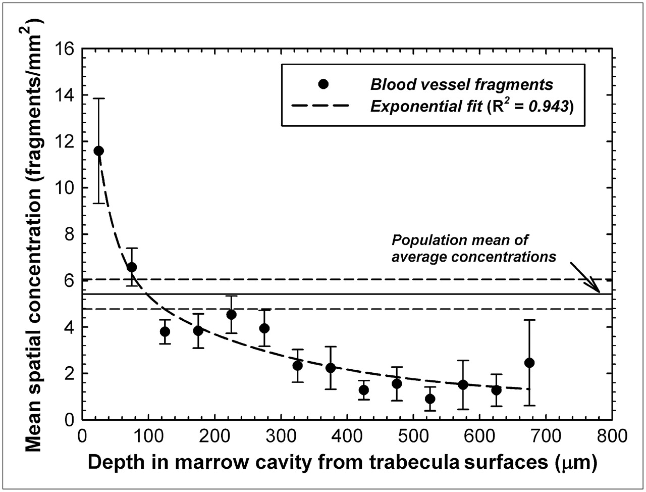
- FIGURE 4.
Mean spatial concentrations of blood vessel fragments as function of distance into bone marrow cavities of iliac crest. Data points indicate specimen-averaged mean values binned every 50 μm, and dashed line represents exponential fit to data. Horizontal lines indicate mean and SD of average concentrations seen across study population.
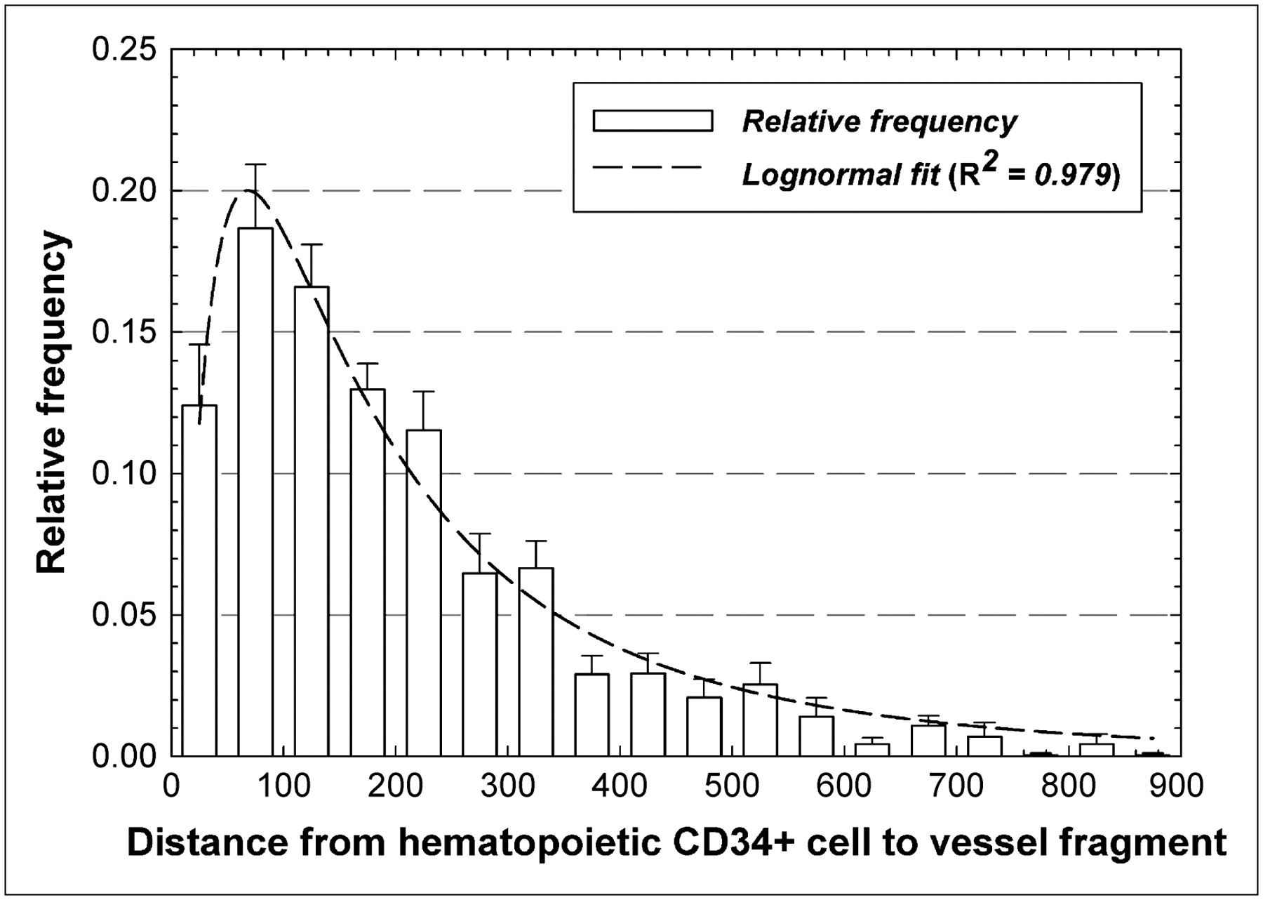
- FIGURE 5.
Frequency distribution of distances separating pairs of hematopoietic CD34+ cells and blood vessels within marrow cavities of iliac crest. Dashed line represents lognormal fit to data.
- FIGURE 6.
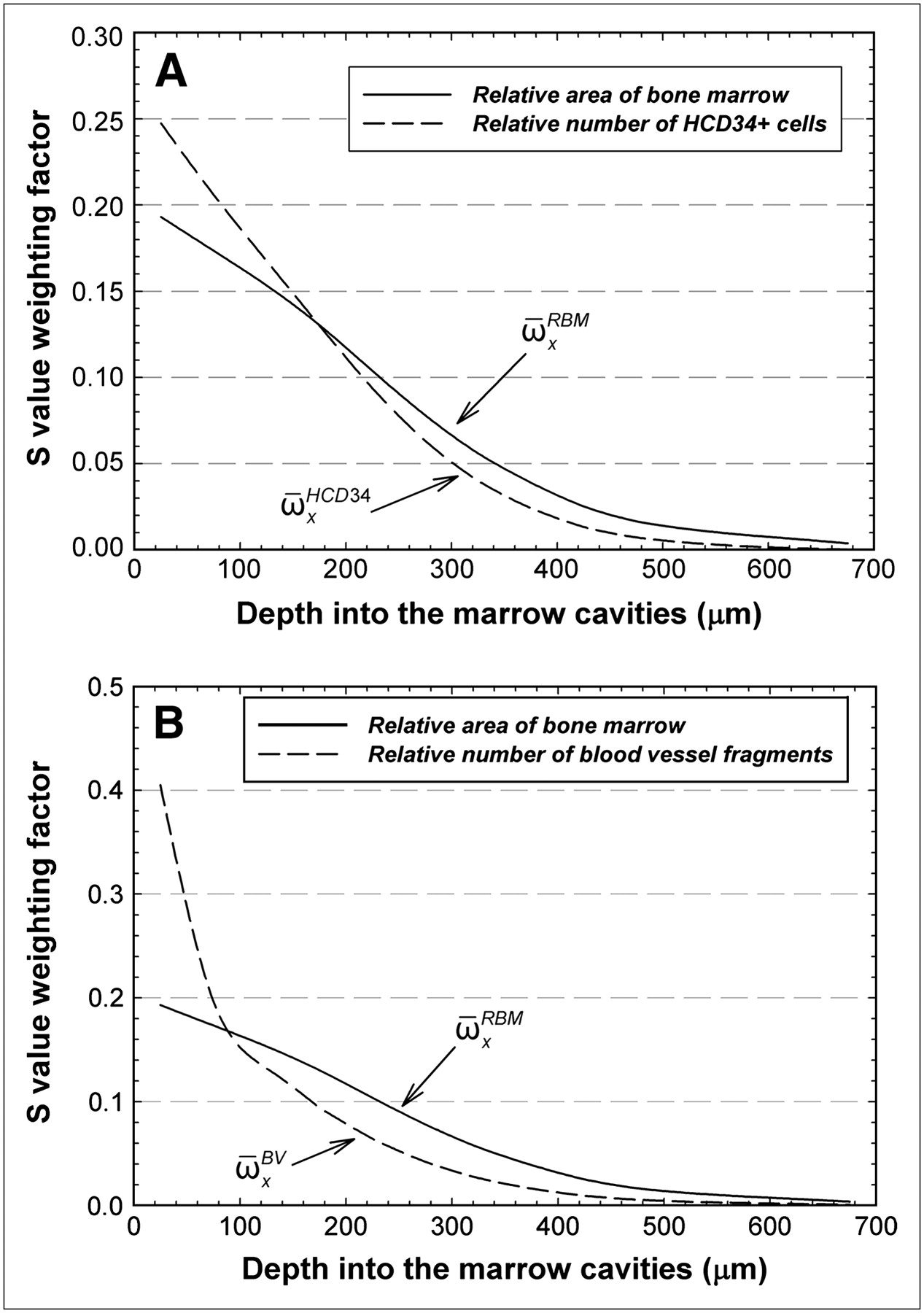
Distance-dependent weighting factors for radionuclide S values given in voxel-based skeletal dosimetry models. Solid lines indicate fraction of total area of bone marrow seen as function of distance into marrow cavities of iliac crest (average for the 14 specimens in current study). Dashed lines indicate relative number of hematopoietic CD34+ cells (A) or blood vessel fragments as function of distance into marrow cavities (B). HCD34+ = hematopoietic CD34+.


Tables
Specimen no. Sex Age (y) Total marrow area (mm2) Specimen weight (fs, in %) Total cell count (HCD34+ cells) Total vessel count 1 M 11 4.93 4 98 23 2 F 35 17.40 13 116 109 3 F 80 14.55 11 128 119 4 F 46 6.42 5 34 22 5 M 19 17.33 13 66 88 6A F 68 3.06 2 33 15 6B F 68 3.39 3 44 9 7A F 7 3.72 3 89 10 7B F 7 1.12 1 44 7 8 F 32 11.24 9 127 54 9 F 69 16.33 13 161 89 10 F 32 11.35 9 113 56 11 M 44 15.34 12 70 47 12 M 2 3.25 3 46 53 Total 129.43 100 1,169 701


Jump to section
Related Articles
Cited By...
- Specific Uptake in the Bone Marrow Causes High Absorbed Red Marrow Doses During [177Lu]Lu-DOTATATE Treatment
- Specific Uptake in the Bone Marrow Causes High Absorbed Red Marrow Doses During [177Lu]Lu-DOTATATE Treatment
- The relationship between bone, hemopoietic stem cells, and vasculature
- Spatial gradients of blood vessels and hematopoietic stem and progenitor cells within the marrow cavities of the human skeleton
- Further Explorations of Cellular Uptake of Radioactivity
- Toward Patient-Friendly Cell-Level Dosimetry