


Article Figures & Data
Figures
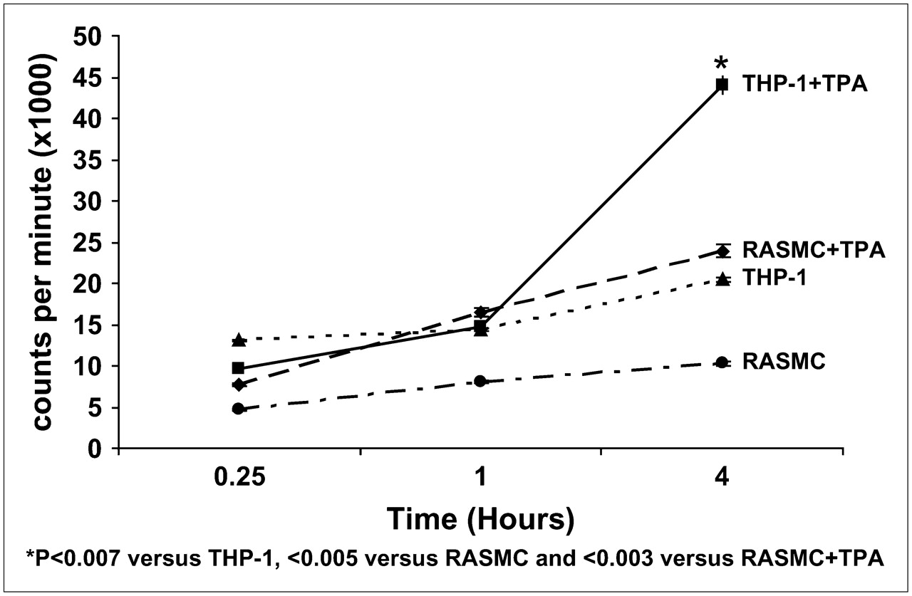
- FIGURE 1.
99mTc MCP-1 uptake in activated and nonactivated SMC and macrophages (in counts/min × 1,000) in culture. Uptake increases significantly when monocytes (THP-1 cells) are activated with PMA. RASMC = rat aortic smooth muscle cells.
- FIGURE 2.
Noninvasive imaging of MCP-1 receptor expression in experimentally induced atherosclerosis with radiolabeled MCP-1. Images were obtained in both atherosclerotic (A−C) and control (nonatherosclerotic, D−F) rabbits. (A−C) Planar γ-images of atherosclerotic rabbit. (A) Image obtained immediately after intravenous MCP-1 administration outlines aortic blood-pool activity (arrows). (B) At 3 h after radiotracer administration, significant radiotracer accumulation is evident in abdominal aorta (arrows). (C) Ex vivo image of explanted aorta confirms in vivo evidence of MCP-1 uptake (arrows). (D−F) Planar γ-images of control rabbit. (D) Aortic blood pool is seen at time of intravenous MCP-1 injection (arrows). (E) Image shows lack of MCP-1 uptake in region of abdominal aorta, as denoted by arrows in normal rabbit with no atherosclerotic lesions. (F) Ex vivo aortic image of control animal demonstrates absence of MCP-1 uptake (arrows). L = liver; S = spleen; K = kidney. (G) Bar graphs show quantitative 99mTc MCP-1 uptake within abdominal aortas of atherosclerotic and control animals represented as mean %ID/g ± SD. 99mTc-MCP-1 uptake (%ID/g) was significantly higher in atherosclerotic animals (P < 0.0001) compared with that of control animals.
- FIGURE 3.
Histologic and immunohistologic characterization of atherosclerotic plaques with increasing severity in comparison with nonatherosclerotic abdominal aorta in control rabbit. (A) Lesion composed predominantly of superficial macrophages overlying acellular area of extracellular matrix. Only a few smooth muscle cells are evident on surface of lesion. There is relatively intense staining for Monocyte Chemotactic Protein 1 (MCP-1) (rose-red reaction product) primarily near luminal border localized to areas rich in macrophages. (B) More advanced plaque consisting of macrophages, SMC, and underlying extracellular matrix. Immunostaining for MCP-1 is intense and localized primarily in deeper layers of lesion. Inset shows cytoplasmic staining of MCP-1 in cells resembling macrophages. (C) Lesion shows medial dissection with intense macrophage infiltration in intimal (I) and medial (M) layers with overlying SMC-rich fibrous cap. Immunostaining for MCP-1 is diffusely seen throughout plaque, corresponding to areas of macrophage infiltration. (D) Control vessel shows absence of macrophages, with MPC-1 present only in occasional cells near lumen and medial/adventitial borders. Histologic stain in each panel is Movat pentachrome. Immunostains are counterstained with Gill's hematoxylin; chromogen is 3-amino-9-ethyl carbazole (AEC). SMC = α-actin smooth muscle; MAC = Ram 11.
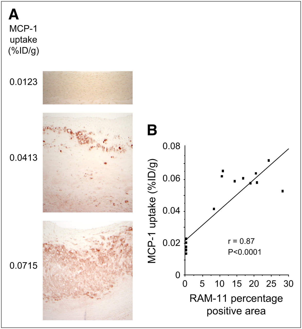
- FIGURE 4.
Extent of MCP-1 uptake and macrophage content in abdominal aorta. (A) Prevalence of macrophages as defined by RAM-11–positive area in neointima (×100) is visibly higher in animal showing high uptake of radiolabeled MCP-1 (bottom). On the other hand, lower RAM-11–positive areas are seen in animal with low quantitative MCP-1 uptake (middle). Control aortic specimens show no RAM 11–positive areas (top). (B) Correlation between MCP-1 uptake and macrophage prevalence shows direct proportional relationship.


Jump to section
Related Articles
Cited By...
- Finding Calcium in Noncalcified Lesions: 18F-Fluoride Offers Insights into Atheroma Evolution
- Atherosclerosis Plaque Heterogeneity and Response to Therapy Detected by In Vivo Molecular Imaging of Matrix Metalloproteinase Activation
- Imaging the Vulnerable Plaque
- Imaging Atherosclerosis and Vulnerable Plaque
- The Year in Molecular Imaging