


Article Figures & Data
Figures
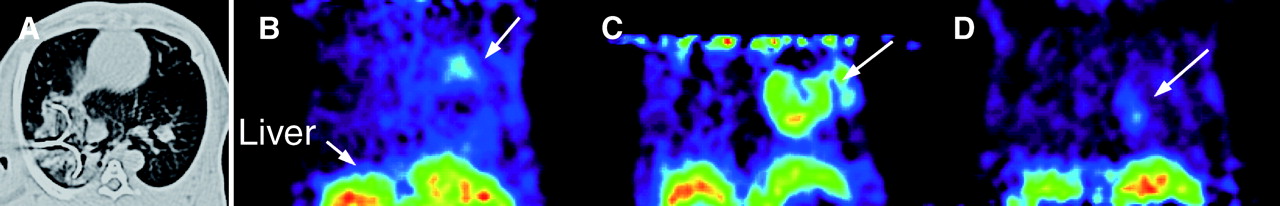
- FIGURE 1.
Follow-up images of normal lung after RFA over time. (A) CT image immediately after RFA shows LeVeen needle positioned in right lower lobe of lung and increased density around needle. (B) Coronal PET image 1 d after RFA shows ring-shaped accumulation of 18F-FDG (arrow) at site of RFA. (C) Coronal PET image 1 wk after RFA shows similar but higher accumulation. (D) Coronal PET image 8 wk after RFA shows reduced 18F-FDG uptake.
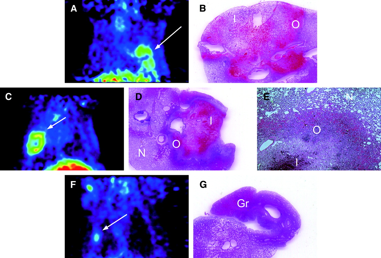
- FIGURE 2.
(A and B) In normal lung 1 d after RFA, coronal PET image (A) shows ring-shaped accumulation of 18F-FDG (arrow) at site of RFA, and histopathologic specimen (B) shows coagulative necrosis in inner zone, relatively fewer morphologic changes in mid zone, congestion and inflammatory cell infiltration in outer zone, and an area of normal lung tissue. Region of 18F-FDG accumulation on PET correlates with outer layer of inflammatory cell infiltration. (C–E) One week after RFA, coronal PET image (C) shows an accumulation similar to but higher than that at 1 d, and histopathologic specimens (normal view [D] and magnified view [×40, E]) show increasing inflammatory cell infiltration and granulomatous changes in outer and inner zones and an area of normal lung tissue. (F and G) Four weeks after RFA, coronal PET image (F) shows reduced 18F-FDG uptake, and histopathologic specimen (G) shows granulomatous tissues, fibrosis, and inner necrotic regions reduced in size. Gr = granulomatous tissues; I = inner zone; N = normal tissue; O = outer zone.
- FIGURE 3.
Graph showing time-dependent changes in 18F-FDG accumulation in normal lungs after RFA, as reflected by changes in early- and delayed-phase RF/M ratios. 18F-FDG uptake was highest at 1 and 2 wk, with a significantly higher RF/M ratio at 1 wk than at 1 d. RF/M ratio was significantly lower after 4 and 8 wk than at 1 wk. These changes corresponded to the histologic changes shown in Figure 2. Up until 2 wk, delayed-phase RF/M ratios remained significantly higher than early-phase RF/M ratios, indicating strong inflammatory responses. After 4 wk, there was no statistical difference between early- and delayed-phase RF/M ratios. †P < 0.05.
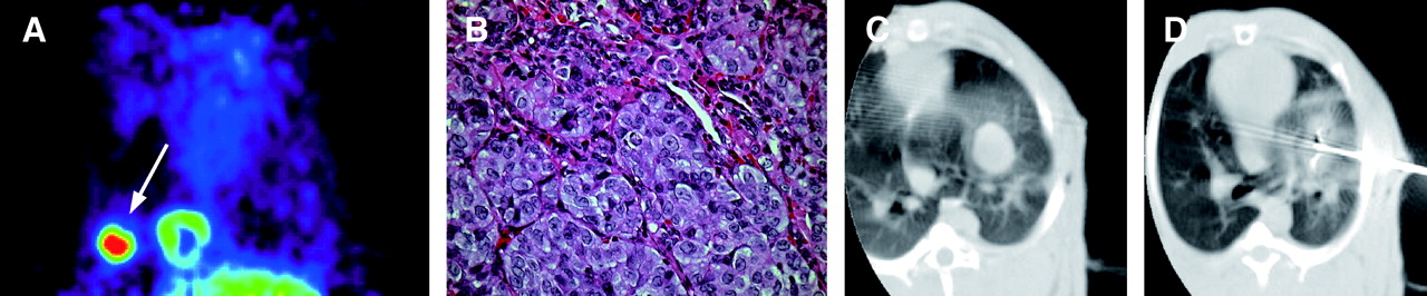
- FIGURE 4.
(A and B) In representative rabbit with untreated VX2, coronal PET image (A) shows extensive 18F-FDG uptake (arrow) in VX2, and histopathologic specimen (×400, B) shows tumor cells with high nuclear-to-cytoplasmic ratio and abnormal nuclear morphology. (C and D) In representative rabbit with RFA-treated VX2, CT image before treatment (C) shows round tumor with clear boundary, and CT image immediately after treatment (D), with LeVeen needle still inserted in tumor, shows increased density around tumor.
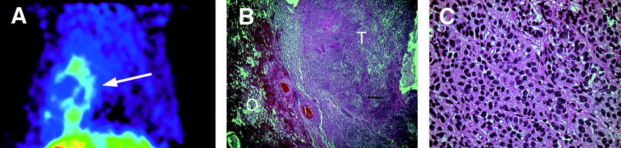
- FIGURE 5.
(A and B) One day after RFA, coronal PET image (A) shows completely ablated VX2 and ring-shaped accumulation of 18F-FDG (arrow) around site of RFA. Histopathologic specimen (×40, B) shows VX2 necrosis and outer-zone tissue congestion and inflammatory cell infiltration. Comparison with PET image indicates that inner necrotic zone corresponds to ablated tumor mass, whereas outer inflammatory layer corresponds to ring-shaped 18F-FDG accumulation. (C) High-power (×400) view of histopathologic specimen shows blurred cytoplasmic borders, pyknotic nuclei, and blurred chromatins—characteristic features of ongoing necrosis. O = outer zone; T = VX2.
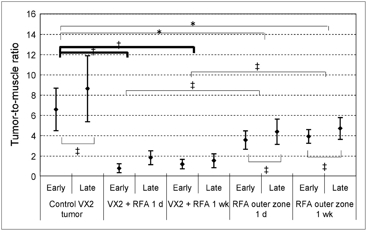
- FIGURE 6.
Graph comparing early- and delayed-phase T/M ratios in treated and untreated VX2s. T/M ratios of RFA-treated VX2s at 1 d and 1 wk are significantly different from those of untreated VX2s on both early-phase and delayed-phase scanning (*P < 0.001). Outer-layer T/M ratios of tissues surrounding treated VX2s at 1 d and 1 wk are significantly different from those of untreated VX2s on both early-phase and delayed-phase scanning (†P < 0.01). Outer-layer T/M ratios of tissues surrounding treated VX2s at 1 d and 1 wk are also significantly different from those of treated VX2s on both early-phase and delayed-phase scanning (‡P < 0.05). In treated VX2s, T/M ratios at early phase do not significantly differ from those at delayed phase (P = 0.57 and 0.48, respectively), but in untreated VX2s and outer layer, which correlated with ring-shaped 18F-FDG accumulation, T/M ratios are higher on delayed-phase images than on early-phase images at both 1 d and 1 wk (‡P < 0.05).


Tables
- TABLE 1
Comparison of Visual and Quantitative Results and Histopathologic Findings at Various Times of Sacrifice
Ratio* Time of sacrifice Visual score Early Delayed Histopathologic findings RFA-treated normal-lung groups 1 d 1.3 ± 0.6 2.9 ± 1.0 3.3 ± 0.8 Inflammation, congestion, and hemorrhage 1 wk 2.2 ± 0.3 4.1 ± 0.6 5.2 ± 0.9 Marked infiltration of neutrophils 2 wk 2.0 ± 0.0 4.1 ± 1.0 5.3 ± 1.5 Inflammatory changes (same as at 1 wk) 4 wk 1.3 ± 0.5 3.1 ± 0.5 3.6 ± 1.1 Fibrosis of granulomatous changes 8 wk 1.0 ± 0.0 1.8 ± 0.1 2.3 ± 0.1 Small granulation; size reduction of necrotic lesions VX2 groups Without RFA 2.9 ± 0.4 6.6 ± 2.1 8.6 ± 3.3 Viable VX2 cells With RFA (ablated tumor), 1 d 0.3 ± 0.5 0.8 ± 0.4 1.1 ± 0.7 Ongoing necrosis of tumor cells With RFA (outer zone), 1 d 1.3 ± 0.5 3.5 ± 0.9 4.4 ± 1.2 Inflammation, congestion, and hemorrhage With RFA (ablated tumor), 1 wk 0.4 ± 0.5 1.2 ± 0.5 1.5 ± 0.7 Ongoing necrosis of tumor cells With RFA (outer zone), 1 wk 1.7 ± 0.5 3.9 ± 0.7 4.7 ± 1.1 Strong infiltration of neutrophils ↵* Data are RF/M for RFA-treated normal-lung groups and T/M for VX2 groups.


Jump to section
Related Articles
Cited By...
- Immediate Postablation 18F-FDG Injection and Corresponding SUV Are Surrogate Biomarkers of Local Tumor Progression After Thermal Ablation of Colorectal Carcinoma Liver Metastases
- 18F-FDG PET/CT Is an Immediate Imaging Biomarker of Treatment Success After Liver Metastasis Ablation
- 18F-FDG PET After Radiofrequency Ablation: Is Timing Everything?