


Article Figures & Data
Figures
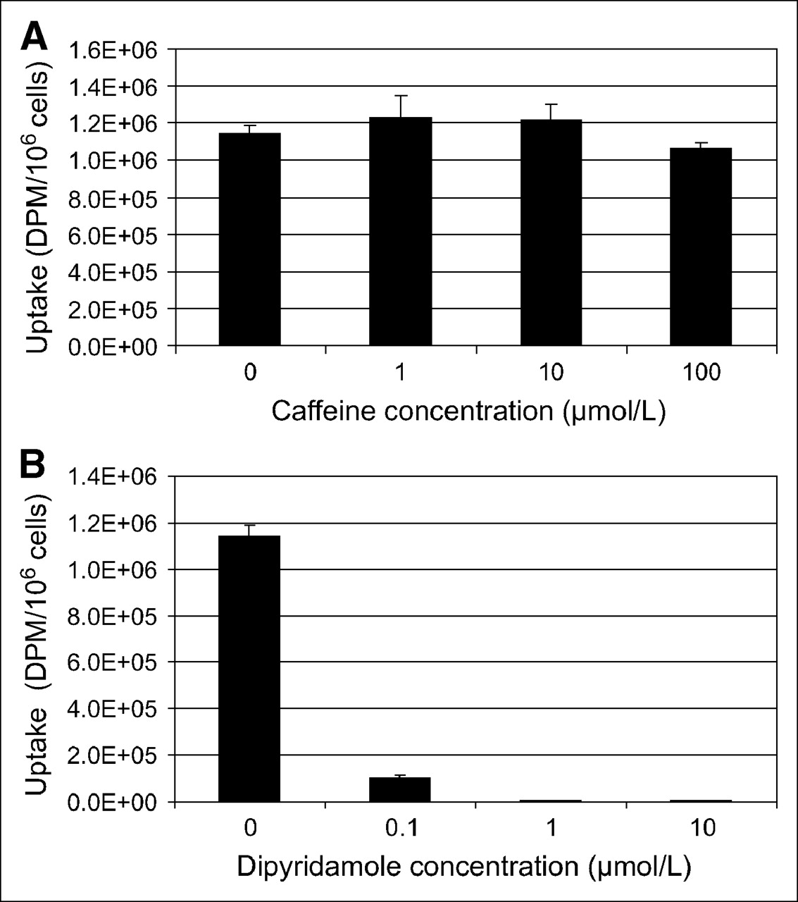
- FIGURE 1.
3H-AMP is accumulated in SKOV-3 cells via nucleoside transporter and not by adenosine receptor binding. Uptake in SKOV-3 cells after exposure to 3H-AMP is not inhibited by caffeine (A), an adenosine receptor antagonist, but is inhibited by dipyridamole (B), an ENT inhibitor, in dose-dependent manner.
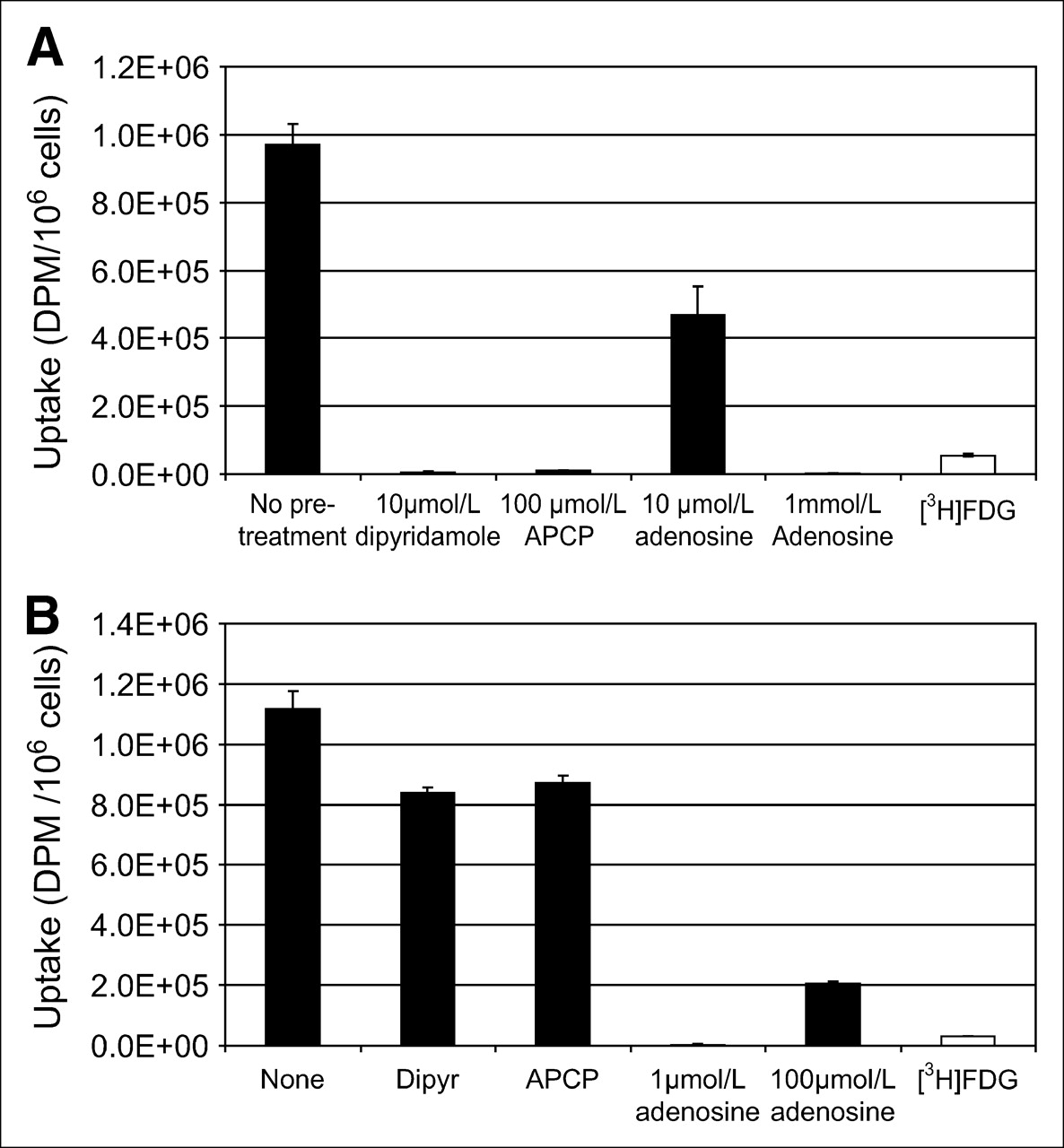
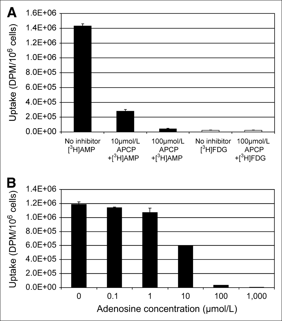
- FIGURE 2.
3H-AMP is converted to 3H-adenosine before tumor cell uptake via nucleoside transporter. (A) Inhibition of CD73 (ecto-5′-nucleotidase) with APCP (α,β-methylene adenosine-5′-diphosphate) blocks uptake of 3H-AMP (black bars), but not same dose of 3H-FDG (white bars), in dose-dependent manner. (B) 3H-AMP uptake in SKOV-3 cells is competitively inhibited by exposure to increasing concentrations of extracellular unlabeled adenosine.
- FIGURE 3.
(A) Significantly more 3H-AMP than 3H-FDG accumulates in U251 human glioblastoma cells (P < 0.05). 3H-AMP is dephosphorylated by CD73, and 3H-adenosine is taken up by ENT nucleoside transporter in U251 cells. Cells were exposed to 3.7 kBq (0.1 μCi) of radiotracer in vitro. (B) Significantly more 3H-AMP than 3H-FDG accumulates in U87 human anaplastic astrocytoma cells (P < 0.05). Dephosphorylation to 3H-adenosine is required, but transport is predominantly via non–dipyridamole-inhibited ENT route. Cells were exposed to 3.7 kBq of radiotracer in vitro. Dipyr = dipyridamole.
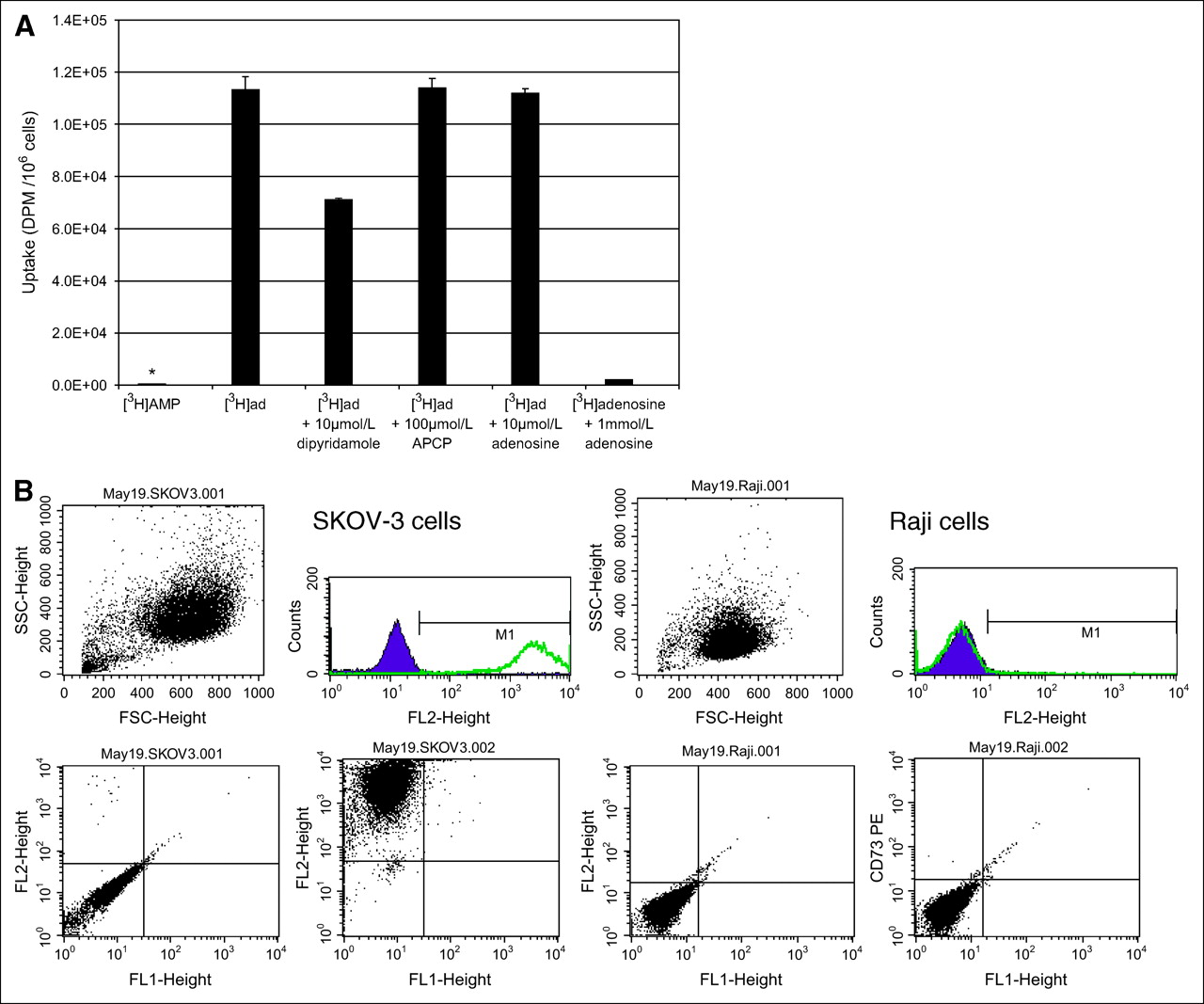
- FIGURE 4.
Low uptake of 3H-AMP by Raji cells because of lack of CD73 expression. (A) In Raji cells, uptake of 3H-AMP (*) is very low and uptake of 3H-adenosine is high (P < 0.05). 3H-Adenosine uptake is only partially inhibited by dipyridamole and is competitively inhibited by unlabeled adenosine. CD73 inhibition by APCP does not affect 3H-adenosine uptake. Cells were exposed to 3.7 kBq of radiotracer in vitro. (Daudi cells also showed similarly low uptake of 3H-AMP and high uptake of 3H-adenosine—data not shown.) (B) Extracellular CD73 is expressed on SKOV-3 cells but not on Raji cells. Viable cells were selected by physical parameters, size, and internal complexity (upper left graphs). Immunophenotyping of these viable cells was compared using cells labeled with phycoerythrin-conjugated CD73 antibody and control cells without antibody labeling. Cell fluorescence intensity is graphed on y-axis and cell physical parameter on x-axis for control cells (lower left graphs) and CD73-labeled cells (lower right graphs). These graphs are also represented as number of cells on y-axis versus fluorescence intensity on x-axis (upper right graphs). Consistent with CD73 expression, intensity of fluorescence is greater on SKOV-3 cells labeled with CD73 than on control cells. However, Raji cells did not show difference in fluorescence intensity between cells labeled with CD73 and control cells. (Daudi cells were also negative for CD73—data not shown.)
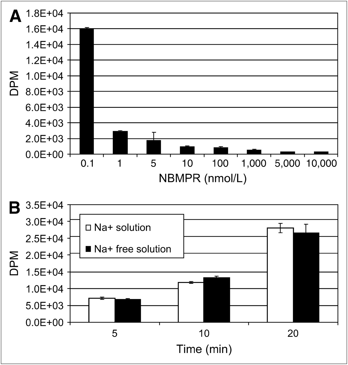
- FIGURE 5.
Nucleoside transporter characterization reveals that SKOV-3 cells possess primarily hENT1s. (A) 3H-Adenosine uptake is inhibited by low nanomolar concentrations of NBMPR in Na+-free physiologic solution. (B) No evidence is seen of concentrative Na+-dependent 3H-adenosine transport in SKOV-3 cells in physiologic or Na+-free physiologic buffer.
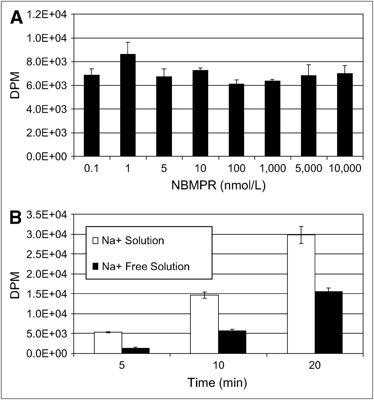
- FIGURE 6.
Nucleoside transporter characterization of U87 cells. U87 cells possess both ENTs and CNTs. ENT appears to be NBMPR resistant. (A) 3H-Adenosine uptake is not inhibited by nanomolar or micromolar concentrations of NBMPR in Na+-free physiologic solution. (B) Evidence is seen of concentrative Na+-dependent 3H-adenosine transport in SKOV-3 cells in physiologic or Na+-free physiologic buffer.
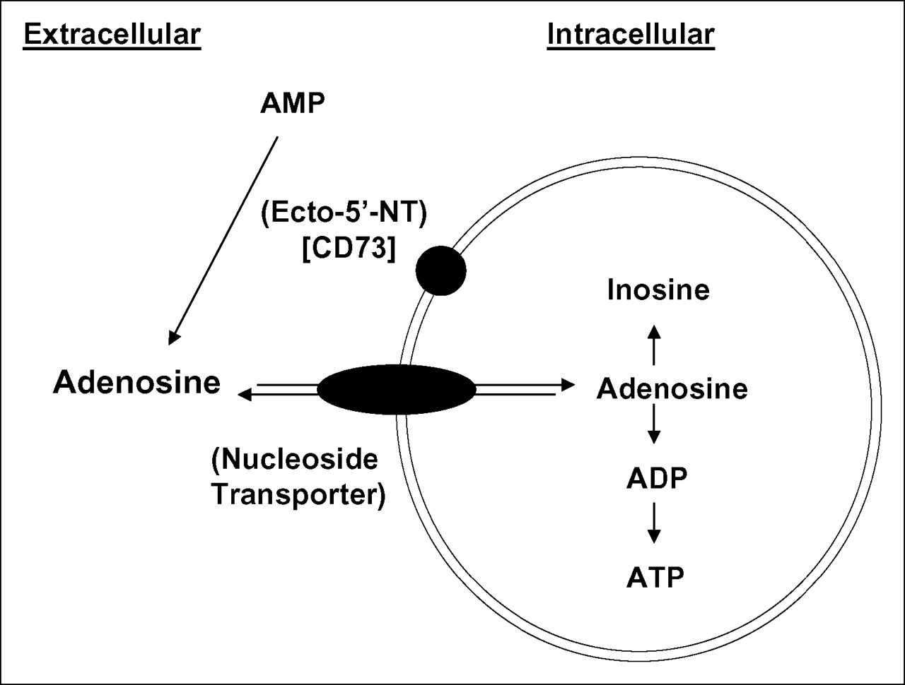
- FIGURE 7.
Schematic diagram of proposed mechanism of intracellular uptake of 3H-AMP based on the in vitro experiments performed in this project.


Tables
- TABLE 1
3H-AMP Retention in SKOV3 Cells After 1 Hour of Uptake Followed by Washout with 3H-AMP–Free Medium
Washout Parameter Uptake (1 h) 30 min 1 h 2 h Average DPM/106 cells ± SD 734,745 ± 13,559 701,518 ± 26,336 584,989 ± 33,927 588,245 ± 10,249 - TABLE 2
High-Performance Liquid Chromatography Determination of Intracellular Adenylate and Inosine Species in SKOV-3 Cells
Species Total intracellular 3H-labeled nucleosides (%) ATP 34.5–38.5 ADP 45.9–52.1 AMP 1.3–1.5 Inosine 6.0–11.7 Data are result of 2 experiments.


