


Article Figures & Data
Figures
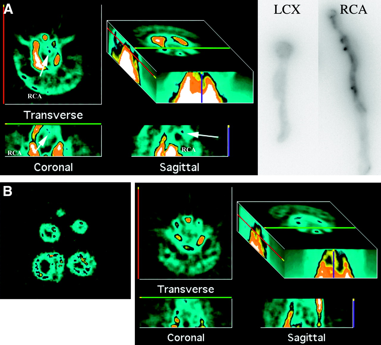
- FIGURE 1.
(A) Example of scan-positive vessel, with in vivo SPECT reconstructions on left and phosphor screen images on right. RCA was injured vessel, and LCX was control vessel. Phosphor screen images show tracer uptake in proximal half of RCA specimen. Transverse, coronal, and sagittal reconstructions of in vivo images show linear uptake of tracer in region of RCA. In 3-dimensional image, upper threshold is turned down to bring out any focal uptake and exaggerate bone and liver activity. (B) Example of scan-negative vessel. Myocardial slices imaged on detector are on left and in vivo SPECT reconstructions on right. Scaling is similar to that in A. No focal uptake of tracer is seen in region of heart.
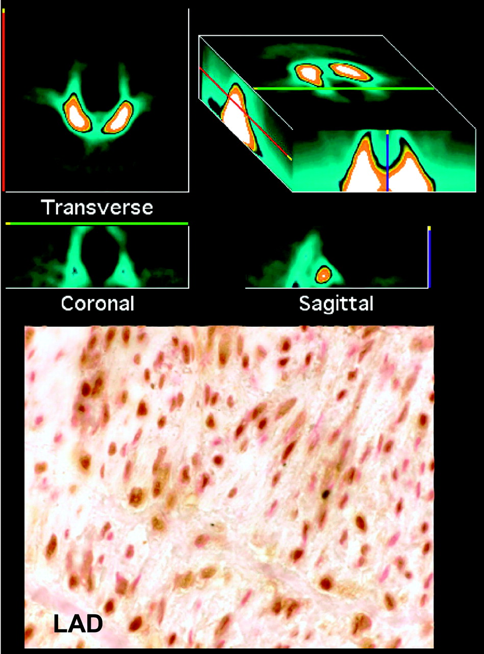
- FIGURE 2.
SPECT reconstructions (left) and ex vivo–imaged myocardial slices (bottom right). This animal had LAD injury and apical infarct. Focal uptake is seen in LAD and apex of left ventricle. Myocardial uptake was confirmed on ex vivo images.
- FIGURE 3.
Photomicrographs of sections of injured coronary artery. Neointimal thickening is seen, comprising predominantly spindle-shaped cells characteristic of class II lesions. Sections are stained with hematoxylin and eosin (H & E) (A), elastic stain (B), and trichrome (C). Vessel was flattened for phosphor screen. IEL = internal elastic membrane; L = lumen.
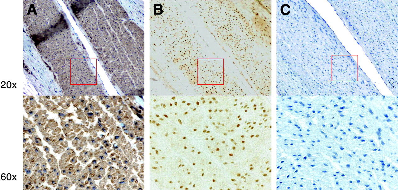
- FIGURE 4.
Photomicrographs of serial sections from injured scan-positive coronary vessel stained for α-actin (A), caspase-3 (B), and macrophages (C). Lesion cells staining positively for caspase-3 (B) were determined to be smooth muscle cells on the basis of brown cytoplasmic staining (A) and negative staining for macrophages (C).
- FIGURE 5.
Photomicrographs of sections stained for caspase-3 (brown nuclear stain) with blue nuclear counterstain. On left is section from scan-positive injured vessel; on right, section from scan-negative injured vessel.
- FIGURE 6.
Animal injected with 99mTc-DTPA (control). SPECT reconstructions are on top, and photomicrograph of section from injured LAD is on bottom. No focal uptake of tracer is seen in region of heart. On immunohistochemical section, caspase chromagen is from a 3,3′-diaminobenzidine substrate kit (Vector Laboratories) and counterstain is nuclear fast red (Vector Laboratories). Section shows abundant brown nuclear and cytoplasmic staining corresponding to high rate of apoptosis in LAD.
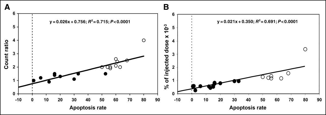
- FIGURE 7.
(A) Plot of count ratio (injured coronary artery to noninjured coronary artery) vs. apoptosis rate as determined from morphometric immunostaining. (B) Plot of tracer uptake vs. apoptosis rate. Closed circles represent experiments that did not show focal uptake in coronary arteries on in vivo SPECT; open circles represent experiments that did show focal uptake.


Tables
Experiment no. Vessel Count ratio Apoptosis rate (%) In vivo scan interpretation 2 LAD 2.6 60 Positive RCA 2.4 61 Positive LCX control Tissue lost Negative 3 LAD Data lost 68 Positive RCA 62 Positive LCX control Tissue lost Negative 4 LAD 2 56 Positive RCA 2 54 Positive LCX control 3 Negative 5 LCX 1.5 33 Negative RCA 1.1 30 Negative LAD control 6 Negative 6 LCX 1.3 20 Negative RCA 2 63 Positive LAD control 8 Negative 7 LCX 0.9 12 Negative RCA 1.5 16 Negative LAD control 6 Negative 8 RCA 2.5 68 Positive LCX control 15 Negative LAD not injected 9 Negative 9 LCX 1.5 52.5 Negative RCA 4 80 Positive LAD 2.7 Tissue lost Positive Distal LAD control 0.7 Negative 10 LCX 2.1 60 Positive RCA 1.2 6 Negative LAD control 2.4 Negative 11 LAD 1.4 16 Negative LCX 1 1 Negative RCA control 1.3 Negative 12 RCA 2 50 Positive LCX 1.9 56 Positive LAD control 2 Negative
In this issue


Jump to section
Related Articles
Cited By...
- Targeted Imaging for Cell Death in Cardiovascular Disorders
- Dual-Energy Computed Tomography Imaging of Atherosclerotic Plaques in a Mouse Model Using a Liposomal-Iodine Nanoparticle Contrast Agent
- Molecular Imaging of Atherosclerosis for Improving Diagnostic and Therapeutic Development
- Imaging Atherosclerosis and Vulnerable Plaque
- Molecular Imaging of Macrophage Cell Death for the Assessment of Plaque Vulnerability
- Clinical Feasibility of Molecular Imaging of Plaque Inflammation in Atherosclerosis
- Targeting of Lectinlike Oxidized Low-Density Lipoprotein Receptor 1 (LOX-1) with 99mTc-Labeled Anti-LOX-1 Antibody: Potential Agent for Imaging of Vulnerable Plaque
- Radionuclide Imaging: A Molecular Key to the Atherosclerotic Plaque
- Broad and Specific Caspase Inhibitor-Induced Acute Repression of Apoptosis in Atherosclerotic Lesions Evaluated by Radiolabeled Annexin A5 Imaging
- Application of 18F-FDG PET for Monitoring the Therapeutic Effect of Antiinflammatory Drugs on Stabilization of Vulnerable Atherosclerotic Plaques
- New Opportunities for Identification and Reduction of Coronary Risk: Treatment of Vulnerable Patients, Arteries, and Plaques