
Main menu

  • Home
  • Content
    • Current
    • Ahead of print
    • Past Issues
    • JNM Supplement
    • SNMMI Annual Meeting Abstracts
    • Continuing Education
    • JNM Podcasts
  • Subscriptions
    • Subscribers
    • Institutional and Non-member
    • Rates
    • Journal Claims
    • Corporate & Special Sales
  • Authors
    • Submit to JNM
    • Information for Authors
    • Assignment of Copyright
    • AQARA requirements
  • Info
    • Reviewers
    • Permissions
    • Advertisers
  • About
    • About Us
    • Editorial Board
    • Contact Information
  • More
    • Alerts
    • Feedback
    • Help
    • SNMMI Journals
  • SNMMI
    • JNM
    • JNMT
    • SNMMI Journals
    • SNMMI

User menu

  • Subscribe
  • My alerts
  • Log in
  • My Cart

Search

  • Advanced search
Journal of Nuclear Medicine
  • SNMMI
    • JNM
    • JNMT
    • SNMMI Journals
    • SNMMI
  • Subscribe
  • My alerts
  • Log in
  • My Cart
Journal of Nuclear Medicine

Advanced Search

  • Home
  • Content
    • Current
    • Ahead of print
    • Past Issues
    • JNM Supplement
    • SNMMI Annual Meeting Abstracts
    • Continuing Education
    • JNM Podcasts
  • Subscriptions
    • Subscribers
    • Institutional and Non-member
    • Rates
    • Journal Claims
    • Corporate & Special Sales
  • Authors
    • Submit to JNM
    • Information for Authors
    • Assignment of Copyright
    • AQARA requirements
  • Info
    • Reviewers
    • Permissions
    • Advertisers
  • About
    • About Us
    • Editorial Board
    • Contact Information
  • More
    • Alerts
    • Feedback
    • Help
    • SNMMI Journals
  • View or Listen to JNM Podcast
  • Visit JNM on Facebook
  • Join JNM on LinkedIn
  • Follow JNM on Twitter
  • Subscribe to our RSS feeds
OtherBasic Science Investigations

Dosimetry and Radiographic Analysis of 131I-Labeled Anti−Tenascin 81C6 Murine Monoclonal Antibody in Newly Diagnosed Patients with Malignant Gliomas: A Phase II Study

Gamal Akabani, David A. Reardon, R. Edward Coleman, Terence Z. Wong, Scott D. Metzler, James E. Bowsher, Daniel P. Barboriak, James M. Provenzale, Kim L. Greer, David DeLong, Henry S. Friedman, Allan H. Friedman, Xiao-Guang Zhao, Charles N. Pegram, Roger E. McLendon, Darell D. Bigner and Michael R. Zalutsky
Journal of Nuclear Medicine June 2005, 46 (6) 1042-1051;
Gamal Akabani
  • Find this author on Google Scholar
  • Find this author on PubMed
  • Search for this author on this site
David A. Reardon
  • Find this author on Google Scholar
  • Find this author on PubMed
  • Search for this author on this site
R. Edward Coleman
  • Find this author on Google Scholar
  • Find this author on PubMed
  • Search for this author on this site
Terence Z. Wong
  • Find this author on Google Scholar
  • Find this author on PubMed
  • Search for this author on this site
Scott D. Metzler
  • Find this author on Google Scholar
  • Find this author on PubMed
  • Search for this author on this site
James E. Bowsher
  • Find this author on Google Scholar
  • Find this author on PubMed
  • Search for this author on this site
Daniel P. Barboriak
  • Find this author on Google Scholar
  • Find this author on PubMed
  • Search for this author on this site
James M. Provenzale
  • Find this author on Google Scholar
  • Find this author on PubMed
  • Search for this author on this site
Kim L. Greer
  • Find this author on Google Scholar
  • Find this author on PubMed
  • Search for this author on this site
David DeLong
  • Find this author on Google Scholar
  • Find this author on PubMed
  • Search for this author on this site
Henry S. Friedman
  • Find this author on Google Scholar
  • Find this author on PubMed
  • Search for this author on this site
Allan H. Friedman
  • Find this author on Google Scholar
  • Find this author on PubMed
  • Search for this author on this site
Xiao-Guang Zhao
  • Find this author on Google Scholar
  • Find this author on PubMed
  • Search for this author on this site
Charles N. Pegram
  • Find this author on Google Scholar
  • Find this author on PubMed
  • Search for this author on this site
Roger E. McLendon
  • Find this author on Google Scholar
  • Find this author on PubMed
  • Search for this author on this site
Darell D. Bigner
  • Find this author on Google Scholar
  • Find this author on PubMed
  • Search for this author on this site
Michael R. Zalutsky
  • Find this author on Google Scholar
  • Find this author on PubMed
  • Search for this author on this site
  • Article
  • Figures & Data
  • Info & Metrics
  • PDF
Loading

Article Figures & Data

Figures

  • Tables
  • FIGURE 1.
    • Download figure
    • Open in new tab
    • Download powerpoint
    FIGURE 1.

    Effect of vasogenic edema in distribution of 131I-mu81C6 mAb in brain parenchyma. MRI (left) and coregistered MRI/SPECT (right) images of patients with vasogenic edema (A–C) and without vasogenic edema (E–G). Average activity ratio between 2-cm cavity margins and SCRC was 0.26 (0.11–0.38) for patients with vasogenic edema, whereas the ratio for patients without vasogenic edema was 0.05 (0.03–0.08). Thus, activity concentrations within regions of vasogenic edema were a factor of 5 higher than in those regions without vasogenic edema. D and H represent a 3-dimensional maximum-intensity-projection view of activity distribution for patients described in C and F, respectively.

  • FIGURE 2.
    • Download figure
    • Open in new tab
    • Download powerpoint
    FIGURE 2.

    (A) Kaplan–Meier survival plot for all patients (n = 33) and for GBM patients (n = 27). Median survival for all patients and GBM patients was 89 wk (95% CI, 61–113 wk) and 79 wk (95% CI, 52–113 wk), respectively. (B) Kaplan–Meier survival plot for GBM patients with reversible or irreversible neurotoxicity. Median survival for GBM patients who received XRT with reversible (n = 14) and irreversible neurotoxicity (n = 10) was 98 wk (95% CI, 39–126 wk) and 73 wk (95% CI, 47–116 wk), respectively.

  • FIGURE 3.
    • Download figure
    • Open in new tab
    • Download powerpoint
    FIGURE 3.

    Sequential registered MRI/PET images from patient 15, who received 35 Gy to 2-cm cavity margins with an initial dose rate of 0.9 Gy h−1. A uniform rim in MRI was observed 1 mo after therapy and persisted unchanged over 59 wk with minimal vasogenic edema and minimal 18F-FDG accumulation. Subsequently, grade III irreversible neurologic toxicity developed and, 118 wk after therapy, a biopsy sample was obtained, which indicated radionecrosis. However, 177 wk after therapy, new intense 18F-FDG hypermetabolic nodularities and MRI enhancements with extensive vasogenic edema were observed on registered MRI/PET images, indicative of tumor progression.
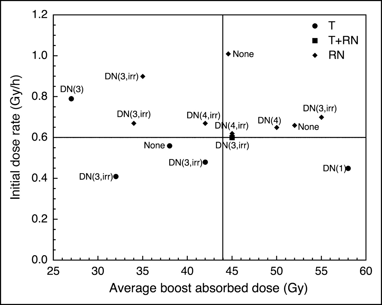
  • FIGURE 4.
    • Download figure
    • Open in new tab
    • Download powerpoint
    FIGURE 4.

    Scatter plot of biopsy results and neurologic toxicity among GBM tumor patients as function of average absorbed dose DCM and maximum dose rate ḊCMmax to 2-cm cavity margins. T = tumor; RN = radionecrosis; DN = delayed neurotoxicity and grade 1, 2, 3, or 4; irr = irreversible.
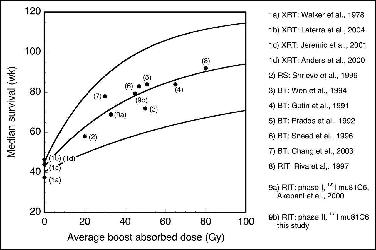
  • FIGURE 5.
    • Download figure
    • Open in new tab
    • Download powerpoint
    FIGURE 5.

    Regression analysis (95% CI) between average boost absorbed dose and median survival for newly diagnosed GBM patients receiving XRT and radiosurgery, brachytherapy, or RIT. Among the different clinical studies, including ours, an asymptotic increase in median survival as a function of average boost absorbed dose after XRT was observed. Dose estimates for Riva et al. (35) were based on an average residence time of 82 h, average cavity size of 19 cm3, and 5 courses of 131I-BC2 and BC4 mAbs for a total administered activity of 9,250 MBq. This relationship among studies confirms the relevance of limiting the absorbed dose to tumor foci to minimize normal brain tissue injury and maximize tumor control. Reoperation rates were between 40% and 60% among brachytherapy studies, whereas rate for RIT using 131I-mu81C6 mAb was 3%. RS = radiosurgery; BT = brachytherapy.

Tables

  • Figures
    • View popup
    TABLE 1

    Absorbed Dose Estimates, Survival, Neurologic and Hematologic Toxicity, and Biopsy Results for 33 Patients with Newly Diagnosed Gliomas Treated with 131I-mu81C6 mAb

    Patient no.DxSex and raceAge (y)Weight (kg)KPSSCRC volume (cm3)VEAdm. activity (MBq)ḊCMMax (Gy/h)DCM (Gy)DCMXRT (Gy)DCMTotal (Gy)DWB (cGy)DBM (cGy)Survival (wk)ToxicityBiopsy
    NTHT
    1GBMMW449210012.5−4,4400.564563108468479S (3)3
    2GBMMW47831008.6−4,4400.665263115468187S (2)3
    3GBMMW38818013.8−4,4400.5443601037799414
    4GBMFW566810023.3−4,4400.43345993638561D (4, irr)3
    5GBMMW43779026.4−4,4400.413259927696116D (3, irr)4T
    6GBMFB4879708.5−4,4400.665259111811003044R, R, T
    7GBMFW39661007.2−4,4400.7055581136585148D (3, irr)3R
    8AAMW398110013.8−4,4000.522960896283291*3
    9GBMFW5872802.1−4,4401.1373—734372393
    10GBMFW35611008.7−4,4400.6550591107989268*D (4)4R
    11AAMW198810012.9−4,4400.5638641024881893T
    12GBMMW36981005.5−4,4400.7740599968941134
    13GBMMW538810020.5−4,4400.455860118539995D (1)4T
    14GBMMW51771002.9−4,4401.01456110677881234R, R, T
    15GBMMW55841003.8−4,4400.9035751104068172D (3, irr)4R
    16GBMMW60751002.2−4,4401.124665111376435D (3)3
    17GBMMW509710017.9+4,4400.484260102257355D (3, irr)3T
    18AAMW38691009.4−4,4400.632561866978219D (2)3
    19GBMMW497610010.9−4,4400.604559105558352D (3, irr)3T + R
    20AOMW68811005.5+4,4400.775064114568395D (4, irr)3
    21AAMW46811005.2−4,4400.79276390748558D (3)4T
    22GBMFW47711008.0−4,4400.674260102567877D (4, irr)3R, T
    23GBMFW597110010.0−4,4400.6245601059410165D (4, irr)4R
    24GBMMW60721008.1+4,4400.67346094487173D (3, irr)3R
    25GBMMW388310016.3−4,4400.50346094841011443
    26GBMMW5510510016.3+4,4400.5039609951911263
    27AOFW355410012.2−4,4400.5751—519696230*D (2)3
    28GBMMW6370801.2+4,4401.35116—116447824A, S, D (3, irr)3
    29GBMMW53901000.5+4,4401.80113—113749797A, S, D (3, irr)4T
    30GBMFW657010012.1−4,4400.5777601376710247D (4, irr)4
    31GBMMW541081004.3+1,3690.2642601021434503
    32GBMMW527210030.5+4,4400.386560125671121094
    33GBMFW547610013.0+4,4400.562760875275273
    • ↵* Censored (alive).

    • Dx = diagnosis; VE = vasogenic edema; Adm. activity = administered activity; ḊCMMax = maximum initial dose rate; DCM = dose from RIT; DCMXRT = dose from XRT; DCMTotal = total dose from RIT and XRT; DWB = WB dose; DBM = bone marrow dose; NT = neurotoxicity; HT = hematologic toxicity (grade: 1, 2, 3, or 4); S = subacute; D = delayed (grade: 1, 2, 3, or 4; irr = irreversible); T = tumor; R = radionecrosis; A = acute.

PreviousNext
Back to top

In this issue

Journal of Nuclear Medicine: 46 (6)
Journal of Nuclear Medicine
Vol. 46, Issue 6
June 1, 2005
  • Table of Contents
  • About the Cover
  • Index by author
Print
Download PDF
Article Alerts
Sign In to Email Alerts with your Email Address
Email Article

Thank you for your interest in spreading the word on Journal of Nuclear Medicine.

NOTE: We only request your email address so that the person you are recommending the page to knows that you wanted them to see it, and that it is not junk mail. We do not capture any email address.

Enter multiple addresses on separate lines or separate them with commas.
Dosimetry and Radiographic Analysis of 131I-Labeled Anti−Tenascin 81C6 Murine Monoclonal Antibody in Newly Diagnosed Patients with Malignant Gliomas: A Phase II Study
(Your Name) has sent you a message from Journal of Nuclear Medicine
(Your Name) thought you would like to see the Journal of Nuclear Medicine web site.
Citation Tools
Dosimetry and Radiographic Analysis of 131I-Labeled Anti−Tenascin 81C6 Murine Monoclonal Antibody in Newly Diagnosed Patients with Malignant Gliomas: A Phase II Study
Gamal Akabani, David A. Reardon, R. Edward Coleman, Terence Z. Wong, Scott D. Metzler, James E. Bowsher, Daniel P. Barboriak, James M. Provenzale, Kim L. Greer, David DeLong, Henry S. Friedman, Allan H. Friedman, Xiao-Guang Zhao, Charles N. Pegram, Roger E. McLendon, Darell D. Bigner, Michael R. Zalutsky
Journal of Nuclear Medicine Jun 2005, 46 (6) 1042-1051;

Citation Manager Formats

  • BibTeX
  • Bookends
  • EasyBib
  • EndNote (tagged)
  • EndNote 8 (xml)
  • Medlars
  • Mendeley
  • Papers
  • RefWorks Tagged
  • Ref Manager
  • RIS
  • Zotero
Share
Dosimetry and Radiographic Analysis of 131I-Labeled Anti−Tenascin 81C6 Murine Monoclonal Antibody in Newly Diagnosed Patients with Malignant Gliomas: A Phase II Study
Gamal Akabani, David A. Reardon, R. Edward Coleman, Terence Z. Wong, Scott D. Metzler, James E. Bowsher, Daniel P. Barboriak, James M. Provenzale, Kim L. Greer, David DeLong, Henry S. Friedman, Allan H. Friedman, Xiao-Guang Zhao, Charles N. Pegram, Roger E. McLendon, Darell D. Bigner, Michael R. Zalutsky
Journal of Nuclear Medicine Jun 2005, 46 (6) 1042-1051;
Twitter logo Facebook logo LinkedIn logo Mendeley logo
  • Tweet Widget
  • Facebook Like
  • Google Plus One
Bookmark this article

Jump to section

  • Article
    • Abstract
    • METHODS AND MATERIALS
    • RESULTS
    • DISCUSSION
    • CONCLUSION
    • Footnotes
    • REFERENCES
  • Figures & Data
  • Info & Metrics
  • PDF

Related Articles

  • THIS MONTH IN JNM
  • PubMed
  • Google Scholar

Cited By...

  • PL1 Peptide Engages Acidic Surfaces on Tumor-Associated Fibronectin and Tenascin Isoforms to Trigger Cellular Uptake
  • Biodistribution and Dosimetry of Intraventricularly Administered 124I-Omburtamab in Patients with Metastatic Leptomeningeal Tumors
  • Development of a Function-Blocking Antibody Against Fibulin-3 as a Targeted Reagent for Glioblastoma
  • Clinical Experience with {alpha}-Particle Emitting 211At: Treatment of Recurrent Brain Tumor Patients with 211At-Labeled Chimeric Antitenascin Monoclonal Antibody 81C6
  • Tenascin-C Stimulates Glioma Cell Invasion through Matrix Metalloproteinase-12
  • Phase I Single-Dose Study of Intracavitary-Administered Iodine-131-TM-601 in Adults With Recurrent High-Grade Glioma
  • Novel Human IgG2b/Murine Chimeric Antitenascin Monoclonal Antibody Construct Radiolabeled with 131I and Administered into the Surgically Created Resection Cavity of Patients with Malignant Glioma: Phase I Trial Results
  • Google Scholar

More in this TOC Section

  • Counting Rate Characteristics and Image Distortion in Preclinical PET Imaging During Radiopharmaceutical Therapy
  • Design and Fabrication of Kidney Phantoms for Internal Radiation Dosimetry Using 3D Printing Technology
  • Synthesis and Biologic Evaluation of Novel 18F-Labeled Probes Targeting Prostate-Specific Membrane Antigen for PET of Prostate Cancer
Show more Basic Science Investigations

Similar Articles

SNMMI

© 2025 SNMMI

Powered by HighWire