


Article Figures & Data
Figures
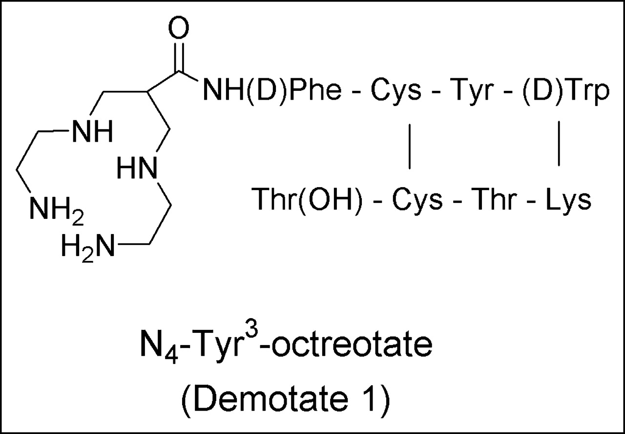
- FIGURE 1.
Structure of Demotate 1.
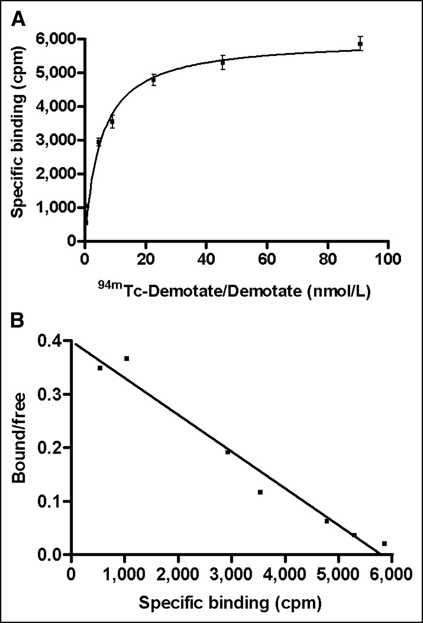
- FIGURE 2.
Representative plot of 94mTc-Demotate 1–Demotate 1 saturation binding curve (A) and Scatchard transformation (B) for membrane preparations from A-427 cells infected with AdHASSTR2 at 10 pfu per cell. It should be noted that 94mTc-Demotate 1 concentrations included unlabeled Demotate 1 and that saturation binding curve represented specific binding (nonspecifically bound subtracted from total bound). Each data point represents mean ± SEM of triplicate measurements.
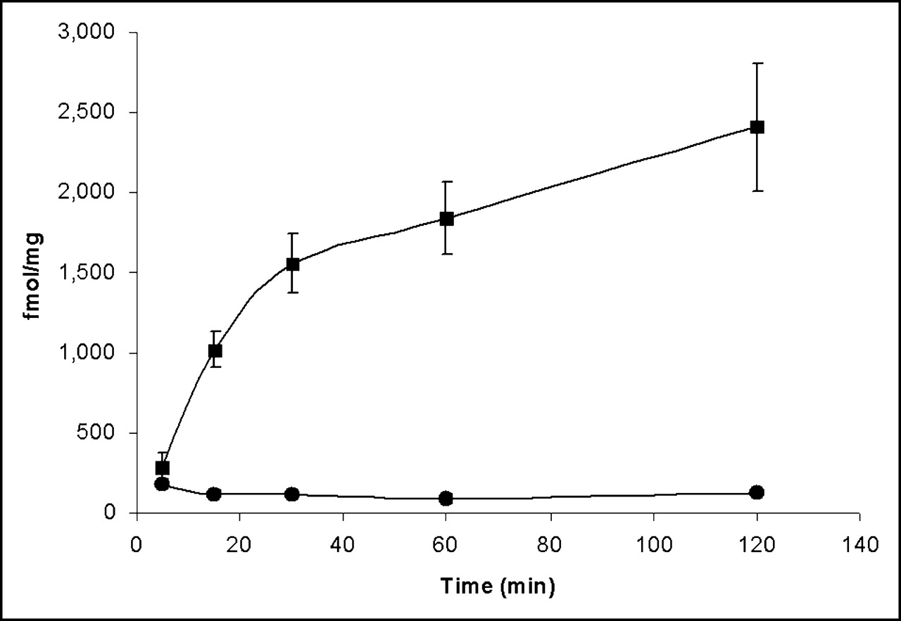
- FIGURE 3.
Specific internalization at 37°C of 94mTc-Demotate 1 into A-427 cells infected with AdHASSTR2 at 10 pfu per cell. 94mTc-Demotate 1 (1 nmol/L) was incubated with cells for various times in presence or absence of inhibitor. Cells were acid washed to remove surface-bound radioactivity and then were harvested to determine internalized radioactivity. Specific internalized radioactivity (internalized with inhibitor subtracted from internalized without inhibitor) (▪) and specific surface-bound radioactivity (surface bound with inhibitor subtracted from surface bound without inhibitor) (•) are shown. Data for each time point are presented as mean ± SEM of 2–5 experiments each performed in triplicate.
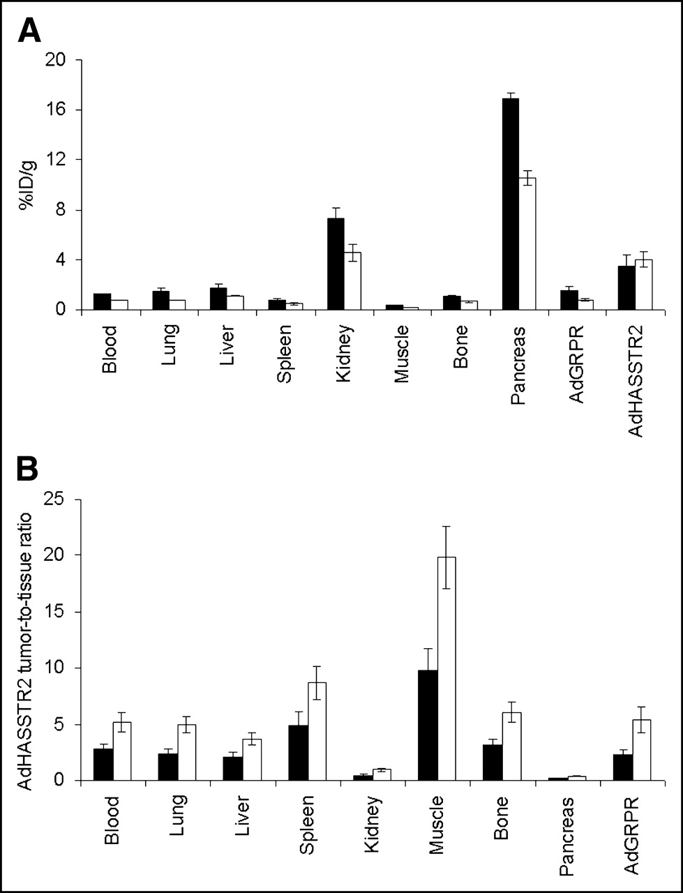
- FIGURE 4.
Biodistribution of 94mTc-Demotate 1 in mice bearing A-427 tumor xenografts. Tumors were injected directly with either AdHASSTR2 or AdGRPR as control, and 94mTc-Demotate 1 was injected via tail vein 2 d later. Mice were sacrificed 1 h (▪) and 2 h (□) later (n = 6 for each group). Data are presented as mean ± SEM %ID/g for each type of tissue (A) and as ratio of AdHASSTR2-injected tumor uptake to tissue uptake at both time points (B).
- FIGURE 5.
(A and B) Coronal (A) and transaxial (B) microPET projection images of A-427 tumor–bearing mice at 1 h after injection of 94mTc-Demotate 1. Mice carried axillary tumors in which left tumor was injected directly with AdHASSTR2 and right tumor was injected with AdGRPR. Coronal image shows uptake of 94mTc-Demotate 1 in AdHASSTR2-injected tumor and clearance through kidneys and bladder but background uptake in AdGRPR-injected tumor. (C) SUVs obtained from microPET images for AdGRPR-injected tumors, AdHASSTR2-injected tumors, and kidneys in 3 mice at 1 h (▪) and 2 h (□). (D) Ratios of uptake in AdHASSTR2-injected tumors to that in AdGRPR-injected tumors and of uptake in AdHASSTR2-injected tumors to that in kidneys, as determined by SUV analysis (red bars; n = 3) and biodistribution analysis (blue bars; n = 11 for 1 h and n = 6 for 2 h). Data in C and D are presented as mean ± SEM.


Tables
- TABLE 1
Biodistribution (n = 5) of 94mTc-Demotate 1 in Mice Bearing A-427 Tumor Xenografts Directly Injected with AdHASSTR2 or AdGRPR*
Tissue Mean ± SEM %ID/g for: Unblocked samples Blocked samples Blood 0.37 ± 0.05 0.41 ± 0.09 Lungs 1.16 ± 0.27 0.59 ± 0.08 Liver 1.37 ± 0.23 0.64 ± 0.06† Spleen 0.56 ± 0.16 0.24 ± 0.02 Kidneys 9.43 ± 1.13 5.90 ± 0.97† Muscle 0.27 ± 0.08 0.33 ± 0.09 Bone 1.09 ± 0.34 0.30 ± 0.04 Pancreas 21.31 ± 1.53 0.22 ± 0.03† AdGRPR-injected tumors 0.82 ± 0.06 1.33 ± 0.18 AdHASSTR2-injected tumors 5.39 ± 0.84 1.56 ± 0.26†
In this issue


Jump to section
Related Articles
Cited By...
- Noninvasive Molecular Imaging Using Reporter Genes
- Imaging Expression of the Human Somatostatin Receptor Subtype-2 Reporter Gene with 68Ga-DOTATOC
- Multimodality Imaging of Gene Transfer with a Receptor-Based Reporter Gene
- Titration of Variant HSV1-tk Gene Expression to Determine the Sensitivity of 18F-FHBG PET Imaging in a Prostate Tumor