


Article Figures & Data
Figures
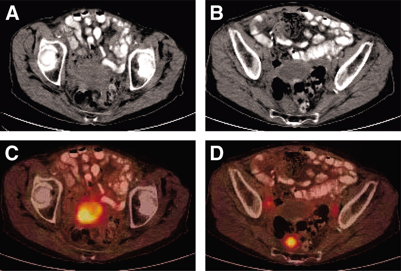
- FIGURE 1.
Images of 84-y-old woman with cervical cancer who was referred for evaluation of recurrent disease and for restaging. (A and B) CT shows mass in pelvis and enlarged right iliac node. (C and D) 18F-FDG PET/CT shows uptake in lower pelvic mass (SUV, 9.2), in both pelvic lymph nodes (SUV, 4.5 on right and 3.2 on left), and as a single focus in right presacral region corresponding to soft-tissue nodule (SUV, 6.8).
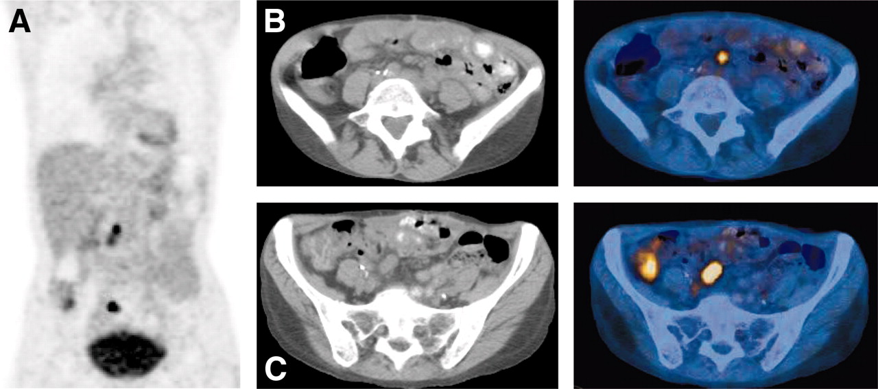
- FIGURE 2.
Images of 62-y-old woman with ovarian cancer who had a rising level of CA-125 and was being evaluated for recurrent disease. (A) Maximum-intensity projection coronal 18F-FDG PET/CT image (A) and transaxial CT images at 2 levels (B and C) show active disease in paraaortic and pelvic nodes (SUV, 4.3 and 5.8) and mesenteric disease.
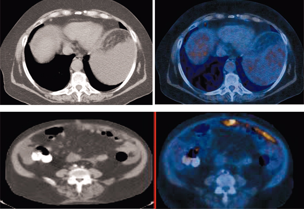
- FIGURE 3.
Images of 72-y-old woman with endometrial carcinoma who was being evaluated for recurrent disease. CT scans (left) show questionable node in supradiaphragmatic node. PET scans (right) show increased 18F-FDG uptake in small right supradiaphragmatic lymph node (SUV, 3.5) and in omentum (SUV, 7.1).
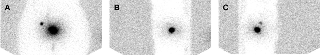
- FIGURE 4.
Images of 52-y-old woman with stage IB1 cervical carcinoma who was referred for sentinel node mapping. Anterior (A), left lateral (B), and right lateral (C) images show uptake in injection site in cervix and focal uptake in right pelvis consistent with sentinel node in iliac region.


