
Main menu

  • Home
  • Content
    • Current
    • Ahead of print
    • Past Issues
    • JNM Supplement
    • SNMMI Annual Meeting Abstracts
    • Continuing Education
    • JNM Podcasts
  • Subscriptions
    • Subscribers
    • Institutional and Non-member
    • Rates
    • Journal Claims
    • Corporate & Special Sales
  • Authors
    • Submit to JNM
    • Information for Authors
    • Assignment of Copyright
    • AQARA requirements
  • Info
    • Reviewers
    • Permissions
    • Advertisers
  • About
    • About Us
    • Editorial Board
    • Contact Information
  • More
    • Alerts
    • Feedback
    • Help
    • SNMMI Journals
  • SNMMI
    • JNM
    • JNMT
    • SNMMI Journals
    • SNMMI

User menu

  • Subscribe
  • My alerts
  • Log in
  • My Cart

Search

  • Advanced search
Journal of Nuclear Medicine
  • SNMMI
    • JNM
    • JNMT
    • SNMMI Journals
    • SNMMI
  • Subscribe
  • My alerts
  • Log in
  • My Cart
Journal of Nuclear Medicine

Advanced Search

  • Home
  • Content
    • Current
    • Ahead of print
    • Past Issues
    • JNM Supplement
    • SNMMI Annual Meeting Abstracts
    • Continuing Education
    • JNM Podcasts
  • Subscriptions
    • Subscribers
    • Institutional and Non-member
    • Rates
    • Journal Claims
    • Corporate & Special Sales
  • Authors
    • Submit to JNM
    • Information for Authors
    • Assignment of Copyright
    • AQARA requirements
  • Info
    • Reviewers
    • Permissions
    • Advertisers
  • About
    • About Us
    • Editorial Board
    • Contact Information
  • More
    • Alerts
    • Feedback
    • Help
    • SNMMI Journals
  • View or Listen to JNM Podcast
  • Visit JNM on Facebook
  • Join JNM on LinkedIn
  • Follow JNM on Twitter
  • Subscribe to our RSS feeds
OtherBasic Science Investigations

Labeling of Cerebral Amyloid β Deposits In Vivo Using Intranasal Basic Fibroblast Growth Factor and Serum Amyloid P Component in Mice

Jiong Shi, George Perry, Marc S. Berridge, Gjumrakch Aliev, Sandy L. Siedlak, Mark A. Smith, Joseph C. LaManna and Robert P. Friedland
Journal of Nuclear Medicine August 2002, 43 (8) 1044-1051;
Jiong Shi
  • Find this author on Google Scholar
  • Find this author on PubMed
  • Search for this author on this site
George Perry
  • Find this author on Google Scholar
  • Find this author on PubMed
  • Search for this author on this site
Marc S. Berridge
  • Find this author on Google Scholar
  • Find this author on PubMed
  • Search for this author on this site
Gjumrakch Aliev
  • Find this author on Google Scholar
  • Find this author on PubMed
  • Search for this author on this site
Sandy L. Siedlak
  • Find this author on Google Scholar
  • Find this author on PubMed
  • Search for this author on this site
Mark A. Smith
  • Find this author on Google Scholar
  • Find this author on PubMed
  • Search for this author on this site
Joseph C. LaManna
  • Find this author on Google Scholar
  • Find this author on PubMed
  • Search for this author on this site
Robert P. Friedland
  • Find this author on Google Scholar
  • Find this author on PubMed
  • Search for this author on this site
  • Article
  • Figures & Data
  • Info & Metrics
  • PDF
Loading

Article Figures & Data

Figures

  • FIGURE 1.
    • Download figure
    • Open in new tab
    • Download powerpoint
    FIGURE 1.

    Biodistribution of 125I-bFGF in 2 groups of experimental animals, Tg(+) and control, given intranasal injection of 125I-bFGF (33.3 MBq/μL per 10 g of body weight). (A) Brain distribution of 125I-bFGF is depicted as mean ± SD (n = 5 in each group). *P < 0.05 vs. control. (B) Whole-body distribution of 125I-bFGF is depicted as mean ± SEM (n = 5 in each group). When SEM is not depicted, it was too small to be shown.

  • FIGURE 2.
    • Download figure
    • Open in new tab
    • Download powerpoint
    FIGURE 2.

    Adjacent serial sections indicate that immunostaining with antisera to AβPP shows localization of amyloid plaques (A) and that intranasally injected SAP binds to amyloid plaques in Tg mouse cortex as detected by antibody to SAP (B). (D) Brain sections from Tg mice receiving bovine serum albumin plus vehicle immunostained for SAP show no reaction in amyloid plaques, although many plaques are seen on adjacent serial sections stained for AβPP (C). Scale bar = 50 μm. Arrows mark same plaques in serial sections (A and B) and (C and D).
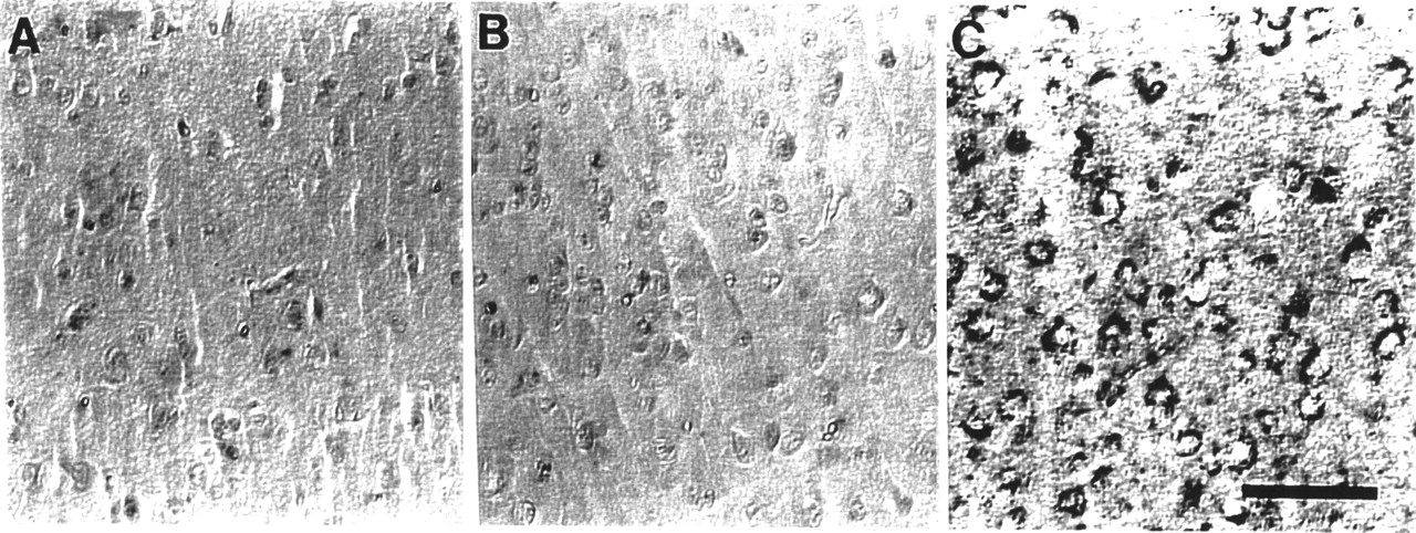
  • FIGURE 3.
    • Download figure
    • Open in new tab
    • Download powerpoint
    FIGURE 3.

    Light microscopic study of bFGF localization in AβPP Tg mouse cortex, with and without intranasal bFGF injection. bFGF is not readily detectable in neurons in Tg(−) mice after receiving bFGF intranasally (A) or in Tg(+) mice without bFGF injection (B). (C) Nasally injected bFGF bound to cytoplasm of neurons around Aβ deposits in frontal, parietal, and occipital regions in Tg(+) mice. Scale bar = 50 μm.

  • FIGURE 4.
    • Download figure
    • Open in new tab
    • Download powerpoint
    FIGURE 4.

    Electron microscopy immunogold staining in neuritic plaques in Tg(+) mouse cortex. (A) Tg(+) mice with intravenous injection of bFGF do not show presence of bFGF in brain (original magnification, ×8,000). (B) Same area as in A under higher magnification (×20,000). (C) Representative electron micrograph shows that peripheral margins of neuritic plaques from animals that received bFGF intranasally contain bFGF immunogold reactivity (original magnification, ×50,000). (D) bFGF is also associated with amyloid fibrils in plaque after intranasal injection (original magnification, ×50,000). Arrows indicate bFGF gold labeling. Scale bars = 10 nm.

PreviousNext
Back to top

In this issue

Journal of Nuclear Medicine: 43 (8)
Journal of Nuclear Medicine
Vol. 43, Issue 8
August 1, 2002
  • Table of Contents
  • Index by author
Print
Download PDF
Article Alerts
Sign In to Email Alerts with your Email Address
Email Article

Thank you for your interest in spreading the word on Journal of Nuclear Medicine.

NOTE: We only request your email address so that the person you are recommending the page to knows that you wanted them to see it, and that it is not junk mail. We do not capture any email address.

Enter multiple addresses on separate lines or separate them with commas.
Labeling of Cerebral Amyloid β Deposits In Vivo Using Intranasal Basic Fibroblast Growth Factor and Serum Amyloid P Component in Mice
(Your Name) has sent you a message from Journal of Nuclear Medicine
(Your Name) thought you would like to see the Journal of Nuclear Medicine web site.
Citation Tools
Labeling of Cerebral Amyloid β Deposits In Vivo Using Intranasal Basic Fibroblast Growth Factor and Serum Amyloid P Component in Mice
Jiong Shi, George Perry, Marc S. Berridge, Gjumrakch Aliev, Sandy L. Siedlak, Mark A. Smith, Joseph C. LaManna, Robert P. Friedland
Journal of Nuclear Medicine Aug 2002, 43 (8) 1044-1051;

Citation Manager Formats

  • BibTeX
  • Bookends
  • EasyBib
  • EndNote (tagged)
  • EndNote 8 (xml)
  • Medlars
  • Mendeley
  • Papers
  • RefWorks Tagged
  • Ref Manager
  • RIS
  • Zotero
Share
Labeling of Cerebral Amyloid β Deposits In Vivo Using Intranasal Basic Fibroblast Growth Factor and Serum Amyloid P Component in Mice
Jiong Shi, George Perry, Marc S. Berridge, Gjumrakch Aliev, Sandy L. Siedlak, Mark A. Smith, Joseph C. LaManna, Robert P. Friedland
Journal of Nuclear Medicine Aug 2002, 43 (8) 1044-1051;
Twitter logo Facebook logo LinkedIn logo Mendeley logo
  • Tweet Widget
  • Facebook Like
  • Google Plus One
Bookmark this article

Jump to section

  • Article
    • Abstract
    • MATERIALS AND METHODS
    • RESULTS
    • DISCUSSION
    • CONCLUSION
    • Acknowledgments
    • Footnotes
    • REFERENCES
  • Figures & Data
  • Info & Metrics
  • PDF

Related Articles

  • Erratum
  • PubMed
  • Google Scholar

Cited By...

  • S100A6 Amyloid Fibril Formation Is Calcium-modulated and Enhances Superoxide Dismutase-1 (SOD1) Aggregation
  • Molecular Targeting with Radionuclides: State of the Science
  • Potential for a Specific Neuroradiologic Diagnosis of Alzheimer's Disease
  • Google Scholar

More in this TOC Section

  • Tumor-Specific Binding of Radiolabeled PEGylated GIRLRG Peptide: A Novel Agent for Targeting Cancers
  • PET/MRI of Hypoxic Atherosclerosis Using 64Cu-ATSM in a Rabbit Model
  • Tumor Uptake of Anti-CD20 Fabs Depends on Tumor Perfusion
Show more Basic Science Investigations

Similar Articles

SNMMI

© 2025 SNMMI

Powered by HighWire