


Article Figures & Data
Figures
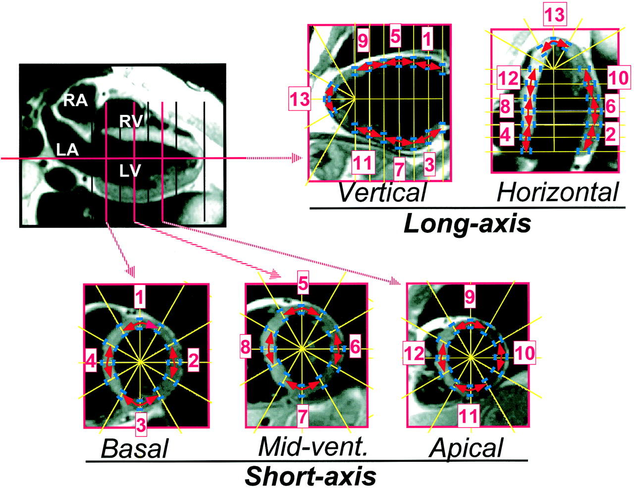
- FIGURE 1.
Example of MRI slices acquired at end-diastole for healthy volunteer with application of point-positioning software. Epicardial and endocardial borders of left ventricular walls are directed (blue marks) along radial and parallel lines (yellow lines), midwall points being then automatically positioned. For each segment, wall curvature (schematized by red arrows) was determined using coordinates of 3 adjacent midwall points on both orthogonal slices that were used to analyze segment. The 13 left ventricular segments are anterobasal (1), laterobasal (2), inferobasal (3), septobasal (4), anteromedial (5), lateromedial (6), inferomedial (7), septomedial (8), anteroapical (9), lateroapical (10), inferoapical (11), septoapical (12), and apical (13). LA = left atrium; LV = left ventricle; RA = right atrium; RV = right ventricle.
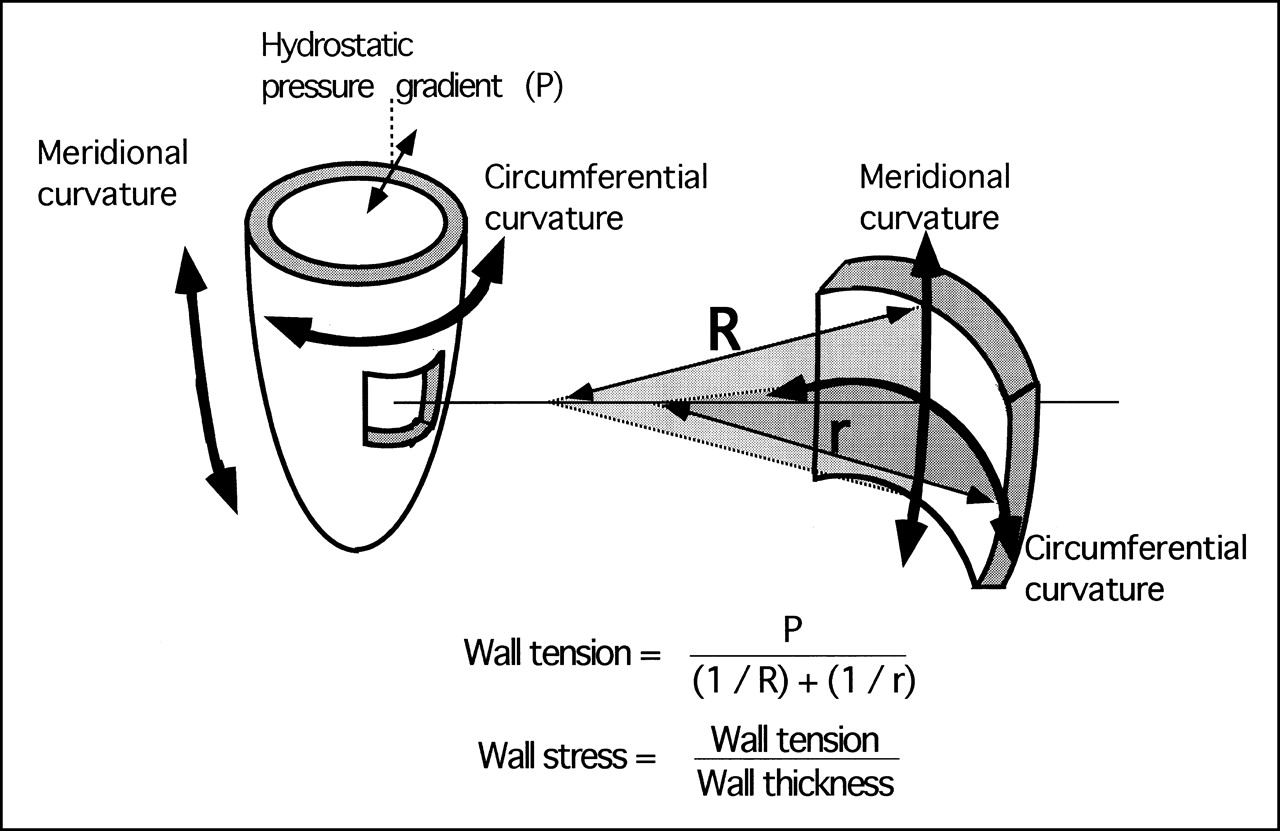
- FIGURE 2.
Schematic representation of parameters involved in segmental determination of left ventricular wall stress and tension using Laplace’s law.
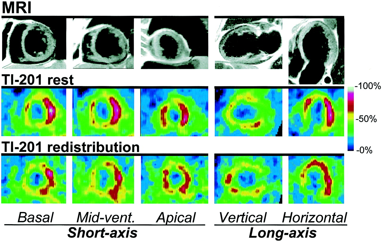
- FIGURE 3.
Example of MRI and SPECT images obtained for 60-y-old patient presenting with severe dilated cardiomyopathy. Rest SPECT defects predominate in inferior, anterior, and apical segments. On MRI slices, these segments correspond to walls that look thinner and more curved than other walls. On this MRI sequence, myocardium is gray, blood cavities are black, and surrounding fat is white.
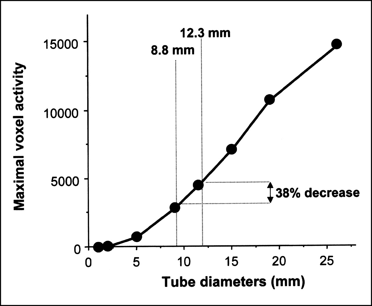
- FIGURE 4.
Relationship between diameters of tubes filled with constant concentration of 210Tl and maximal voxel activity within tubes after cardiac SPECT. A 38% decrease in maximal voxel activity was associated with the decreased tube size corresponding to the difference in myocardial thickness between segments without and with SPECT defects (from 12.3 to 8.8 mm).
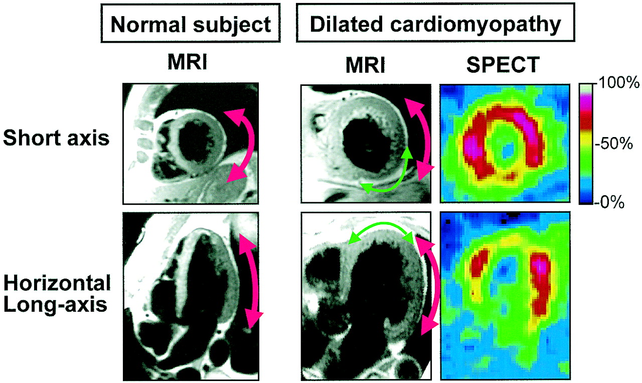
- FIGURE 5.
Schematic representation of global meridional and circumferential left ventricular curvatures in healthy volunteer and in patient with dilated cardiomyopathy and of segmental curvatures within patient segments showing SPECT defects. Compared with left ventricular walls from healthy volunteer, those from patient are less curved in circumferential direction (red arrows on short-axis slices) but more curved in meridional direction (red arrows on long-axis slices). In addition, in patient segments showing SPECT defects, walls are more curved (green arrows) and thinner than in other segments from this patient.


Tables
- TABLE 1
MRI Variables in Segments from Healthy Volunteers and in Overall Segments from Patients
Variable Healthy volunteers (n = 74) Patients (n = 88) Thickness (cm) 0.99 ± 0.15 1.12 ± 0.36* Curvature radii (cm)† Circumferential 3.2 ± 1.2 4.0 ± 2.0* Meridional 20 ± 41 11 ± 17* Diastolic wall tension index‡ (N · m−1 · mm Hg−1) 3.2 ± 1.2 3.1 ± 1.1 Diastolic wall stress index‡ (hN · m−2 · mm Hg−1) 3.2 ± 1.6 2.9 ± 1.3 -
↵* P < 0.05 for comparisons between overall segments from patients and segments from healthy volunteers.
-
↵ † Apical segments, which are not analyzed using meridional and circumferential curvature radii, are excluded.
-
↵ ‡ End-diastolic wall tension and wall stress are calculated for standard value of 1 mm Hg of left ventricular hydrostatic pressure at end-diastole.
-
- TABLE 2
MRI Variables in Segments from Healthy Volunteers and in Patient Segments With and Without 201TI Defects
Variable Healthy volunteers (n = 74) Patients No 201TI defects (n = 67) 201TI defects (n = 21) Thickness (cm) 0.99 ± 0.15* 1.23 ± 0.33* 0.88 ± 0.30 Curvature radii (cm)† Circumferential 3.2 ± 1.2 4.2 ± 1.7 3.3 ± 1.6 Meridional 20 ± 41* 13 ± 21 6 ± 4 Diastolic wall tension index‡ (N · m−1 · mm Hg−1) 3.2 ± 1.2* 3.3 ± 1.1* 2.5 ± 1.0 Diastolic wall stress index‡ (hN · m−2 · mm Hg−1) 3.2 ± 1.6 2.8 ± 1.2 3.0 ± 1.4 -
↵* P < 0.05 for comparisons with segments that have 201TI SPECT defects.
-
↵ † Apical segments, which are not analyzed using meridional and circumferential curvature radii, are excluded.
-
↵ ‡ End-diastolic wall tension and wall stress were calculated for standard value of 1 mm Hg of left ventricular hydrostatic pressure at end-diastole.
-


