


Article Figures & Data
Figures
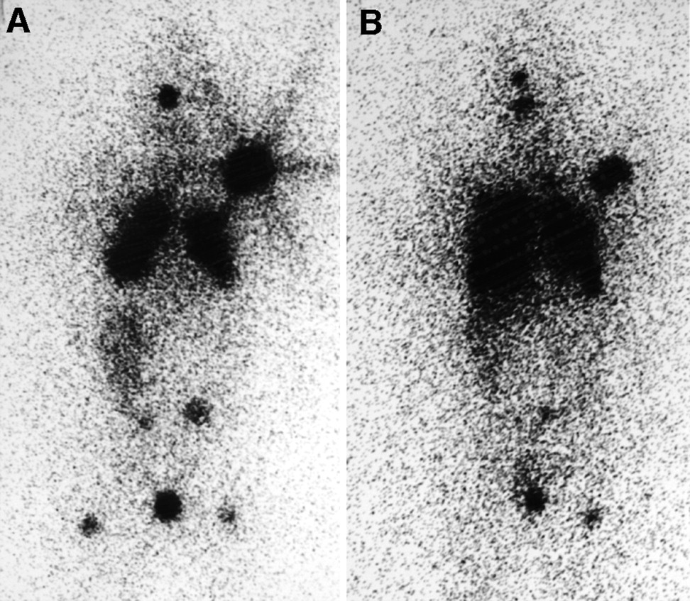
- FIGURE 1.
Anterior scintigraphic images of patient 1 made 48 h after 18.5 MBq of diagnostic 131I were administered. (A) Before first 131I therapy, radioactivity concentrations are seen irregularly throughout both lungs, in left shoulder, and in skull and as small foci in spine, pelvis, and femurs. Activity in liver is contiguous with and below that in right lung. (B) Before fifth 131I therapy, although lungs appear more prominent, radioactivity concentration is overall less and absent from some regions of small foci.
- FIGURE 2.
Anterior radiographs of chest of patient 1. (A) Before first 131I therapy, macronodules are readily visible in upper lungs and are widespread. Heart failure is evident. (B) After all 5 131I therapies, nodules appear slightly smaller and heart failure has disappeared although cardiomegaly persists.
- FIGURE 3.
Time–activity curves from patient 1 after diagnostic 131I. Radioactivity declines in first 2 studies but then rises and plateaus as radioiodide is converted into secreted radiothyroxine. Initial declines in radioactivity are slower before treatments 2, 4, and 5 than before treatment 1 because radioiodide is being sequestered less rapidly by thyroid tumors. Rx = treatment.
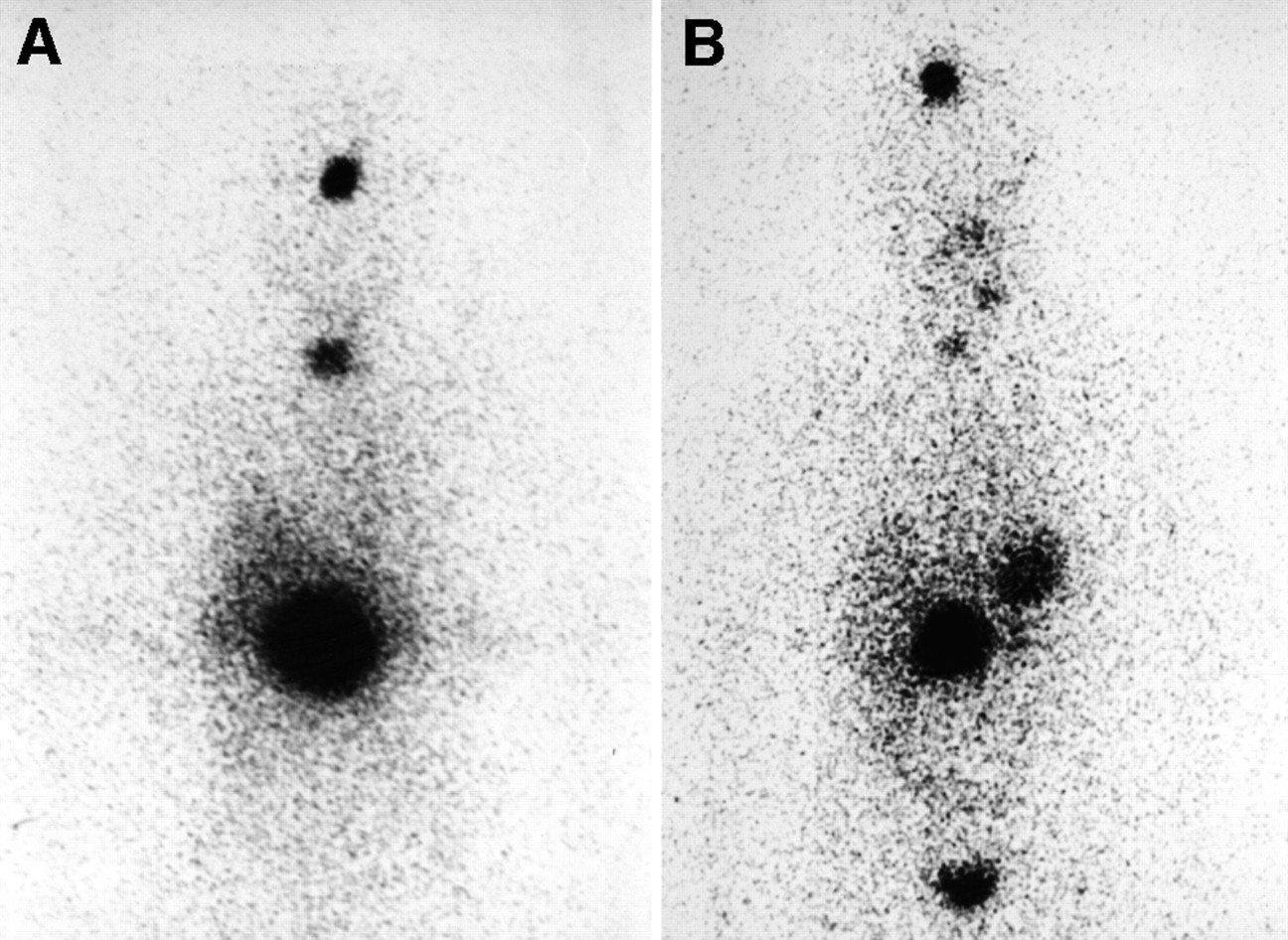
- FIGURE 4.
Anterior scintigraphic images of patient 2 obtained 48 h after 35 MBq of diagnostic 131I were administered. (A) Before first 131I therapy, largest concentration of radioactivity is in region of tumor in upper lumbar spine. Other foci of metastases are in skull and mid neck. Radioactivity in liver is above and to right of spine tumor. (B) Before third 131I therapy, radioactivity concentrations in spine and skull are less intense. Foci in separate neck node metastases are visible. Stomach is visible, and bladder activity is at bottom.
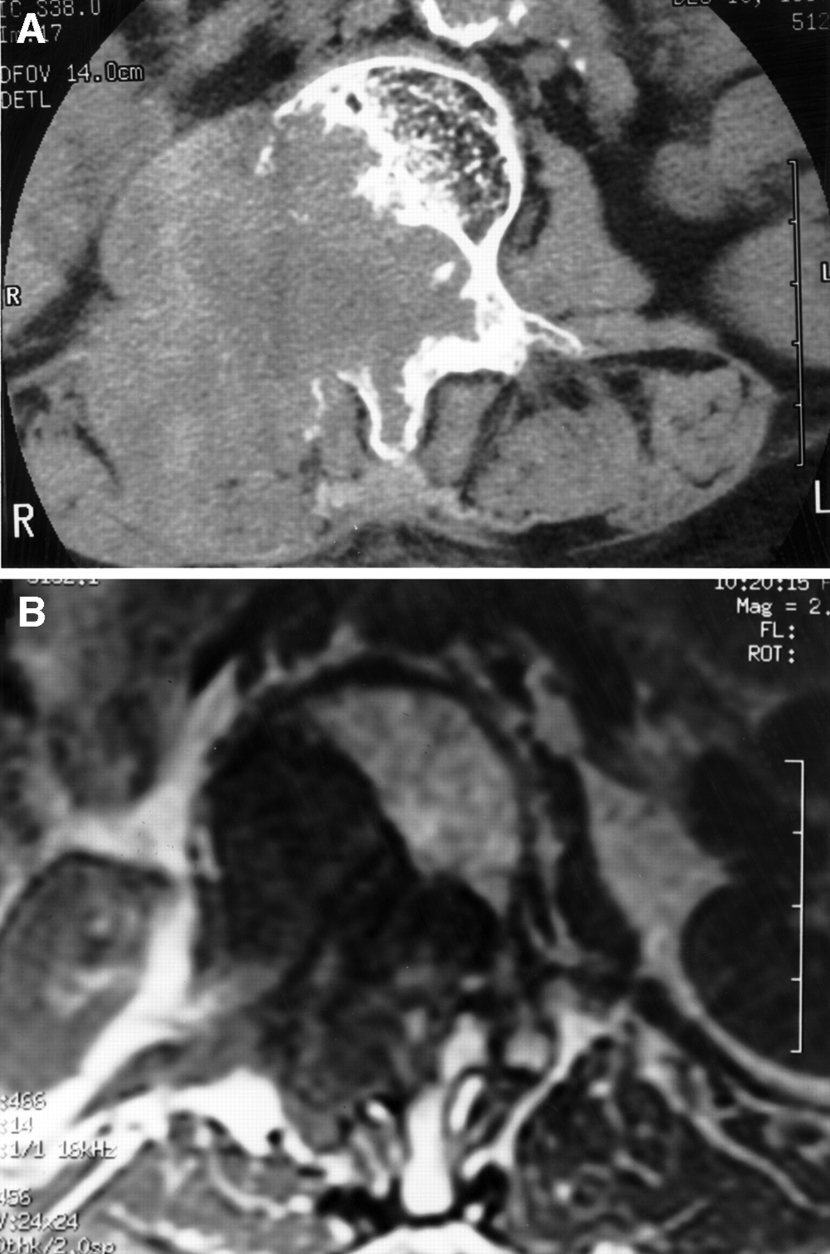
- FIGURE 5.
CT (A) and MR (B) images of patient 2. Before first 131I therapy, CT section shows partial destruction of L1 vertebra and soft-tissue mass to right. Before third therapy, MR section shows similar destruction of L1 but markedly reduced soft-tissue mass.


Tables
Date Thyroxine (μg/dL) fT4 (ng/dL) TSH (mU/L) Tg (ng/mL) TSI index Body retention (fraction of activity) Body dose (cGy) Blood dose (cGy) 131I treatment (GBq) Other treatment Patient 1 Jan-98 13.1 <0.01 >30,000 6.2 0.87 150 194 1.85 Restart antithyroid drug* Apr-98 1.6 0.06 0.76 207 275 2.59 Restart antithyroid drug* Sep/Oct-98 0.7 11.1 >30,000 0.77 309 3.15 Oct-98 13 Begin thyroxine at 0.05 mg/d Nov-98 2.2 Jan-99 0.9 17.8 4 0.29 219 311 5.7 Restart thyroxine at 0.1 mg/d Apr-99 1.4 0.8 28,193 Jul-99 0.4 48 >30,000 0.58 344 416 4.81 Restart thyroxine at 0.1 mg/d Oct/Nov-99 1.3 1.8 12,120 Change thyroxine to 0.125 mg/d Jan-00 0.57 27,300 3.1 Continue thyroxine Mar/Apr-00 1.6 0.23 17,800 Continue thyroxine Patient 2 Dec-97 6.7 18 4,800 0.58 144 222 2.2 Begin thyroxine at 0.125 mg/d Apr-98 0.9 480 Continue thyroxine Sep-98 7.6 8 1,310 0.39 202 4.6 Restart thyroxine Nov-98 1.6 21 110 Feb-00 9.7 11.2 150 Stop thyroxine Mar-00 0.67 34 247 0.23 161 5.6 Restart thyroxine at 0.137 mg/d Apr-00 1 ↵* Before treatments with 131I, antithyroid drug was stopped at least 1 wk and thyroxine for 6 wk.
fT4 = free thyroxine; Tg = thyroglobulin.
Reference ranges are 4.4–12.4 μg/dL for thyroxine, 0.7–1.79 ng/dL for fT4, 0.3–5.5 mU/L for TSH, and ≤1.3 for TSI.
Date Hemoglobin (g/dL) Leukocytes (1,000/μL) Neutrophils (1,000/μL) Platelets (1,000/μL) 131I treatment (GBq) Other treatment Patient 1 Jan-98 11 6 3.3 228 1.85 Feb-98 12.1 4.7 159 Antithyroid drug Apr-98 10.1 3.7 2.5 186 2.59 May-98 10.4 3.3 2.4 241 Antithyroid drug Sep/Oct-98 10.4 4.2 3 201 3.15 Nov-98 11.6 3.1 2.2 160 Thyroxine Jan-99 11.2 3.7 2.5 184 5.7 Mar-99 9.8 2.5 1.7 150 Thyroxine Jul-99 10.2 3.8 2.3 176 4.8 Aug-99 9.8 2.3 1.7 73 Thyroxine Sep-99 7.2 1.9 1.2 59 Thyroxine plus transfusion of 2 units Dec-99 9.6 2.8 1.7 108 Thyroxine Mar-00 10 3 1.8 144 Thyroxine Patient 2 Dec-97 11 3.9 2.3 372 2.2 Begin thyroxine Jan-98 11.3 4 383 Continue thyroxine Sep-98 9.6 3.8 2.6 281 4.6 Restart thyroxine Nov-98 9.8 3.2 2 287 Continue thyroxine Feb-00 10.4 4.9 3.6 302 Stop thyroxine Mar-00 9.9 2.9 1.7 267 5.6 Restart thyroxine at 0.137 mg/d Apr-00 9.6 2.1 1.2 195 Continue thyroxine Hematologic indices were obtained just before and 5–6 wk after 131I therapies except as noted; additional measurements were made but did not show marked differences.
In this issue


Jump to section
Related Articles
Cited By...
- Complications of Radioactive Iodine Treatment of Thyroid Carcinoma
- Uncommon Causes of Thyrotoxicosis
- Differentiated thyroid cancer presenting with thyrotoxicosis due to functioning metastases
- Increasing Efficacy and Safety of Treatments of Patients with Well-Differentiated Thyroid Carcinoma by Measuring Body Retentions of 131I