


Article Figures & Data
Figures
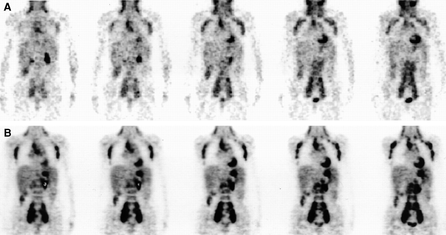
- FIGURE 1.
Patient 10. Coronal images obtained by FDG hybrid PET (A) and dedicated PET (B). Bilateral cervical to axillary, mediastinal, and para-aortic to bilateral iliac sites are detected on both sets of images. Clearer images and higher lesion-to-background contrast are achieved with dedicated PET than with hybrid PET.
- FIGURE 2.
Patient 4. Coronal images obtained by FDG hybrid PET (A) and dedicated PET (B). Left supraclavicular site is clearly depicted by dedicated PET. Hybrid PET, on other hand, shows only faint uptake at corresponding site (arrow), which is not considered pathologic.
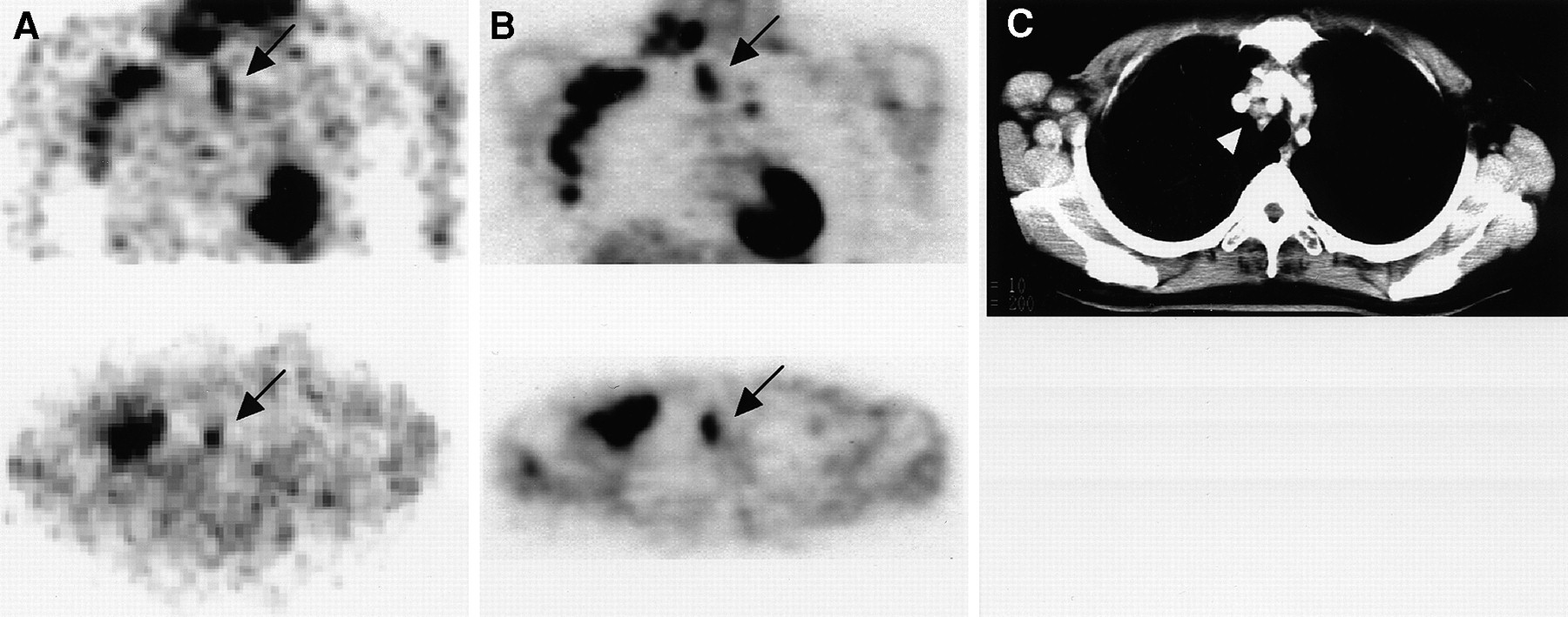
- FIGURE 3.
Patient 24. Diffuse spleen involvement (arrows) is detected by both FDG hybrid PET (A) and dedicated PET (B) (top: coronal; bottom: axial). (C) CT fails to detect involvement, showing only splenomegaly.
- FIGURE 4.
Patient 1. Images obtained by FDG hybrid PET (A) and dedicated PET (B) (top: coronal; bottom: axial) show hot uptake at mediastinal site (arrows). Eight-millimeter lesion is seen on CT (arrowhead) but was diagnosed as negative for NHL because of size criteria.
- FIGURE 5.
Patient 12, with indolent, low-risk lymphoplasmacytic lymphoma. Images obtained by both coronal FDG hybrid PET (A) and dedicated PET (B) show only faint uptake at sites that CT (C) shows to have numerous bulky lesions. Lesions extend from cervical to inguinal regions, including mediastinal (arrow) and para-aortic (arrowheads).


Tables
Patient no. Age (y) Sex Clinical group* Stage† 1 48 F Indolent II 2 41 F Indolent III 3 49 F Indolent III 4 49 F Indolent III 5 64 M Indolent III 6 31 F Indolent IV 7 37 M Indolent IV 8 39 F Indolent IV 9 54 M Indolent IV 10 54 F Indolent IV 11 55 F Indolent IV 12 66 M Indolent IV 13 73 M Indolent IV 14 64 M Aggressive I 15 79 M Aggressive I 16 79 F Aggressive I 17 45 F Aggressive II 18 70 M Aggressive II 19 71 M Aggressive II 20 75 M Aggressive II 21 82 M Aggressive II 22 62 M Aggressive III 23 63 M Aggressive III 24 66 F Aggressive IV 25 66 F Aggressive IV 26 69 M Aggressive IV 27 72 F Aggressive IV 28 75 F Aggressive IV 29 14 M Very aggressive IV 30 20 M Very aggressive IV - TABLE 2
Distribution of True-Positive Disease Sites Based on FDG Hybrid PET and Dedicated PET, CIS, and Reference Standard
Site Hybrid PET Dedicated PET CIS Reference standard Cervical 26 29 28 33 Supraclavicular 23 27 22 29 Axillary 15 18 20 22 Mediastinal and hilar 12 14 13 16 Subtotal: supradiaphragmatic 76 88 83 100 Para-aortic 13 14 13 15 Abdominal 9 9 11 11 Iliac 21 25 20 28 Inguinal 19 20 22 24 Subtotal: infradiaphragmatic 62 68 66 78 Splenic 4 4 3 4 Bone and bone marrow 7 7 1 12 Other extranodal 10 12 11 12 Subtotal: extranodal 21 23 15 28 Total 159 179 164 206 - TABLE 3
Comparison of Disease Stage Based on FDG Hybrid PET and Dedicated PET, CIS, and Reference Standard
Stage Hybrid PET Dedicated PET CIS Reference standard I 4 (3) 4 (3) 4 (3) 3 II 7 (6) 6 (6) 6 (6) 6 III 9 (5) 9 (5) 15 (6) 6 IV 10 (10) 11 (10) 5 (5) 15 Numbers in parentheses are numbers concordant with reference standard.


