


Article Figures & Data
Figures
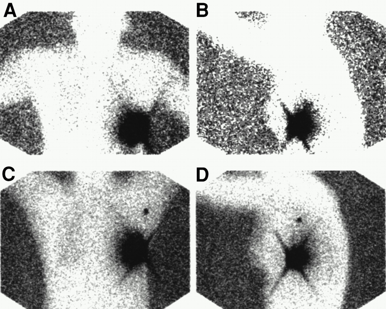
- FIGURE 1.
Anterior (A and C) and left lateral (B and D) transmission images show injection site in left breast and focal uptake in sentinel node in left axilla, seen only on 2-h delayed images. (A and B) Early images at 30 min. (C and D) Delayed images at 2 h.
Tables
Parameter 1-d protocol 2-d protocol No. of cases 514 152 Mean age* (y) 57.3 (22–88) 55.2 (31–87) Size of primary† (mean ± SD) 1.4 ± 0.9 1.6 ± 1.1 Histology Ductal 387 (75.3%) 117 (77.0%) Lobular 44 (8.6%) 10 (6.6%) Other‡ 83 (16.0%) 89 (16.4%) Positive SLN (%) 30 37 Prior excisional biopsy 235 (45.7%) 89 (58.6%) Site Upper outer quadrant 270 (52.5%) 91 (59.9%) Upper inner quadrant 87 (16.9%) 12 (7.9%) Lower outer quadrant 49 (9.5%) 25 (16.4%) Lower inner quadrant 31 (6.0%) 6 (3.9%) Central 63 (12.3%) 13 (8.6%) Unknown or no cancer found 14 (2.7%) 5 (3.3%) ↵* Range in parentheses.
↵† Invasive component only. Patients with unknown size or diagnosis of ductal carcinoma in situ (no invasive component) or patients with no cancer found at time of resection were not included.
↵‡ Includes 11 cases with no cancer found in 1-d protocol and 3 cases with no cancer found in 2-d protocols.
Image 1-d protocol 2-d protocol Early (30 min) 352 (69%) 104 (68%) Late (2 h) Not done 130 (86%) Overall 352 (69%) 130 (86%) Sentinel nodes found 1-d protocol 2-d protocol By isotope (% of total) 1084 (85.1) 367 (88.4) By blue dye (% of total) 881 (69.2) 291 (70.1) Total 1274 415 Average no. of nodes/axilla 2.5 2.8 Isotope/dye concordance* 97% 95% ↵* At least one lymph node with both isotope and dye, in patients in whom both isotope and dye were found in axilla.
Surgeon No. of prior lymphatic mappings No. of 1-d protocols (% of total) No. of 2-d protocols (% of total) A 346 131 (25.5) 71 (46.7) B 239 95 (18.5) 18 (11.8) C 16 16 (3.1) 1 (0.7) D 4 108 (21.0) 14 (9.2) E 167 31 (6.0) 1 (0.7) F 38 61 (11.9) 16 (10.5) H 100 59 (11.5) 28 (18.4) I 49 13 (2.5) 3 (2.0) Total 959 514 (100) 152 (100) Surgeon G did not do any Sentinel node mapping during the study period.
In this issue


Jump to section
Related Articles
Cited By...
- Organ and Fetal Absorbed Dose Estimates from 99mTc-Sulfur Colloid Lymphoscintigraphy and Sentinel Node Localization in Breast Cancer Patients
- Fusion of SPECT and Multidetector CT Images for Accurate Localization of Pelvic Sentinel Lymph Nodes in Prostate Cancer Patients
- Reverse echelon node and a lymphatic ectasia in the same patient during breast lymphoscintigraphy: the importance of injection and imaging technique
- Patient effective dose from sentinel lymph node lymphoscintigraphy in breast cancer: a study using a female humanoid phantom and thermoluminescent dosemeters
- Radioguided Sentinel Lymph Node Biopsy in Breast Cancer Surgery