
Main menu

  • Home
  • Content
    • Current
    • Ahead of print
    • Past Issues
    • JNM Supplement
    • SNMMI Annual Meeting Abstracts
    • Continuing Education
    • JNM Podcasts
  • Subscriptions
    • Subscribers
    • Institutional and Non-member
    • Rates
    • Journal Claims
    • Corporate & Special Sales
  • Authors
    • Submit to JNM
    • Information for Authors
    • Assignment of Copyright
    • AQARA requirements
  • Info
    • Reviewers
    • Permissions
    • Advertisers
  • About
    • About Us
    • Editorial Board
    • Contact Information
  • More
    • Alerts
    • Feedback
    • Help
    • SNMMI Journals
  • SNMMI
    • JNM
    • JNMT
    • SNMMI Journals
    • SNMMI

User menu

  • Subscribe
  • My alerts
  • Log in
  • Log out
  • My Cart

Search

  • Advanced search
Journal of Nuclear Medicine
  • SNMMI
    • JNM
    • JNMT
    • SNMMI Journals
    • SNMMI
  • Subscribe
  • My alerts
  • Log in
  • Log out
  • My Cart
Journal of Nuclear Medicine

Advanced Search

  • Home
  • Content
    • Current
    • Ahead of print
    • Past Issues
    • JNM Supplement
    • SNMMI Annual Meeting Abstracts
    • Continuing Education
    • JNM Podcasts
  • Subscriptions
    • Subscribers
    • Institutional and Non-member
    • Rates
    • Journal Claims
    • Corporate & Special Sales
  • Authors
    • Submit to JNM
    • Information for Authors
    • Assignment of Copyright
    • AQARA requirements
  • Info
    • Reviewers
    • Permissions
    • Advertisers
  • About
    • About Us
    • Editorial Board
    • Contact Information
  • More
    • Alerts
    • Feedback
    • Help
    • SNMMI Journals
  • View or Listen to JNM Podcast
  • Visit JNM on Facebook
  • Join JNM on LinkedIn
  • Follow JNM on Twitter
  • Subscribe to our RSS feeds
Research ArticleCLINICAL INVESTIGATIONS

Factors Affecting Sentinel Node Localization During Preoperative Breast Lymphoscintigraphy

Philip I. Haigh, Nora M. Hansen, Armando E. Giuliano, G. Keith Edwards, Wei Ye and Edwin C. Glass
Journal of Nuclear Medicine October 2000, 41 (10) 1682-1688;
Philip I. Haigh
  • Find this author on Google Scholar
  • Find this author on PubMed
  • Search for this author on this site
Nora M. Hansen
  • Find this author on Google Scholar
  • Find this author on PubMed
  • Search for this author on this site
Armando E. Giuliano
  • Find this author on Google Scholar
  • Find this author on PubMed
  • Search for this author on this site
G. Keith Edwards
  • Find this author on Google Scholar
  • Find this author on PubMed
  • Search for this author on this site
Wei Ye
  • Find this author on Google Scholar
  • Find this author on PubMed
  • Search for this author on this site
Edwin C. Glass
  • Find this author on Google Scholar
  • Find this author on PubMed
  • Search for this author on this site
  • Article
  • Figures & Data
  • Info & Metrics
  • PDF
Loading

Article Figures & Data

Figures

  • Tables
  • FIGURE 1.
    • Download figure
    • Open in new tab
    • Download powerpoint
    FIGURE 1.

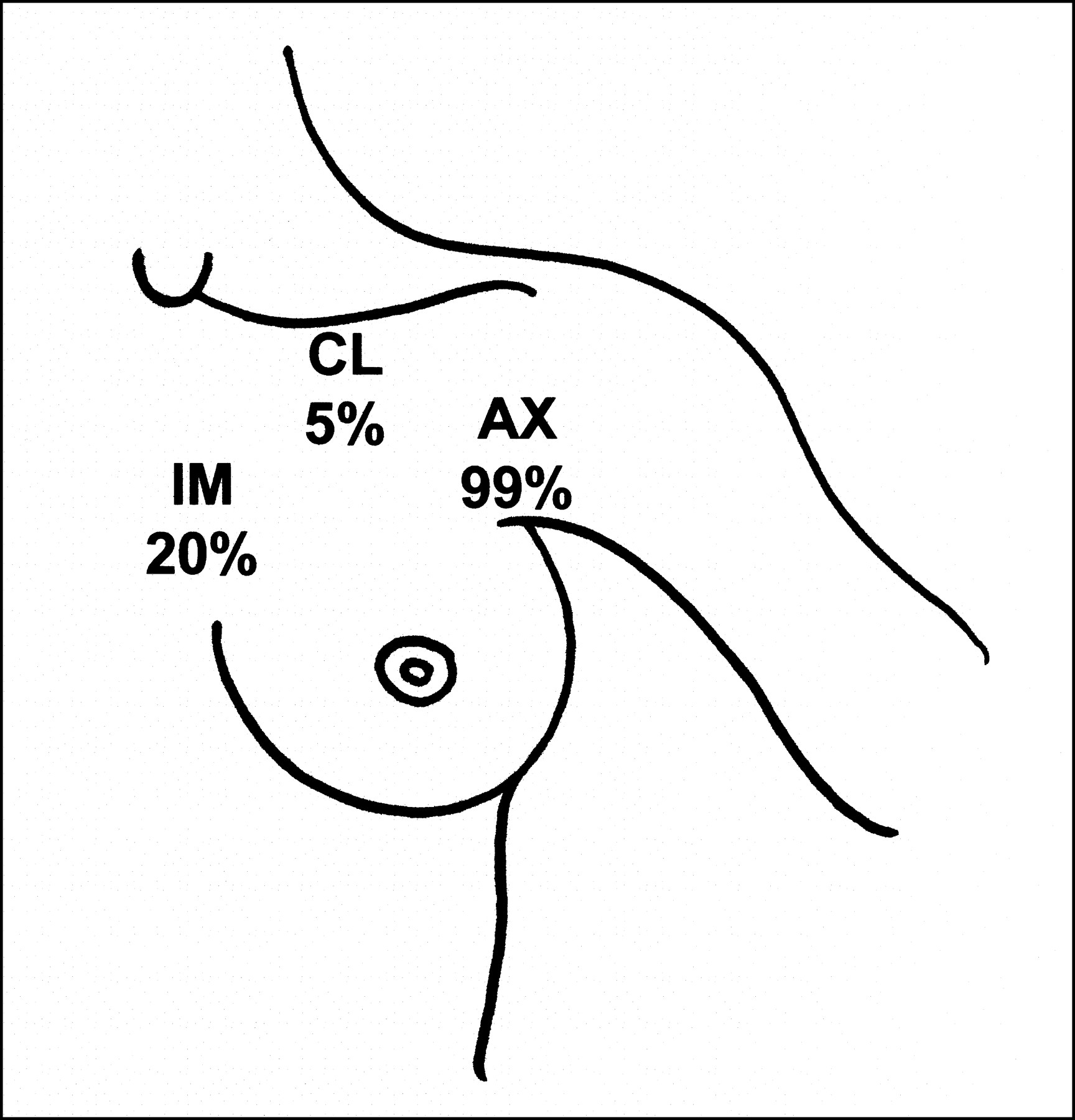
    Frequency of AX, IM, and CL sentinel nodes found on preoperative lymphoscintigraphy in all cases. No correlation was found between inner versus outer quadrant primary tumors and location of sentinel node.

  • FIGURE 2.
    • Download figure
    • Open in new tab
    • Download powerpoint
    FIGURE 2.

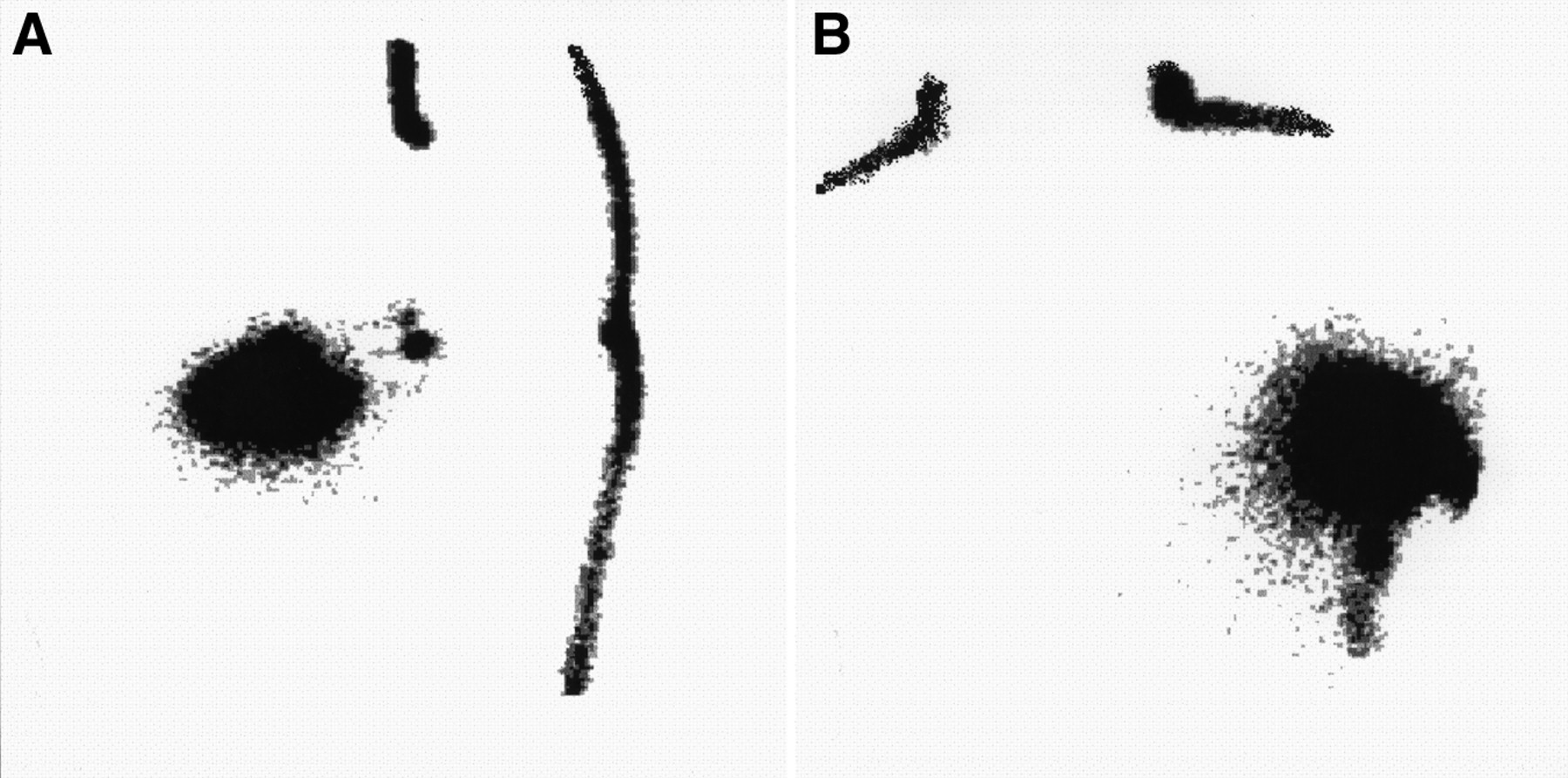
    Lymphoscintigrams from patient with upper outer quadrant tumor in left breast 30 min after injection at primary tumor site. MOVA shows distinct AX sentinel nodes (A) that are not seen on anterior view (B). MOVA revealed AX sentinel nodes in all patients, whereas anterior view missed 50% of AX sentinel nodes.

  • FIGURE 3.
    • Download figure
    • Open in new tab
    • Download powerpoint
    FIGURE 3.

    (A) Patient positioned on foam wedge with arm extended and abducted overhead to obtain MOVA image. (B) Schematic representation of craniocaudal view of breast at time of lymphoscintigraphy for outer quadrant tumor. (Left) In anterior view, distance projected onto scintillation camera (y) from injection site to AX sentinel node (SN) is short. (Right) Using MOVA, patient positioning on 45° wedge allows breast and injection site to shift medially from AX sentinel node, increasing distance y, and brings sentinel node closer to gamma camera (x), thereby improving AX sentinel node identification.

  • FIGURE 4.
    • Download figure
    • Open in new tab
    • Download powerpoint
    FIGURE 4.

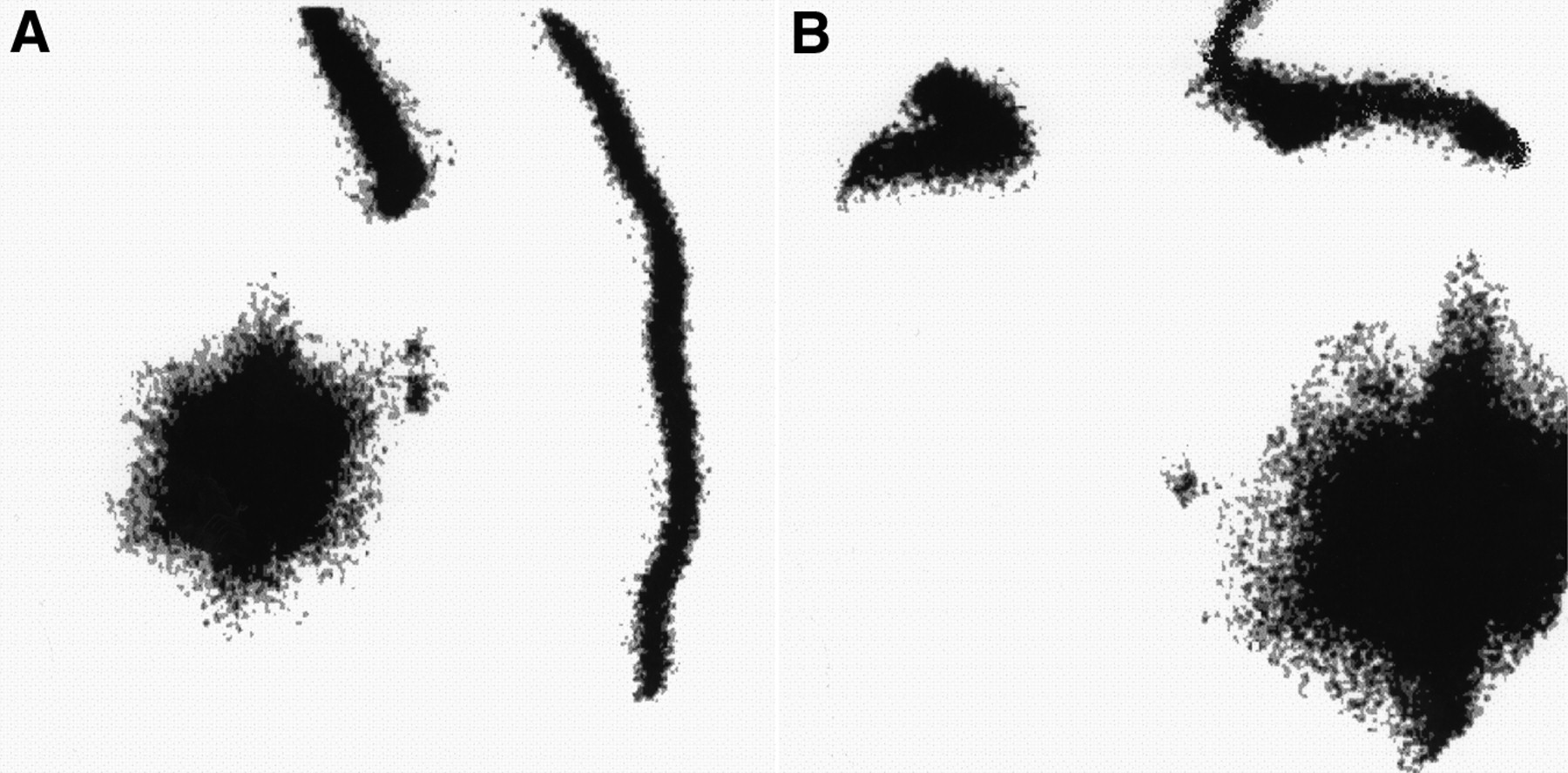
    Lymphoscintigrams from patient with upper outer quadrant tumor in left breast 15 min after injection at primary tumor site. MOVA reveals AX sentinel nodes (A), whereas anterior view shows distinct IM sentinel node, but AX sentinel nodes are not visualized (B). Both MOVA and anterior images must be obtained to evaluate all regional drainage sites.

Tables

  • Figures
    • View popup
    TABLE 1.

    Patient Demographics and Tumor Characteristics

    Parametern*
    Age (y)
     Median59
     Range29–81
    Primary tumor size (cm)
     Median1.5
     Range0.4–7.1
    Primary tumor type
     Invasive71 (93)
      Infiltrating ductal63
      Infiltrating lobular6
      Other2
     Noninvasive5 (7)
      Ductal carcinoma in situ5
    Location
     Outer quadrants37 (49)
      Upper27
      Lower10
     Inner quadrants38 (50)
      Upper25
      Lower13
     Subareolar1 (1)
    Method of biopsy
     Fine-needle aspiration21 (28)
     Core needle19 (25)
     Excision36 (47)
    • ↵* Values in parentheses are percentages.

    • View popup
    TABLE 2.

    Primary and Secondary Drainage Patterns in 76 Breast Lymphoscintigrams

    nPrimary drainageSecondary drainage
    58AXNone
    8AXIM
    2AXCL
    1AX + IMNone
    1AX + IM + CLNone
    1IMNone
    4IMAX
    1CLAX
    • View popup
    TABLE 3.

    Effect of 6 Factors on Radiopharmaceutical Transit Time Determined by Logistic Regression

    FactorP
    Inner quadrants vs. outer quadrants*0.91
    Interval from biopsy to lymphoscintigraphy (d)0.77
    Method of biopsy (FNA vs. core vs. excision)0.45
    Age (y, continuous variable)0.45
    Weight (kg, continuous variable)0.31
    Breast size
     Small vs. medium0.93
     Large vs. small0.01
     Large vs. medium0.03
    • ↵* 1 subareolar case excluded.

    • FNA = fine-needle aspiration.

    • View popup
    TABLE 4.

    Pathology of Sentinel Nodes

    Axillary SLNDn*
    Total procedures76
    Sentinel node identified76 (100)
     Metastases absent48 (63)
     Metastases present28 (37)
      >2 mm8 (29)
      <2 mm, H&E12 (43)
      <2 mm, IHC only8 (29)
    • ↵* Values in parentheses are percentages.

    • H&E = hematoxylin and eosin; IHC = immunohistochemistry.

    • View popup
    TABLE 5.

    Summary of Techniques and Drainage Patterns in SLND Series Using Breast Lymphoscintigraphy

    StudynAgentTotal MBqTotal injection volume* (mL)Early/late scan timing after injection (h)ViewsOverall sentinel node detection (%)AX drainage† (%)IM drainage† (%)
    TotalExclusiveTotalExclusive
    Uren et al. (12)34ASC50–700.2–0.40/2.5A, L, O919458396
    Roumen et al. (10)83CA6024/18A, L809786113
    De Cicco et al. (9)250CA or ASC70.4 or 0.5‡0.25–0.5/3A, AO98989822
    Borgstein et al. (7)130CA4042/18A, L919881182
    O'Hea et al. (8)56SC114 0.8–1§   A, L7510086140
    van der Ent et al. (11)70NC370416§A, ALNR97NR34NR
    Current series76SC12–163–80/¶A, MOVA1009979201
    • ↵* All injections were peritumoral except for series from De Cicco (9).

    • ↵† Values represent drainage patterns after scan failures were excluded.

    • ↵‡ 0.4 mL for subdermal injection (175 patients) and 0.5 mL for peritumoral injection (75 patients).

    • ↵§ Only 1 scan obtained at these times.

    • ↵¶ Images obtained continuously until sentinel node visualized, unless no uptake after 4 h.

    • A = anterior; L = lateral; O = oblique; CA = colloidal albumin; AO = antero-oblique; SC = sulfur colloid; NC = nanocolloid; AL = anterolateral; NR = not recorded.

PreviousNext
Back to top

In this issue

Journal of Nuclear Medicine
Vol. 41, Issue 10
October 1, 2000
  • Table of Contents
  • Table of Contents (PDF)
  • Index by author
Print
Download PDF
Article Alerts
Sign In to Email Alerts with your Email Address
Email Article

Thank you for your interest in spreading the word on Journal of Nuclear Medicine.

NOTE: We only request your email address so that the person you are recommending the page to knows that you wanted them to see it, and that it is not junk mail. We do not capture any email address.

Enter multiple addresses on separate lines or separate them with commas.
Factors Affecting Sentinel Node Localization During Preoperative Breast Lymphoscintigraphy
(Your Name) has sent you a message from Journal of Nuclear Medicine
(Your Name) thought you would like to see the Journal of Nuclear Medicine web site.
Citation Tools
Factors Affecting Sentinel Node Localization During Preoperative Breast Lymphoscintigraphy
Philip I. Haigh, Nora M. Hansen, Armando E. Giuliano, G. Keith Edwards, Wei Ye, Edwin C. Glass
Journal of Nuclear Medicine Oct 2000, 41 (10) 1682-1688;

Citation Manager Formats

  • BibTeX
  • Bookends
  • EasyBib
  • EndNote (tagged)
  • EndNote 8 (xml)
  • Medlars
  • Mendeley
  • Papers
  • RefWorks Tagged
  • Ref Manager
  • RIS
  • Zotero
Share
Factors Affecting Sentinel Node Localization During Preoperative Breast Lymphoscintigraphy
Philip I. Haigh, Nora M. Hansen, Armando E. Giuliano, G. Keith Edwards, Wei Ye, Edwin C. Glass
Journal of Nuclear Medicine Oct 2000, 41 (10) 1682-1688;
Twitter logo Facebook logo LinkedIn logo Mendeley logo
  • Tweet Widget
  • Facebook Like
  • Google Plus One
Bookmark this article

Jump to section

  • Article
    • Abstract
    • MATERIALS AND METHODS
    • RESULTS
    • DISCUSSION
    • CONCLUSION
    • Acknowledgments
    • Footnotes
    • REFERENCES
  • Figures & Data
  • Info & Metrics
  • PDF

Related Articles

  • No related articles found.
  • PubMed
  • Google Scholar

Cited By...

  • A Practical Technique to Improve Visualization of Sentinel Nodes in the Axillary Region on Breast Lymphoscintigraphy: Medial Breast Traction by Patient
  • Scintigraphic Investigations of the Lymphatic System: The Influence of Injected Volume and Quantity of Labeled Colloidal Tracer
  • Usefulness of Medium-Energy Collimator for Sentinel Node Lymphoscintigraphy Imaging in Breast Cancer Patients.
  • Continuing Evolution in Breast Cancer Surgical Management
  • Reducing Exposure from 57Co Sources During Breast Lymphoscintigraphy by Optimizing Energy Windows and Other Suggested Enhancements of Acquisition and the Display of Images
  • Reverse echelon node and a lymphatic ectasia in the same patient during breast lymphoscintigraphy: the importance of injection and imaging technique
  • Factors Affecting Visualization Rates of Internal Mammary Sentinel Nodes During Lymphoscintigraphy
  • Nonvisualization of Axillary Sentinel Node During Lymphoscintigraphy: Is There a Pathologic Significance in Breast Cancer?
  • Google Scholar

More in this TOC Section

  • Feasibility of Ultra-Low-Activity 18F-FDG PET/CT Imaging Using a Long–Axial-Field-of-View PET/CT System
  • Cardiac Presynaptic Sympathetic Nervous Function Evaluated by Cardiac PET in Patients with Chronotropic Incompetence Without Heart Failure
  • Validation and Evaluation of a Vendor-Provided Head Motion Correction Algorithm on the uMI Panorama PET/CT System
Show more Clinical Investigations

Similar Articles

SNMMI

© 2025 SNMMI

Powered by HighWire