
Main menu

  • Home
  • Content
    • Current
    • Ahead of print
    • Past Issues
    • JNM Supplement
    • SNMMI Annual Meeting Abstracts
    • Continuing Education
    • JNM Podcasts
  • Subscriptions
    • Subscribers
    • Institutional and Non-member
    • Rates
    • Journal Claims
    • Corporate & Special Sales
  • Authors
    • Submit to JNM
    • Information for Authors
    • Assignment of Copyright
    • AQARA requirements
  • Info
    • Reviewers
    • Permissions
    • Advertisers
  • About
    • About Us
    • Editorial Board
    • Contact Information
  • More
    • Alerts
    • Feedback
    • Help
    • SNMMI Journals
  • SNMMI
    • JNM
    • JNMT
    • SNMMI Journals
    • SNMMI

User menu

  • Subscribe
  • My alerts
  • Log in
  • Log out
  • My Cart

Search

  • Advanced search
Journal of Nuclear Medicine
  • SNMMI
    • JNM
    • JNMT
    • SNMMI Journals
    • SNMMI
  • Subscribe
  • My alerts
  • Log in
  • Log out
  • My Cart
Journal of Nuclear Medicine

Advanced Search

  • Home
  • Content
    • Current
    • Ahead of print
    • Past Issues
    • JNM Supplement
    • SNMMI Annual Meeting Abstracts
    • Continuing Education
    • JNM Podcasts
  • Subscriptions
    • Subscribers
    • Institutional and Non-member
    • Rates
    • Journal Claims
    • Corporate & Special Sales
  • Authors
    • Submit to JNM
    • Information for Authors
    • Assignment of Copyright
    • AQARA requirements
  • Info
    • Reviewers
    • Permissions
    • Advertisers
  • About
    • About Us
    • Editorial Board
    • Contact Information
  • More
    • Alerts
    • Feedback
    • Help
    • SNMMI Journals
  • View or Listen to JNM Podcast
  • Visit JNM on Facebook
  • Join JNM on LinkedIn
  • Follow JNM on Twitter
  • Subscribe to our RSS feeds
Meeting ReportInstrumentation & Data Analysis Track

Artificial neural network based outcome prediction in DAT SPECT imaging of Parkinson's Disease

Jing Tang, Bao Yang, Nikolay Shenkov, Sima Fotouhi, Esmaeil Davoodi-Bojd, Lijun Lu, Hamid Soltanian-Zadeh, Vesna Sossi and Arman Rahmim
Journal of Nuclear Medicine May 2017, 58 (supplement 1) 292;
Jing Tang
5Electrical and Computer Engineering Oakland University Rochester MI United States
  • Find this author on Google Scholar
  • Find this author on PubMed
  • Search for this author on this site
Bao Yang
5Electrical and Computer Engineering Oakland University Rochester MI United States
  • Find this author on Google Scholar
  • Find this author on PubMed
  • Search for this author on this site
Nikolay Shenkov
1Vancouver BC Canada
  • Find this author on Google Scholar
  • Find this author on PubMed
  • Search for this author on this site
Sima Fotouhi
4Johns Hopkins University Baltimore MD United States
  • Find this author on Google Scholar
  • Find this author on PubMed
  • Search for this author on this site
Esmaeil Davoodi-Bojd
3Electrical and Computer Engineering Henry Ford Health Systems Detroit MI United States
  • Find this author on Google Scholar
  • Find this author on PubMed
  • Search for this author on this site
Lijun Lu
6Southern Medical University Guangzhou China
  • Find this author on Google Scholar
  • Find this author on PubMed
  • Search for this author on this site
Hamid Soltanian-Zadeh
2Henry Ford Health System Detroit MI United States
  • Find this author on Google Scholar
  • Find this author on PubMed
  • Search for this author on this site
Vesna Sossi
7UBC Vancouver BC Canada
  • Find this author on Google Scholar
  • Find this author on PubMed
  • Search for this author on this site
Arman Rahmim
4Johns Hopkins University Baltimore MD United States
  • Find this author on Google Scholar
  • Find this author on PubMed
  • Search for this author on this site
  • Article
  • Figures & Data
  • Info & Metrics
Loading

Abstract

292

Objectives: Dopamine transporter (DAT) SPECT imaging has been widely used in the diagnosis of Parkinson’s disease (PD). Compared to visual interpretation of DAT SPECT images, quantitative analysis has the significant potential to enable early stage detection of disease and improve progression tracking. Our past radiomic efforts focused on seeking the correlations between DAT SPECT radiomic measures and clinical assessments. By contrast, the goal of this study is to predict clinical outcome using DAT SPECT radiomic features and non-imaging features at baseline (year 0). We accomplish this goal through developing an artificial neural network (ANN) based prediction technique.

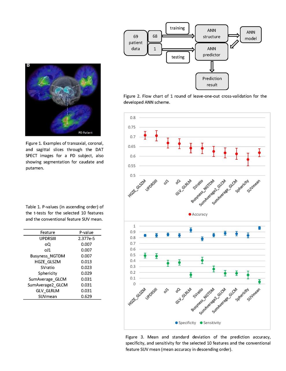
Methods: Sixty-nine subjects in the Parkinson’s Progression Markers Initiative database were included in our study. The subjects were imaged by SPECT 4+-0.5 h following injection of 111-185 MBq of 123I-Ioflupane, and also underwent high-resolution 3T MRI scans. The MR images were segmented to obtain the structures of caudate, putamen, and occipital cortex which were then overlaid on the co-registered SPECT images. Outcome was set as motor part (III) of the unified Parkinson’s disease rating scale (UPDRS) at year 4. From the more affected caudate in the SPECT image at baseline, we extracted a total of 92 image features, including 14 first-order intensity features, 21 shape features, and 57 second- and higher-order textural features. Six non-imaging features were also included in the study, i.e. gender, age, disease duration (DD)_sympt, DD_diag, UPDRSIII, and Montreal cognitive assessment (MoCA). We performed a t-test for each of the 98 features to classify the patients to group 1 (UPDRSIII of year 4 < 30) and group 2 (UPDRSIII of year 4 > =30). Through sorting the p-values from the t-tests in ascending order, we selected the top 10 features. The mean standardized uptake value (SUV mean), as a conventional feature, was also used to predict outcome at year 4 for a comparison purpose. Using the 10 selected features and the SUV mean at baseline as the inputs, we applied ANNs with one hidden layer to predict which group each patient belonged to. The number of neurons in the hidden layer was set to 1, 3, and 5, respectively. Having limited number of patients, we assessed the prediction accuracy of different individual features and different ANN architectures via 6900 rounds of leave-one-out cross-validation. In each round, one subject was left for testing and the remaining subjects for training. The means and standard deviations for prediction accuracy, specificity, and sensitivity were calculated from the rounds. For every feature used, the final prediction result was optimized from the three ANNs of different neuron numbers.

Results: After sorting the t-test p-values, 1 non-imaging feature (UPDRSIII at baseline), 4 shape features, and 5 textual features were selected as the top 10 features. Among the selected individual features and the SUV mean, the highest prediction accuracy of 70.9% was achieved by the textural feature HGZE_GLSZM, and the second highest accuracy of 70.7% by the UPDRSIII at baseline. By contrast, the conventional feature SUV mean gave the prediction accuracy of 61.8% and the sensitivity of 3.7%, meaning that it barely recognized subjects that belonged to group 2. Among all the image features, the HGZE_GLSZM showed performance superior to all the intensity and shape features, especially the conventional feature, in predicting outcome.

Conclusion: Using the non-imaging feature UPDRSIII and the imaging features extracted from the DAT SPECT images at baseline, we predicted year 4 outcome using the developed ANN scheme. In doing so, we established an accuracy of 70% for this challenging task and demonstrated the feasibility of applying ANNs on textural and non-imaging features for clinical prediction. We expect that continuing efforts will augment diagnostic accuracy and improve outcome prediction in PD. Research Support: NSF ECCS-1454552 and M. J. Fox Foundation ID 9036.01

Figure
  • Download figure
  • Open in new tab
  • Download powerpoint
Previous
Back to top

In this issue

Journal of Nuclear Medicine
Vol. 58, Issue supplement 1
May 1, 2017
  • Table of Contents
  • Index by author
Article Alerts
Sign In to Email Alerts with your Email Address
Email Article

Thank you for your interest in spreading the word on Journal of Nuclear Medicine.

NOTE: We only request your email address so that the person you are recommending the page to knows that you wanted them to see it, and that it is not junk mail. We do not capture any email address.

Enter multiple addresses on separate lines or separate them with commas.
Artificial neural network based outcome prediction in DAT SPECT imaging of Parkinson's Disease
(Your Name) has sent you a message from Journal of Nuclear Medicine
(Your Name) thought you would like to see the Journal of Nuclear Medicine web site.
Citation Tools
Artificial neural network based outcome prediction in DAT SPECT imaging of Parkinson's Disease
Jing Tang, Bao Yang, Nikolay Shenkov, Sima Fotouhi, Esmaeil Davoodi-Bojd, Lijun Lu, Hamid Soltanian-Zadeh, Vesna Sossi, Arman Rahmim
Journal of Nuclear Medicine May 2017, 58 (supplement 1) 292;

Citation Manager Formats

  • BibTeX
  • Bookends
  • EasyBib
  • EndNote (tagged)
  • EndNote 8 (xml)
  • Medlars
  • Mendeley
  • Papers
  • RefWorks Tagged
  • Ref Manager
  • RIS
  • Zotero
Share
Artificial neural network based outcome prediction in DAT SPECT imaging of Parkinson's Disease
Jing Tang, Bao Yang, Nikolay Shenkov, Sima Fotouhi, Esmaeil Davoodi-Bojd, Lijun Lu, Hamid Soltanian-Zadeh, Vesna Sossi, Arman Rahmim
Journal of Nuclear Medicine May 2017, 58 (supplement 1) 292;
Twitter logo Facebook logo LinkedIn logo Mendeley logo
  • Tweet Widget
  • Facebook Like
  • Google Plus One
Bookmark this article

Jump to section

  • Article
  • Figures & Data
  • Info & Metrics

Related Articles

  • No related articles found.
  • Google Scholar

Cited By...

  • No citing articles found.
  • Google Scholar

More in this TOC Section

Instrumentation & Data Analysis Track

  • Deep Learning Based Kidney Segmentation for Glomerular Filtration Rate Measurement Using Quantitative SPECT/CT
  • Comparison of 22 partial volume correction methods for amyloid PET imaging with 11C-PiB
  • The Benefit of Time-of-Flight in Digital Photon Counting PET Imaging: Physics and Clinical Evaluation
Show more Instrumentation & Data Analysis Track

Advanced Image Processing and Analysis

  • Simplified approach for measuring image noise in reconstructed nuclear medicine images using frequency domain masking.
  • A metric to quantify DaTSCAN tracer uptake in subjects with Parkinson’s disease computed without MRI-based regions of interest
Show more Advanced Image Processing and Analysis

Similar Articles

SNMMI

© 2025 SNMMI

Powered by HighWire