
Main menu

  • Home
  • Content
    • Current
    • Ahead of print
    • Past Issues
    • JNM Supplement
    • SNMMI Annual Meeting Abstracts
    • Continuing Education
    • JNM Podcasts
  • Subscriptions
    • Subscribers
    • Institutional and Non-member
    • Rates
    • Journal Claims
    • Corporate & Special Sales
  • Authors
    • Submit to JNM
    • Information for Authors
    • Assignment of Copyright
    • AQARA requirements
  • Info
    • Reviewers
    • Permissions
    • Advertisers
  • About
    • About Us
    • Editorial Board
    • Contact Information
  • More
    • Alerts
    • Feedback
    • Help
    • SNMMI Journals
  • SNMMI
    • JNM
    • JNMT
    • SNMMI Journals
    • SNMMI

User menu

  • Subscribe
  • My alerts
  • Log in
  • My Cart

Search

  • Advanced search
Journal of Nuclear Medicine
  • SNMMI
    • JNM
    • JNMT
    • SNMMI Journals
    • SNMMI
  • Subscribe
  • My alerts
  • Log in
  • My Cart
Journal of Nuclear Medicine

Advanced Search

  • Home
  • Content
    • Current
    • Ahead of print
    • Past Issues
    • JNM Supplement
    • SNMMI Annual Meeting Abstracts
    • Continuing Education
    • JNM Podcasts
  • Subscriptions
    • Subscribers
    • Institutional and Non-member
    • Rates
    • Journal Claims
    • Corporate & Special Sales
  • Authors
    • Submit to JNM
    • Information for Authors
    • Assignment of Copyright
    • AQARA requirements
  • Info
    • Reviewers
    • Permissions
    • Advertisers
  • About
    • About Us
    • Editorial Board
    • Contact Information
  • More
    • Alerts
    • Feedback
    • Help
    • SNMMI Journals
  • View or Listen to JNM Podcast
  • Visit JNM on Facebook
  • Join JNM on LinkedIn
  • Follow JNM on Twitter
  • Subscribe to our RSS feeds
Meeting ReportInstrumentation & Data Analysis Track

A metric to quantify DaTSCAN tracer uptake in subjects with Parkinson’s disease computed without MRI-based regions of interest

Nikolay Shenkov, Ivan Klyuzhin, Sima Fotouhi, Esmaeil Davoodi-Bojd, Hamid Soltanian-Zadeh, Arman Rahmim and Vesna Sossi
Journal of Nuclear Medicine May 2017, 58 (supplement 1) 291;
Nikolay Shenkov
6Physics University of British Columbia Vancouver BC Canada
  • Find this author on Google Scholar
  • Find this author on PubMed
  • Search for this author on this site
Ivan Klyuzhin
5University of British Columbia Vancouver BC Canada
  • Find this author on Google Scholar
  • Find this author on PubMed
  • Search for this author on this site
Sima Fotouhi
2Johns Hopkins Hospital Salisbury MD United States
  • Find this author on Google Scholar
  • Find this author on PubMed
  • Search for this author on this site
Esmaeil Davoodi-Bojd
1Henry Ford Health System Detroit MI United States
  • Find this author on Google Scholar
  • Find this author on PubMed
  • Search for this author on this site
Hamid Soltanian-Zadeh
1Henry Ford Health System Detroit MI United States
  • Find this author on Google Scholar
  • Find this author on PubMed
  • Search for this author on this site
Arman Rahmim
3Johns Hopkins University Baltimore MD United States
  • Find this author on Google Scholar
  • Find this author on PubMed
  • Search for this author on this site
Vesna Sossi
4UBC Vancouver BC Canada
  • Find this author on Google Scholar
  • Find this author on PubMed
  • Search for this author on this site
  • Article
  • Figures & Data
  • Info & Metrics
Loading

Abstract

291

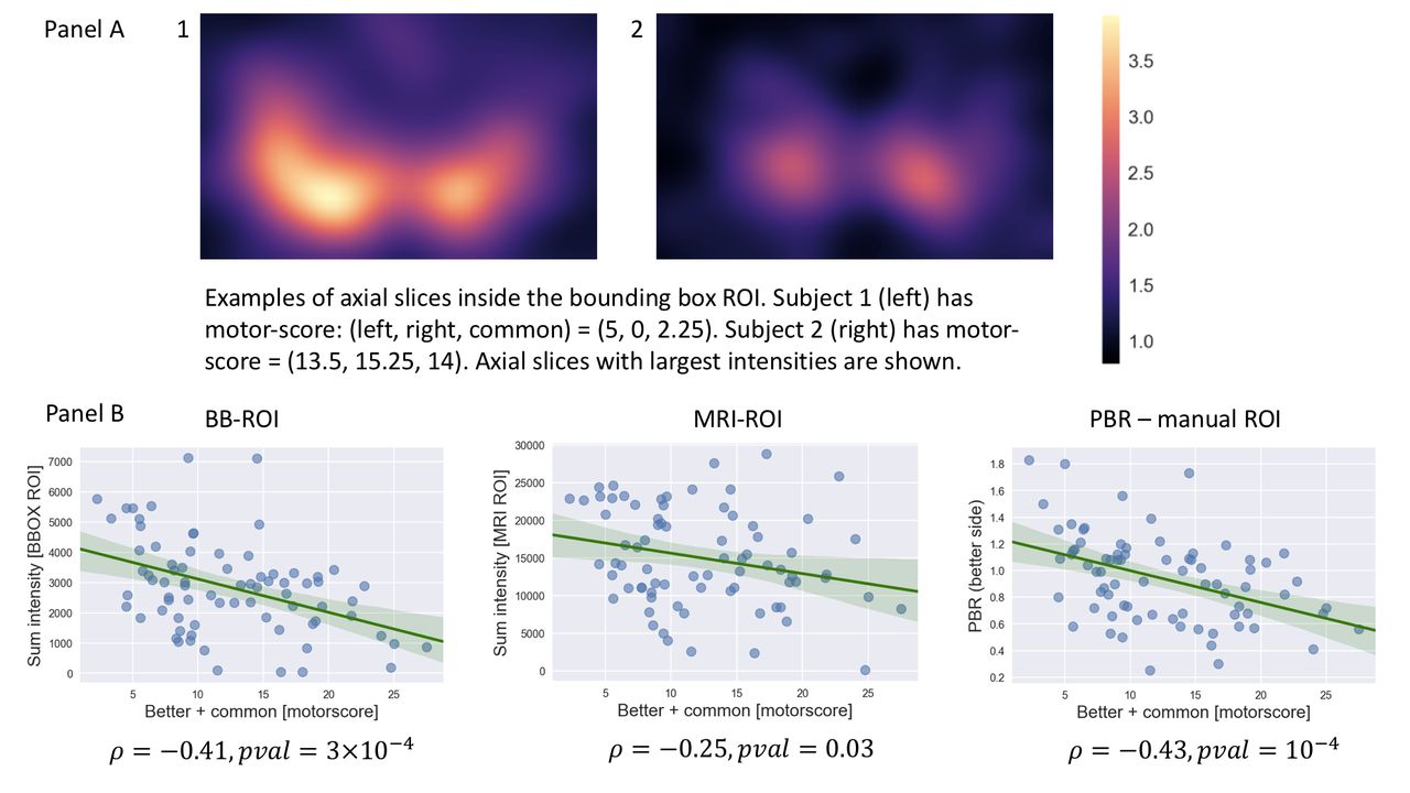
Objectives: Several image metrics can be used to quantify DaTSCAN binding from SPECT scans of subjects with Parkinson’s disease (PD). These metrics are typically calculated on regions of interest (ROIs) that require either coregistration with MR structural images or manual placement. We propose a new approach comprised of a new metric, sum intensity (SI), evaluated over a bounding box (BB) that is automatically placed on SPECT images, thus eliminating the need of MR images or manual ROI placement. We compute the correlation of the SI values with UPDRS motor score of PD subjects. This correlation is compared with the ones obtained (i) by the same metric evaluated over MRI-defined ROIs and (ii) by the commonly used putamen binding ratio (PBR) evaluated over manually placed ROIs, as used in the standard analysis of the Parkinson’s Progression Markers Initiative (PPMI) DaTSCAN data.

Methods: We compute all image metrics on DaTSCAN SPECT images of 76 PD subjects from the PPMI database. SI is computed on the voxels in the more- and less-affected side of the striatum separately. SI is defined as the sum of all voxel intensities in the region that have normalized intensity larger than 2, where 1 is the reference region (occipital cortex) intensity. The following metrics are calculated: (1) SI using a BB ROI, (2) SI using a putamen MRI ROI and (3) putamen binding ratio (PBR) using manual ROI selection. For the SI-BB calculation all SPECT scans are registered to an average healthy control SPECT template. The box-shaped ROI is automatically fitted by finding the region in the image with the highest tracer activity. The box size is (84 x 52 x 24) mm^3 and it encloses the entire striatum (panel A in the figure). For the SI - MRI ROI calculation, each SPECT image is coregistered with the corresponding MR structural image. Automatic segmentation of the MR scan is used to obtain the MR-based striatum ROIs used for the metric calculation. The PBRs were obtained from the PPMI database. These metrics are calculated on manually-placed ROIs from the 8 slices with highest activity of the striatal region. The PBR is the ratio between the average tracer binding in the putamen and that in the occipital cortex. For each subject the motor scores from all clinical exams within 1 year of the imaging date are averaged to reduce clinical exam variability.

Results: Scatterplots of each image metric and motor scores are shown in panel B in the figure. The SI metric achieves stronger correlation with motor score when computed on the BB ROI (Spearman rho = -0.41 for SI-BB and rho = -0.27 for SI-MRI). The SI-BB metric performs very similarly to the PBR which uses manually placed ROIs (rho = -0.43). The strongest correlation across all metrics is found when regressing imaging data from each side against the contralateral motor score components (less affected side + common) and (more affected side + common). Averaging the motor scores of each subject from clinical examinations over a pre-determined time period increases the correlation between them and imaging data by 10-15%, indicating an intrinsic noise in the clinical assessments.

Conclusion: The new SI metric computed on BB ROI performs very similarly to the reference PBR metric, in terms of correlation with motor score. The SI-BB metric might significantly simplify the quantification of SPECT DaTSCAN binding in patients with PD, since it is automatically computed and does not require anatomical guidance provided by MR images. Using averages from multiple UPDRS score assessments may be very beneficial when comparing imaging to clinical disease progression outcomes.

Figure
  • Download figure
  • Open in new tab
  • Download powerpoint
Previous
Back to top

In this issue

Journal of Nuclear Medicine
Vol. 58, Issue supplement 1
May 1, 2017
  • Table of Contents
  • Index by author
Article Alerts
Sign In to Email Alerts with your Email Address
Email Article

Thank you for your interest in spreading the word on Journal of Nuclear Medicine.

NOTE: We only request your email address so that the person you are recommending the page to knows that you wanted them to see it, and that it is not junk mail. We do not capture any email address.

Enter multiple addresses on separate lines or separate them with commas.
A metric to quantify DaTSCAN tracer uptake in subjects with Parkinson’s disease computed without MRI-based regions of interest
(Your Name) has sent you a message from Journal of Nuclear Medicine
(Your Name) thought you would like to see the Journal of Nuclear Medicine web site.
Citation Tools
A metric to quantify DaTSCAN tracer uptake in subjects with Parkinson’s disease computed without MRI-based regions of interest
Nikolay Shenkov, Ivan Klyuzhin, Sima Fotouhi, Esmaeil Davoodi-Bojd, Hamid Soltanian-Zadeh, Arman Rahmim, Vesna Sossi
Journal of Nuclear Medicine May 2017, 58 (supplement 1) 291;

Citation Manager Formats

  • BibTeX
  • Bookends
  • EasyBib
  • EndNote (tagged)
  • EndNote 8 (xml)
  • Medlars
  • Mendeley
  • Papers
  • RefWorks Tagged
  • Ref Manager
  • RIS
  • Zotero
Share
A metric to quantify DaTSCAN tracer uptake in subjects with Parkinson’s disease computed without MRI-based regions of interest
Nikolay Shenkov, Ivan Klyuzhin, Sima Fotouhi, Esmaeil Davoodi-Bojd, Hamid Soltanian-Zadeh, Arman Rahmim, Vesna Sossi
Journal of Nuclear Medicine May 2017, 58 (supplement 1) 291;
Twitter logo Facebook logo LinkedIn logo Mendeley logo
  • Tweet Widget
  • Facebook Like
  • Google Plus One
Bookmark this article

Jump to section

  • Article
  • Figures & Data
  • Info & Metrics

Related Articles

  • No related articles found.
  • Google Scholar

Cited By...

  • No citing articles found.
  • Google Scholar

More in this TOC Section

Instrumentation & Data Analysis Track

  • Deep Learning Based Kidney Segmentation for Glomerular Filtration Rate Measurement Using Quantitative SPECT/CT
  • Comparison of 22 partial volume correction methods for amyloid PET imaging with 11C-PiB
  • The Benefit of Time-of-Flight in Digital Photon Counting PET Imaging: Physics and Clinical Evaluation
Show more Instrumentation & Data Analysis Track

Advanced Image Processing and Analysis

  • Simplified approach for measuring image noise in reconstructed nuclear medicine images using frequency domain masking.
  • Artificial neural network based outcome prediction in DAT SPECT imaging of Parkinson's Disease
Show more Advanced Image Processing and Analysis

Similar Articles

SNMMI

© 2025 SNMMI

Powered by HighWire