


Abstract
123I-5-IA-85380 (123I-5-IA; [123I]-5-iodo-3-[2(S)-azetidinylmethoxy]pyridine) is a promising SPECT radiotracer for imaging β2-containing nicotinic acetylcholine receptors (β2-nAChRs) in brain. β2-nAChRs are the initial site of action of nicotine and are implicated in various neuropsychiatric disorders. The feasibility and reproducibility of the bolus-plus-constant-infusion paradigm for equilibrium modeling of 123I-5-IA using SPECT in healthy nonsmokers was studied. Methods: Ten healthy nonsmokers (mean age ± SD, 43.7 ± 9.9 y) underwent two 123I-5-IA SPECT scans within 4 wk. 123I-5-IA was administered as a bolus (125.8 ± 14.6 MBq) plus constant infusion (18.1 ± 1.5 MBq/h). SPECT acquisitions (30 min) and venous blood sampling were performed every 60 min throughout the infusion (10–14 h). The test–retest variability and reliability of plasma activity (kBq/mL), the regional brain activity reflected by units of kBq/mL and %ID/mL (injected dose/mL brain tissue), and the equilibrium outcome measures VT′ (ratio of total uptake to total plasma parent concentration) and VT (ratio of total uptake to free plasma parent concentration) were evaluated in 4 brain areas, including thalamus, striatum, cortex, and cerebellum. Results: Linear regression analysis revealed that time–activity curves for both plasma and brain 123I-5-IA activity stabilized by 5 h, with an average change of [2.5%/h between 6 and 8 h of infusion, permitting equilibrium modeling. The plasma free fraction (f1), total parent, and clearance demonstrated good test–retest variability (mean, 10.9%–12.5%), whereas the variability of free parent was greater (mean, 24.3%). Regional brain activity (kBq/mL) demonstrated good test–retest variability (11.1%–16.4%) that improved when corrected for infusion rate (mean, 8.2%–9.9%) or for injected dose (mean, 9.5%–13.3%). VT′ demonstrated better test–retest variability (mean, 7.0%–8.9%) than VT (mean, 12.9%–14.6%). Reliability assessed by the intraclass correlation coefficient (ICC) was superior for kBq/mL (ICC = 0.83–0.90) and %ID/mL (ICC = 0.93–0.96) compared with VT′ (ICC = 0.30–0.64) and VT (ICC = 0.28–0.60). The lower reliability of VT was attributed to the poor reliability of the free fraction (ICC = 0.35) and free parent (ICC = 0.68). Conclusion: These results support the feasibility and reproducibility of equilibrium imaging with 123I-5-IA for measurement of β2-nAChRs in human brain.
Many properties necessary for in vivo imaging of nicotinic acetylcholine receptors (nAChRs) with SPECT—including high affinity (dissociation constant [Kd] = 11 pmol/L), rapid entry into brain, low nonspecific binding, and minimal toxicity—are demonstrated by 123I-5-IA-85380 (123I-5-IA; [123I]-5-iodo-3-[2(S)-azetidinylmethoxy]pyridine) (1–12). In brain, the highest uptake is in thalamus, with moderate to low levels in striatum, hippocampus, cerebellum, and cortex (10). The pharmacologic specificity of 123I-5-IA for β2-nAChRs was confirmed by the lack of binding in brain of β2 (−/−) knockout mice (5). 123I-5-IA predominantly labels α4β2-nAChRs in brain. 123I-5-IA uptake is displaced by endogenous acetylcholine (9) and the agonists (−)-nicotine and cytisine but not by noncompetitive nAChR antagonists mecamylamine and bupropion or the muscarinic antagonist (−)-scopolamine (1,2,4,5,13), demonstrating that 123I-5-IA binds to the agonist and not the noncompetitive antagonist site on β2-nAChRs.
123I-5-IA may be administered safely to human subjects at doses as high as 555 MBq on one occasion up to 5 times per year (8). This margin of safety is better than the corresponding PET radiotracer 2-18F-fluoro-3-(2-(S)azetidinylmethoxy)-pyridine, where only 185 MBq may be administered on one occasion up to 4 times per year (14). SPECT radioligands are advantageous because they have longer half-lives and longer imaging time frames (e.g., peak uptake at 50–150 min may be problematic for 11C- and 18F-labeled compounds with shorter half-lives), which provide the capability to measure dynamic processes at the receptor for many hours. SPECT is often criticized for poorer spatial resolution and limited quantitation; however, recent advances in instrumentation and corrections for photon attenuation (15) and scatter (11,16) have established the validity of neuroreceptor SPECT. In particular, paradigms using a constant infusion provide a robust method for determining the density of binding sites comparable to PET (17).
In humans, 123I-5-IA uptake peaks in brain by 2 h, with a gradual return to baseline levels over at least 8 h (10). The accuracy of 123I-5-IA SPECT for β2-nAChR quantitation was validated by good agreement between nAChR density in postmortem human brain and regional distribution volumes obtained by compartmental modeling of 123I-5-IA brain uptake and corresponding arterial input function (12) and also by the high correlation (r = 0.98) between distribution volumes measured using 123I-5-IA SPECT and 6-18F-fluoro-3-(2-azetidinylmethoxy)pyridine (18F-6-FA) PET (18). In the present study, the feasibility of a constant infusion paradigm for equilibrium modeling of 123I-5-IA with SPECT and the reproducibility of quantification of nAChR for 123I-5-IA SPECT were examined.
MATERIALS AND METHODS
Subjects
Ten healthy subjects including 3 men (32–50 y) and 7 women (20–60 y) provided written informed consent provided written informed consent. This study was approved by the Yale University School of Medicine Human Investigation Committee, Radiation Safety Committee, the West Haven Veteran Affairs Connecticut Healthcare System Human Subjects Subcommittee, and the Food and Drug Administration. Eligibility for the study was determined by a physical examination; laboratory blood tests, including complete blood counts, liver and thyroid function; urine drug screen; and an electrocardiogram. Study participants had no history or presence of a serious medical, neurologic, or psychiatric illness and had no implanted metallic devices. Female participants of child-bearing potential had negative pregnancy tests. Each subject participated in two 123I-5-IA SPECT studies separated by 10.7 ± 6.8 d (range, 5–28 d).
Radiopharmaceutical Preparation and Plasma Measurements
123I-5-IA was prepared as described previously (19) to give a product with radiochemical purity of 95.8% ± 2.2% and 97.2% ± 1.6% and an overall yield of 44.0% ± 20.0% and 45.0% ± 18.5% for the test and retest studies, respectively. Specific activity was >185 MBq/nmol. The plasma parent, metabolite fractions, and free fraction (f1) were measured in venous blood samples (19).
Clearance of 123I-5-IA in L/h was calculated as R0/Css, where R0 is the infusion rate of the radiotracer in kBq/h and Css is the total (free and protein bound) tracer concentration in kBq/mL in plasma at equilibrium (20).
Data Acquisition
Subjects were pretreated with stable supersaturated potassium iodide (800 mg). Two antecubital venous catheters were placed and 123I-5-IA was administered by a bolus (125.8 ± 14.8 MBq) and continuous infusion (18.1 ± 1.5 MBq/h) at a constant rate using a computer-controlled pump (Gemini PC-1; IMED pump) for a bolus-to-infusion ratio of 6 h in subject 1 and 7 h in the remaining 9 subjects (Table 1). Before scanning, 5 external fiducial markers containing 0.04–0.19 MBq of 123I were placed on the head along the canthomeatal line.
Radiotracer Doses, Variability, and Reliability of Plasma Activity
The pharmacologic safety of 123I-5-IA administration was assessed by standard laboratory blood tests and urinalysis within 2 mo before the test scan and 24 h after each scan. Vital signs, including electrocardiogram, blood pressure, heart rate, and respiration rate, were measured 30–60 min before, within 5 min, and 30–60 min after 123I-5-IA administration.
Simultaneous transmission and emission scans were acquired with a line source containing 740 MBq 57Co on a Picker PRISM 3000XP, 3-head camera (Philips). SPECT acquisitions (30 min) and venous blood sampling were performed every 60 min starting at 2 h after the bolus and continuing throughout the infusion (14 h for subjects 1 and 2 and 10 h for subjects 3–9). The sensitivity of the PRISM 3000XP equipped with a low-energy, ultra-high-resolution fanbeam collimator (photopeak window, 159 keV ± 10%; matrix, 128 × 128) is uniform across the field of view. To control for day-to-day variation in camera sensitivity, a 57Co-distributed source was measured with each experiment. A sealed aliquot of this solution (18.5 kBq) was also measured using the γ-counter (1282 Compugamma; Wallac Inc.) used for measuring the plasma activity for cross-calibration of SPECT counts and plasma counts. This system provides a transaxial resolution and an axial resolution of 14.3- and 18.1-mm full width at half maximum, respectively, measured with 123I line sources and water in a cylindric phantom.
MRI
To identify brain regions on the 123I-5-IA SPECT scan, sagittal MR images were obtained on a 1.5-T Signa camera (GE Healthcare) with a spoiled GRASS (gradient recall acquisition in the steady state) sequence with echo time = 5 ms, repetition time = 24 ms, number of excitations = 1, matrix = 256 × 192, and field of view = 34 cm.
Image Analysis
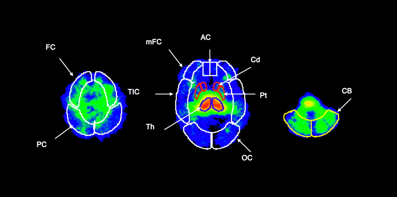
SPECT emission images were filtered using a 3-dimensional (3D) Butterworth filter (order = 10, cutoff frequency = 0.24 cycle/pixel) and reconstructed using a filtered backprojection algorithm with a ramp filter on a 128 × 128 matrix to obtain 50 slices with a pixel size of 2.06 × 2.06 × 3.56 mm in the x-, y-, and z-axes. Nonuniform attenuation correction was performed (15). The MR image was coregistered to the SPECT image using the coregistration function in SPM99 (Statistical Parametric Mapping, version 99). A coregistered MR image was used to guide the placement of standard 2-dimensional ROI templates. A 3D volume of interest (VOI) was generated for each region and transferred to the coregistered SPECT image to determine regional radioactive densities (cpm/pixel) (Fig. 1). Test–retest analyses were performed on the mean of all cortical brain areas, including frontal, parietal, anterior cingulate, temporoinsular, and occipital cortices (cortex) and also on the mean of the caudate and putamen (striatum).

Two-dimensional standardized regions of interest are placed on 3 representative transverse images of 123I-5-IA uptake. FC = frontal cortex; PC = parietal cortex; mFC = medial frontal cortex; TIC = temporoinsular cortex; AC = anterior cingulate; OC = occipital cortex; Cd = caudate; Th = thalamus; Pt = putamen; CB = cerebellum.
Outcome Measures
Regional SPECT activity measurements (cpm/pixel) were converted to absolute units of activity (kBq/mL) as described previously (10). The percentage change per hour was calculated by dividing the slope for the activity values (6–8 h, the earliest time steady state was apparent) by the mean activity (6–8 h) (21). The test–retest reproducibility of the following was evaluated: plasma (clearance, f1, total parent, and free parent) and regional brain activity (kBq/mL, uncorrected and corrected for infusion rate); %ID/mL (percentage injected dose/mL brain tissue); VT′ (ratio of total uptake to total plasma parent concentration, calculated as [regional brain activity]/total plasma parent activity between 6 and 8 h) and VT (ratio of total uptake to free plasma parent concentration, calculated as [regional brain activity]/free plasma parent. Regional brain activity (kBq/mL) was divided by the infusion rate (kBq/h) of the radiotracer to normalize differences in brain activity caused by different infusion rates for test–retest comparisons.
The equilibrium volume of distribution of a compartment is defined as the ratio of the tracer concentration in the compartment to the free tracer in plasma at equilibrium (Vi = Ci/f1Ca). VT is the tissue equilibrium volume of distribution equal to the sum of V2 (nondisplaceable) and V3 (receptor bound) at equilibrium of a tracer dose. Bound/Free = maximum density of binding sites/affinity of radiotracer = binding potential or (B/F = Bmax/Kd = BP). VT is measured directly by measuring the ratio of brain activity (average values between 6 and 8 h) divided by the plasma free activity (kBq/mL brain/kBq/mL blood) (17,22).
Statistical Analysis
Within-subject variability between test and retest conditions was calculated as the absolute value of the difference of the test and retest measure divided by the mean of the test and retest and expressed as a percentage. Repeated-measures ANOVA was used to evaluate within- and between-subject differences for each plasma and brain measure for the 10 pairs of scans on the basis of time. The reliability of the outcome measures was determined relative to between-subject variance by calculation of the intraclass correlation coefficient (ICC or ρ) as described (23). Interrater reliability was determined for 4 raters who analyzed 6 scans by the ICC.
RESULTS
Pharmacologic Effects
All subjects completed each scan session without symptoms from the injection. There were no changes in electrocardiogram, blood pressure, heart rate, and respiration rate or in blood or urinalysis measures relative to baseline measurements in any of the study subjects after 123I-5-IA administration.
Feasibility of Bolus-Plus-Constant-Infusion Paradigm
Time–activity curves for total and free plasma 123I-5-IA activity stabilized after 3 h with rates of change (1.9%/h ± 4.0%/h) between 6 and 8 h of infusion. Time–activity curves for brain 123I-5-IA activity stabilized after 3 h in striatum, cortex, and cerebellum and 5 h in thalamus. Rates of change were low for cerebellum (0.2%/h ± 1.9%/h), cortex (0.6%/h ± 2.2%/h), striatum (1.2%/h ± 3.0%/h), and thalamus (2.5%/h ± 2.7%/h) and supported equilibrium modeling between 6 and 8 h of infusion.
Variability and Reliability of Clearance, f1, and Plasma Parent
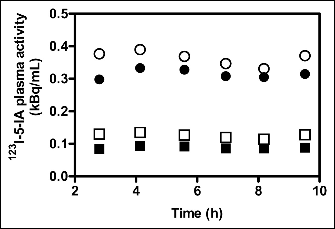
Clearance of plasma parent demonstrated good test–retest variability (12.3%) and reasonable reliability (ICC = 0.71, Table 1; Fig. 2). Variability was good for total parent and the f1 (12.5% and 10.9%, respectively) and poor (24.3%) for free parent. Within-subject variability was not significantly different for any measure. Reliability was better for total parent (ICC = 0.81) than for free parent (ICC = 0.68) because of the poor reliability of the f1 measurement (ICC = 0.35).

Total plasma parent (•, ○) and free parent (▪, □) 123I-5-IA activity are illustrated for test (filled symbols) and retest (open symbols) of representative subject. 123I-5-IA was administered using bolus-plus-constant-infusion paradigm (bolus-to-infusion ratio = 6.94 and 6.96 for test and retest studies, respectively).
Variability and Reliability of Regional Brain Activity and Outcome Measures
Mean regional activity values (kBq/mL) ranged from 5.3 to 13.5 kBq/mL and mean %ID/mL ranged from 0.0014 to 0.0038 %ID/mL for low-density (cortex) and high-density (thalamus) regions, respectively (Fig. 3; Table 2). The test–retest variability was 11.1%–16.4% and the reliability (ICC) ranged from 0.83 to 0.90 for regional brain activity (kBq/mL). The variability improved (range, 8.2%–9.9%) when the regional brain activity (kBq/mL) was corrected for the infusion rate, whereas the reliability (ICC) did not change. The %ID/mL (<13.3% mean test–retest variability) demonstrated good variability (range, 9.5%–13.3%) and excellent reliability (ICC = 0.93–0.96) because of the correction for injected dose. There were no statistically significant differences in any measure between the test and retest studies.
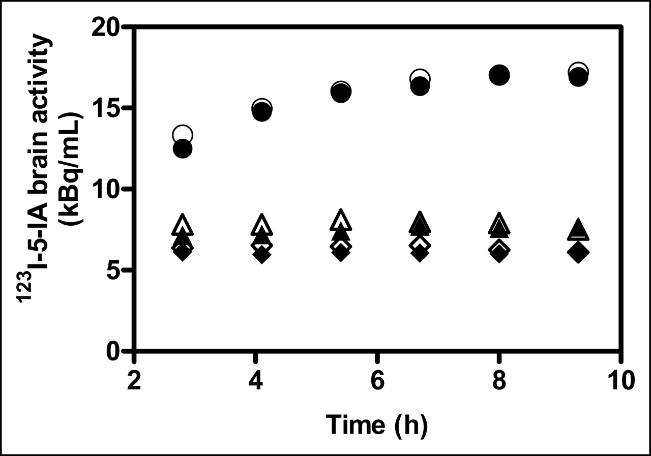
123I-5-IA activity in thalamus (•, ○), striatum (▴, ▵), and cortex (♦, ⋄) is illustrated for test (filled symbols) and retest (open symbols) of representative subject.
Variability and Reliability of Brain Activity
The mean VT′ and VT values calculated between 6 and 8 h of infusion ranged from 18.2 to 47.1 and 49.2 to 139.4 for low-density (cortex) and high-density (thalamus) brain regions, respectively (Table 3). The test–retest variability (range, 7.0%–8.9%) and the reliability (ICC = 0.30–0.64) for VT′ were good and better than the variability (range, 12.9–14.6) and reliability (ICC = 0.28–0.60) observed for VT. The greater variability for VT relative to VT′ is attributed to higher variability of the f1. There was no significant difference in VT′ and VT between test and retest studies.
Variability and Reliability of Brain Outcome Measures
Interrater Reliability of Region-of-Interest Measurements
Interrater differences in image analysis may also alter reliability. The accuracy of the analysis for regional brain 123I-5-IA uptake measurements between 4 different raters for the test and retest 123I-5-IA images from 3 subjects (subjects 2, 6, and 7) was evaluated. Calculation of the ICCs indicated that measures of each region of interest were highly reliable, with ICC values > 0.95 for thalamus, striatum, cortex, and cerebellum. These findings indicate excellent reproducibility between raters for the image-processing steps, including MR coregistration, reorientation to the anterior commissure–posterior commissure line, and also region-of-interest analyses.
DISCUSSION
Equilibrium Imaging of 123I-5-IA SPECT
This study evaluated the feasibility and reproducibility of the bolus-plus-constant-infusion (B/I) paradigm for equilibrium imaging and associated outcome measures using 123I-5-IA SPECT. In the equilibrium model, a steady state of radiotracer in blood is established and maintained by constant infusion so that steady state between free radiotracer in blood and brain and between the specifically bound compartment and free radiotracer in brain is established (17,22,24,25). The equilibrium paradigm offers several advantages over the more commonly used kinetic compartmental model, including the following: (a) equilibrium measures are not influenced by differences in blood flow that may differ between clinical populations; (b) the calculation of a volume of distribution equivalent does not require derivation of transfer rate constants that are difficult to identify because of the greater variability inherent in quantitation of nonlinear kinetics; (c) equilibrium outcome measures are insensitive to the ratio of peripheral clearance and brain washout because the terminal half-life is zero at equilibrium; (d) no arterial blood sampling is required; and (e) computation procedures are more straightforward, simpler, and readily amenable to within-subject receptor occupancy studies (17,25). This paradigm has been implemented successfully for other SPECT radiotracers—123I-iomazenil (17,26,27), 123I-iodobenzamide (28), and 123I-epidepride (20)—and PET radiotracers—18F-cyclofoxy (22), 18F-altanserin (29,30), 18F-deuteroaltanserin (21,30,31), 18F-fallypride (32), and 11C-raclopride (33).
To evaluate the B/I paradigm for 123I-5-IA binding to β2-nAChRs, 4 parameters were assessed: (a) time required for radiotracer binding to reach stable levels throughout all brain regions, including low-density (cortex) and high-density (thalamus) areas; (b) percentage change per hour of regional brain uptake across the infusion time frame; (c) accuracy of measurement of activity levels in low-density (cortex) regions during equilibrium; and (d) reproducibility of plasma and brain activity and the outcome measures (VT′ and VT) during equilibrium. Stable 123I-5-IA levels in plasma and brain were established by approximately 5 h of infusion in all subjects. Equilibrium defined as <5% change per hour in 123I-5-IA activity was established earlier (<3 h) in lower density cortical brain areas and later (∼5 h) in the high-density thalamus. The time frame of 6–8 h was chosen to evaluate equilibrium measures because both plasma and regional brain activities appeared to have achieved steady state, as demonstrated by a mean change of <2.5%/h, and because this interval is reasonable for clinical studies. Brain regions with lower receptor densities (e.g., cerebellum and cortex) all demonstrated <1%/h differences, whereas the high-density thalamus demonstrated a change of 2.5%/h between 6 and 8 h of infusion. This change per hour was less than our a priori criterion of <5%/h and is similar to equilibrium studies with other radiotracers (17,20,21,27,31). This finding differs from an earlier study that stated stable levels of 123I-5-IA uptake did not occur until 12–14 h (10). This conclusion was based on a smaller number of subjects (n = 6) for whom different B/I ratios (range, 4–11.4 h) were used. The extreme range in B/I ratios was chosen based on simulation of the kinetic bolus data and represents the optimal B/I ratio for equilibrium imaging of high-density (B/I = 11.4 h) and low-density (B/I = 4.0 h) brain regions. The wide-ranging B/I ratios resulted in highly variable times that equilibrium was achieved and, in some cases, it was not achieved. In the present study, a constant B/I ratio (7.0 h) was used for most subjects (9/10) and equilibrium was routinely established by 5–6 h across the 10 subjects.
The time (6–8 h) to reach stable levels of binding throughout the brain may present the caveat that counts in low-density brain areas (cortex) may be too low to be measured with high reproducibility (10). Our analyses demonstrated very stable activity measures throughout the cortex, with variability and reliability in the same range as other PET and SPECT radiotracers (20,31,34), suggesting that counts are adequate to reliably measure cortical nAChR using 123I-5-IA SPECT. This conclusion differs from the earlier study (10) that claimed that cortical counts were too low for reliable measurements. The different conclusions may be due to the time that cortical activity was evaluated (e.g., 6–8 h in the present study and 12–14 h in the earlier study) or also may be due to the application of a scatter correction in the earlier study that resulted in low counts in low-density cortical brain areas. Though, theoretically, scatter correction increases the accuracy of SPECT measurements, this correction may introduce error in the precision of the measurement of low-density regions by decreasing “good” counts and increasing variability. However, because scatter correction enhances the theoretical accuracy, it needs to be evaluated in a test–retest study.
Reproducibility of Plasma and Brain Outcome Measures
The test–retest variability and reliability of plasma activity and regional brain activity and outcome measures were evaluated for 123I-5-IA SPECT. The plasma total parent, f1, and clearance of 123I-5-IA all demonstrated good variability (<12.5%), whereas the variability for the free parent was not as good (24.3%). The reliability of the total parent was good (ICC = 0.81), whereas the reliability of the f1 was poor (ICC = 0.35) because of high within-subject variability associated with this measurement that has been observed for other radiotracers (20,31,35). Moreover, the poor reliability of the f1 measurement likely contributed to the lower reliability of free parent measurements (ICC = 0.68). Improved methods for measurement of free parent would significantly decrease the variability associated with VT.
Regional brain activity values (kBq/mL) demonstrated good test–retest variability (<16.4%) that improved after correction for infusion rate (<9.9%). Overall reliability for all brain activity measures was good (ICC > 0.82). The %ID/mL also demonstrated good test–retest variability (<13.3%) and excellent reliability (ICC > 0.93), likely because of the correction for differences in injected dose. The test–retest variability of VT′ was excellent (<8.9%); however, the reliability was not as good (ICC = 0.30–0.64). The test–retest variability for VT was also good (<14.6%), and the reliability was less notable (ICC = 0.28–0.60). The lower variability and reliability observed for VT is in keeping with the increased variability that occurs due to the assessment of the percentage of plasma protein binding. Thus, VT′ demonstrated the best reproducibility for assessment of 123I-5-IA binding to β2-nAChRs in brain. VT′ and VT are proportional to Bmax/Kd under equilibrium conditions and the assumption that plasma protein binding and nondisplaceable uptake are uniform between individuals. If group differences in plasma protein binding exist, VT is the appropriate outcome measure and will require larger sample sizes to override the higher variability and poorer reliability.
Overall, the test–retest variability of the outcome measures studied was very good (<16%, with the exception of the free parent at 24%), whereas the reliability was not as impressive. Variability is a measure of the absolute difference between the test and retest scans. In contrast, reliability assessed by the ICC compares within-subject differences and between-subject differences and is more robust. This phenomenon of good variability and poor reliability has been noted previously (34) and occurs when the within-subject variability is similar to the between-subject variability. Given the narrow age range and relatively homogeneous sample studied herein, this is not surprising. Importantly, the variability observed for plasma and brain measures is within the same range as other PET and SPECT radiotracers (31,34,35).
VT′ and VT do not correct for nondisplaceable uptake. Preclinical studies in nonhuman primates have indicated that there is no gray matter background region for 123I-5-IA (1,4,5,13). The cerebellum, which is often used as a background region, demonstrated at least 35% displacement of 123I-5-IA uptake by nicotine or the nicotinic agonist cytisine in nonhuman primates (4). This finding has recently been confirmed in human subjects, where up to 70% of 123I-5-IA cerebellar uptake was displaced by the nicotine delivered from smoking 2 consecutive cigarettes (E. Mitsis and J.K. Staley, unpublished data). White matter may be used to measure nondisplaceable uptake, but the related PET radiotracer 18F-6-FA demonstrated specific binding in white matter tracts but not the corpus callosum (36). This question will be addressed with 123I-5-IA SPECT in human subjects by measuring the absolute nondisplaceable uptake (V2) after the administration of saturating doses of nicotine. In summary, in the absence of a background region, VT′ and VT may be used as outcome measures under the assumptions that nondisplaceable uptake is uniform between individuals.
CONCLUSION
A constant infusion paradigm with 123I-5-IA and SPECT in healthy human nonsmokers yielded stable time–activity curves for both plasma and brain by 5 h, supporting equilibrium modeling. This paradigm also demonstrated low test–retest variability and reasonable reliability across several outcome measures. These findings support the feasibility of equilibrium imaging with 123I-5-IA SPECT for measurement of β2-nAChRs in subcortical and cortical brain areas. Further studies of the reproducibility of these measures with scatter correction would be useful.
Acknowledgments
We thank Eileen Smith, Gina Morano, Andrea Perez, Stacey Ross, Jane Bartosik, Louis Amici, Nina Sheung, and Suzanne Giddings for technical support. This research was supported by National Institute on Alcohol Abuse and Alcoholism K01AA00288, National Institute on Drug Abuse P50 DA13334, Veterans Affairs Research Enhancement Award Program (REAP), and Veterans Affairs Mental Illness Research Education and Clinical Centers (MIRECC).
Footnotes
Received Mar. 10, 2005; revision accepted May 25, 2005.
For correspondence or reprints contact: Julie K. Staley, PhD, Yale University and Veterans Affairs Connecticut Healthcare System 116A2, 950 Campbell Ave., West Haven, CT 06516.
E-mail: julie.staley{at}yale.edu
REFERENCES
In this issue


Jump to section
Related Articles
Cited By...
- Imaging Changes in Synaptic Acetylcholine Availability in Living Human Subjects
- Quantification of Smoking-Induced Occupancy of {beta}2-Nicotinic Acetylcholine Receptors: Estimation of Nondisplaceable Binding
- 123I-5-IA-85380 SPECT Imaging of Nicotinic Receptors in Alzheimer Disease and Mild Cognitive Impairment
- 123I-5-IA-85380 SPECT Imaging of Nicotinic Acetylcholine Receptor Availability in Nonsmokers: Effects of Sex and Menstrual Phase
- Human Tobacco Smokers in Early Abstinence Have Higher Levels of beta2* Nicotinic Acetylcholine Receptors than Nonsmokers.