


Abstract
Previous studies have shown that vertebral bone metastases (BM) not seen on planar bone scintigraphy (BS) might be present on 18F-fluoride PET scans or at MRI. Therefore, we evaluated the effect of SPECT or 18F-labeled NaF PET (18F PET) imaging on the management of patients with newly diagnosed lung cancer. Methods: Fifty-three patients with small cell lung cancer or locally advanced non–small cell lung cancer were prospectively examined with planar BS, SPECT of the vertebral column, and 18F PET. MRI and all available imaging methods, as well as the clinical course, were used as reference methods. BS with and without SPECT and 18F PET were compared using a 5-point scale for receiver operating characteristic (ROC) curve analysis. Results: Twelve patients had BM. BS produced 6 false-negatives, SPECT produced 1 false-negative, and 18F PET produced no false-negatives. The area under the ROC curve was 0.779 for BS, 0.944 for SPECT, and 0.993 for 18F PET. The areas under the ROC curve of 18F PET and BS complemented by SPECT were not significantly different, and both tomographic methods were significantly more accurate than planar BS. As a result of SPECT or 18F PET imaging, clinical management was changed in 5 patients (9%) or 6 patients (11%), respectively. Conclusion: As indicated by the area under the ROC curve analysis, 18F PET is the most accurate whole-body imaging modality for screening for BM. Routinely performed SPECT imaging is practicable, is cost-effective, and improves the accuracy of BS.
In lung cancer, bone metastases (BM) are present in 20%–30% of patients at initial diagnosis and in 35%–66% at autopsy (1–3). Non–small cell lung cancer (NSCLC) without distant metastases is potentially curable. Approximately 20%–25% of lung cancers are of the small cell type (SCLC). Although the therapy of SCLC remains palliative, the selection of the appropriate therapy regimen depends on pretherapeutic staging. Hence, accurate staging of the skeleton is crucial in all patients with lung cancer and increased probability of having BM.
MRI was reported to reveal vertebral BM earlier than does conventional planar bone scintigraphy (BS) (3,4). PET using 18F-labeled NaF (18F PET) has been shown to be significantly more accurate in detecting BM than is BS (5,6). An evaluation of the effect of the superior accuracy of 18F PET or MRI on patient management has not yet been reported. Consequently, 18F PET and MRI are not currently recommended for routine use. Recent studies have suggested that the sensitivity of BS might be improved by the routine performance of additional SPECT imaging (7–9). Hence, complementing planar BS with SPECT imaging of the vertebral column in all patients with increased risk of metastatic bone disease might be an accurate and cost-effective alternative to 18F PET or MRI. The aim of this prospective study was to compare the diagnostic accuracy of 18F PET and BS with and without SPECT at the initial staging of lung cancer and to determine the effect on patient management.
The vertebral column is the most commonly affected region in patients with BM. Although destruction of the pedicles is a common sign of BM on plain films, the disease begins at the vertebral body (10,11). MRI is accepted as the most accurate imaging modality in detecting BM at the vertebral body (12–14). Therefore, MRI of the vertebral column, complemented by the panel of all available imaging methods and the clinical course, was used as the gold standard.
Materials and Methods
Patients
Our study consisted of 53 patients. Patients were included when SCLC (n = 12) or locally advanced NSCLC (stage III, n = 41) were diagnosed through bronchoscopy and CT. We studied 53 patients (42 men, 11 women; age range, 43–78 y; median age, 63 y; mean age, 63.2 y). A history of extrapulmonary cancer, known metastatic bone disease, NSCLC at stages lower than stage III of the Union Internationale Contre le Cancer, pregnancy, or an age of <18 y were exclusion criteria. All patients gave written consent to participate in this prospective study. The study was approved by the local ethical committee.
Bone Scanning
Two modern double-head gamma cameras (ECAM and Bodyscan; Siemens, Erlangen, Germany) were used. The axial field of view was 40 cm for both cameras. Low-energy, high-resolution collimators (1,024 × 256 matrix) were used for planar BS and for SPECT. Data acquisition was started 3 h after intravenous injection of 740–1,000 MBq 99mTc-methylene diphosphonate. At least 1.5 million counts were required for each gamma camera detector for planar imaging.
Two additional SPECT acquisitions of the cervicothoracic and thoracolumbar spine were performed on all patients. For SPECT imaging, a double-head gamma camera (ECAM; 128 × 128 matrix; 64 steps; 150,000–200,000 counts per step; Butterworth filter; cutoff level, 0.5) was used. The total acquisition time ranged from 25 to 35 min for planar BS and from 120 to 150 min for the combination of BS and SPECT. The bone-scanning procedure was performed in accordance with procedure guidelines published by the Society of Nuclear Medicine (15).
18F PET Imaging
18F PET imaging was performed using a modern PET camera (ECAT EXACT HR+; Siemens/CTI, Knoxville, TN). The emission scan was started 75–180 min after intravenous injection of 370–555 MBq 18F-labeled NaF. Attenuation correction was not performed. An iterative algorithm (16) was used for image reconstruction. The 18F PET scans included 6–7 bed positions (12-min acquisition time per bed position; total acquisition time, 72–84 min) covering the skull, neck, arms, thorax, pelvis, and proximal femora. Coronal, transverse, and sagittal sections and maximum intensity projection images were documented in hard-copy form.
MRI Protocol
MRI examinations of the cervicothoracic spine, thoracolumbar spine, and lumbar spine/sacrum (MR Vision; Siemens, Erlangen, Germany) were performed on all patients. Each region was imaged in 2 perpendicular planes with a T1-weighted spin-echo sequence (Body Array [Siemens]; repetition time, 532 ms; echo time, 15 ms; 5-mm slices; gap, 0.5 mm) and a fat-suppressed T2-weighted sequence (Turbo Inversion Recovery TIRM [Siemens]; repetition time, 5,000 ms; echo time, 60 ms; inversion time, 140 ms; flip angle, 180°; 5-mm slices; gap, 0.1 mm). In lesions indicative of BM, one of the spin-echo sequences was repeated after intravenous application of 0.2 mmol per kilogram of body weight gadolinium (Magnevist; Schering, Berlin, Germany) to verify typical contrast enhancement of BM.
Interpretation of BS, SPECT, and 18F PET
Two nuclear medicine physicians interpreted 18F PET, and 2 other nuclear medicine physicians interpreted BS complemented by SPECT. Planar BS was interpreted without SPECT by 2 other nuclear medicine physicians. The experienced readers of BS, SPECT, and 18F PET were unaware of the findings of each other. The results of all imaging methods were made available to the 2 diagnostic radiologists who interpreted MRI results.
With 18F PET, BS, and SPECT, lesions were classified as arthritis when they were located at joints. Increased tracer uptake on the edge of vertebral bodies adjacent to disk spaces was interpreted as indicating osteophytes. Lesions not located at joints or showing typical linear tracer uptake of fractured endplates were interpreted as BM. Interpretation of BS and SPECT was performed following the criteria described by Krasnow et al. (17).
Definition of Metastatic Bone Disease
Patients were defined as having no BM when BS, SPECT, 18F PET, or MRI did not show BM. Typical gadolinium enhancement at hyperintense lesions in fat-suppressed, T2-weighted images was defined as BM. Lesions not detectable on planar BS but showing the typical pattern of BM from SPECT or 18F PET and from MRI were defined as metastases. Lesions that were unclear at MRI but negative according to each scintigraphic method were assessed with FDG PET and with spiral CT. In the case of negative FDG PET and spiral CT results, these patients underwent curative surgery and the results of MRI were assessed by autopsy (1 patient) or evaluated by the clinical course (1 patient).
Data Analysis
PET and BS with and without SPECT were compared on a patient basis. All patients were judged on a 5-point scale as definitively having BM (score of 1), probably having BM (score of 2), being equivocal (score of 3), probably not having BM (score of 4), and definitively not having BM (score of 5). Receiver operating characteristic (ROC) curve analysis (18) was performed, and the area under the curve was used to test for statistically significant differences between BS, SPECT, and 18F PET in staging patients to be M1 or M0 on the bone site. A probability value of <0.05 was defined as statistically significant (18).
Results
Accuracy of BS With and Without SPECT and 18F PET Imaging
Twelve patients (23%) had metastatic bone disease. With planar BS, only 5 patients were classified correctly as having BM. Six patients were falsely interpreted as negative and 5 patients as equivocal, 2 of whom had BM. Thirty-five patients were defined correctly as being free of BM. Two patients with degenerative lesions were falsely interpreted as having BM.
The sensitivity in detecting BM was significantly improved by SPECT images because vertebral BM were detected in 5 of the 6 patients that were false-negative according to planar BS. Fifty-two patients were correctly interpreted with 18F PET and 1 patient with a single rib metastasis was interpreted as equivocal with SPECT, BS, and 18F PET. The results of planar BS, BS complemented with SPECT, and 18F PET are summarized in Table 1.
Results of Planar BS With and Without SPECT and 18F PET
The areas under the ROC curve were 0.779 (SD, 0.078) for planar BS, 0.944 (SD, 0.043) for BS complemented with SPECT, and 0.993 (SD, 0.008) for 18F PET. The diagnostic accuracy of both tomographic imaging modalities was significantly higher than that of planar BS alone (P < 0.05). The difference between the areas under the ROC curves for 18F PET and for SPECT was statistically not significant.
Changes in Patient Management
As a result of the improved imaging performance of 18F PET and MRI, staging of 3 patients with SCLC and of 3 patients with NSCLC who had BM and normal planar BS was changed (Figs. 1 and 2). Therapy was changed from curative surgery to palliative chemotherapy in the 3 patients with NSCLC. In the 3 patients with SCLC, another chemotherapy regimen was indicated because staging was changed from limited disease to extended disease. Using SPECT instead of 18F PET, BM would have been missed in only 1 of the patients. Compared with the results obtained with 18F PET, the extent of metastatic bone disease was underestimated in 7 of 12 patients (58%) with the combination of BS and SPECT. However, this had no influence on patient management.

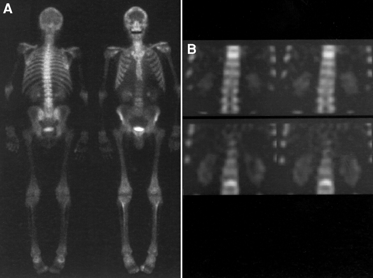
A 53-y-old man with NSCLC. (A) Planar BS was interpreted as normal (score 5). (B) Coronal SPECT images presented lesions at lumbar and lower thoracic spine probably indicative of BM (score 2).

Same patient as shown in Figure 1. 18F PET (left, maximum intensity projection images; middle, sagittal; right, coronal) presented BM in vertebral bodies L3 and T10. Both BM were confirmed by MRI.
In summary, as a result of 18F PET and MRI, the clinical management was changed in 6 of the 12 patients with BM (50%; 11% of all patients). Complementary SPECT imaging of the cervicothoracic and thoracolumbar spine altered patient management in 5 of the 12 patients with BM (42%; 9% of all patients).
Discussion
Radionuclide bone scanning using technetium-labeled polyphosphonates was introduced into clinical practice in the 1970s and was shown to detect BM several months earlier than plain radiographs. Therefore, BS has become one of the most frequently performed nuclear medicine procedures in Europe and the United States. However, the number of BS procedures used in oncology has been reduced because the prevalence of BM in patients with early tumor stages is low and early treatment of metastatic bone disease does not necessarily improve the survival rate. Furthermore, several studies that compared the sensitivity of planar BS with that of MRI have shown that planar BS is less sensitive than previously accepted (3,4,12–14,19).
The limited accuracy of planar BS was confirmed in our study because planar BS produced false-negatives in 50% of the patients with BM. However, complementing BS with routinely performed SPECT images improved the sensitivity of BS significantly (Table 1). This altered treatment in 5 patients. Only 1 patient had BM that were missed with that combination but were present on MRI and 18F PET. Whole-body imaging with 18F PET and the combination of BS with SPECT were significantly more accurate than planar BS, as indicated by the ROC curve analysis. 18F PET and MRI revealed more metastatic lesions than the combination of planar BS and SPECT in 7 patients. However, this had no influence on patient treatment.
Two recent studies indicated that the sensitivity of planar BS depends on the anatomic localization of the BM (5,20). Steinborn et al. (20) reported that whole-body MRI was more sensitive than planar BS in the spine and pelvis, whereas BS revealed more BM in the skull and ribs. A lesion-based comparison with 18F PET indicated that the sensitivity of planar BS in detecting vertebral BM was as low as 40%. In contrast, the sensitivity ranged from 80% to 90% in the skull, thorax, and extremities (5).
Several studies reported a low sensitivity of planar BS in detecting BM when comparing planar BS with MRI of the vertebral column (12–14). However, the interpretation of this finding is limited because a comparison was performed between an anatomic region with the lowest sensitivity using BS and an anatomic region with the highest sensitivity using MRI (5,20).
In our study, SPECT imaging increased the sensitivity of BS significantly by detecting vertebral BM missed by planar BS (Figs. 1 and 2). Because of the low prevalence of BM at initial diagnosis, the American Society of Clinical Oncology does not recommend BS at initial staging of all asymptomatic patients with lung cancer. In 32 patients with SCLC, the use of MRI at initial staging did not indicate the need for a change in therapy (21). In contrast to that study, our series consisted of patients with increased risk of BM. Furthermore, most of the patients had NSCLC. Hence, detection of BM provided very important information that changed the therapy regimen in 6 patients.
At present, FDG is the most commonly used PET tracer for primary staging of lung cancer. Compared with traditional staging methods, FDG PET can result in more accurate classification of the stage of disease (22). FDG PET has been reported to be as sensitive as planar BS in detecting BM of lung cancer (23). Cook et al. (24) suggested that FDG might be generally less sensitive in detecting osteoblastic metastases but more sensitive in detecting osteolytic metastases. In contrast, 18F PET has been shown to be highly sensitive in detecting both osteolytic and osteoblastic lesions.
The combination of planar BS with SPECT is currently more available and less expensive than 18F PET. However, 2 SPECT acquisitions were necessary for assessment of the entire vertebral column. The total acquisition time was 120–150 min for BS/SPECT, compared with 72–84 min for 18F PET. Along with the 2-fold-longer acquisition time of SPECT, there was lower compliance and an increased risk of movement during acquisition. These factors can cause a spatial localization that is lower with SPECT than with 18F PET. Hence, 18F PET should become more and more attractive in the future, although the accuracies of 18F PET and of SPECT were not statistically significant in our series.
Conclusion
The results of this study suggest the use of at least 1 tomographic technique when staging patients with lung cancer and increased risk of metastatic bone disease. 18F PET enables performance of whole-body imaging in a single examination but is costly and not readily available. A practicable and cost-effective strategy that had a significant effect on patient management in our study was the combination of planar BS with SPECT, complemented by MRI in unclear lesions.
Footnotes
Received May 10, 2001; revision accepted Aug. 20, 2001.
For correspondence or reprints contact: Holger Schirrmeister, MD, Department of Nuclear Medicine, University of Ulm, Robert-Koch Strasse 8, D-89070 Ulm, Germany.
References
In this issue


Jump to section
Related Articles
Cited By...
- 18F-Sodium Fluoride PET: History, Technical Feasibility, Mechanism of Action, Normal Biodistribution, and Diagnostic Performance in Bone Metastasis Detection Compared with Other Imaging Modalities
- The Role of 18F-Sodium Fluoride PET/CT Bone Scans in the Diagnosis of Metastatic Bone Disease from Breast and Prostate Cancer
- Repeatability of Quantitative 18F-NaF PET: A Multicenter Study
- Impact of 18F-Fluoride PET on Intended Management of Patients with Cancers Other Than Prostate Cancer: Results from the National Oncologic PET Registry
- PET/MR in Oncology: Non-18F-FDG Tracers for Routine Applications
- PET/CT with Sodium 18F-Fluoride for Management of Patients with Prostate Cancer
- Comparison of 18F-fluoride PET/CT, 18F-FDG PET/CT and bone scintigraphy (planar and SPECT) in detection of bone metastases of differentiated thyroid cancer: a pilot study
- Unmet Needs in the Prediction and Detection of Metastases in Prostate Cancer
- The Kinetics and Reproducibility of 18F-Sodium Fluoride for Oncology Using Current PET Camera Technology
- SNM Practice Guideline for Sodium 18F-Fluoride PET/CT Bone Scans 1.0
- The Role of Radiotracer Imaging in the Diagnosis and Management of Patients with Breast Cancer: Part 1--Overview, Detection, and Staging
- Magnetic Resonance Imaging Versus Bone Scan in High-Risk Prostatic Carcinoma: Some Methodological Considerations
- Skeletal PET with 18F-Fluoride: Applying New Technology to an Old Tracer
- The Detection of Bone Metastases in Patients with High-Risk Prostate Cancer: 99mTc-MDP Planar Bone Scintigraphy, Single- and Multi-Field-of-View SPECT, 18F-Fluoride PET, and 18F-Fluoride PET/CT
- Imaging of Malignant Bone Involvement by Morphologic, Scintigraphic, and Hybrid Modalities
- Assessment of Malignant Skeletal Disease: Initial Experience with 18F-Fluoride PET/CT and Comparison Between 18F-Fluoride PET and 18F-Fluoride PET/CT
- American Society of Clinical Oncology Treatment of Unresectable Non-Small-Cell Lung Cancer Guideline: Update 2003
- PET Imaging of Osteosarcoma
- Molecular Imaging of Protein-Protein Interactions: Controlled Expression of p53 and Large T-Antigen Fusion Proteins in Vivo