
Main menu

  • Home
  • Content
    • Current
    • Ahead of print
    • Past Issues
    • JNM Supplement
    • SNMMI Annual Meeting Abstracts
    • Continuing Education
    • JNM Podcasts
  • Subscriptions
    • Subscribers
    • Institutional and Non-member
    • Rates
    • Journal Claims
    • Corporate & Special Sales
  • Authors
    • Submit to JNM
    • Information for Authors
    • Assignment of Copyright
    • AQARA requirements
  • Info
    • Reviewers
    • Permissions
    • Advertisers
  • About
    • About Us
    • Editorial Board
    • Contact Information
  • More
    • Alerts
    • Feedback
    • Help
    • SNMMI Journals
  • SNMMI
    • JNM
    • JNMT
    • SNMMI Journals
    • SNMMI

User menu

  • Subscribe
  • My alerts
  • Log in
  • Log out
  • My Cart

Search

  • Advanced search
Journal of Nuclear Medicine
  • SNMMI
    • JNM
    • JNMT
    • SNMMI Journals
    • SNMMI
  • Subscribe
  • My alerts
  • Log in
  • Log out
  • My Cart
Journal of Nuclear Medicine

Advanced Search

  • Home
  • Content
    • Current
    • Ahead of print
    • Past Issues
    • JNM Supplement
    • SNMMI Annual Meeting Abstracts
    • Continuing Education
    • JNM Podcasts
  • Subscriptions
    • Subscribers
    • Institutional and Non-member
    • Rates
    • Journal Claims
    • Corporate & Special Sales
  • Authors
    • Submit to JNM
    • Information for Authors
    • Assignment of Copyright
    • AQARA requirements
  • Info
    • Reviewers
    • Permissions
    • Advertisers
  • About
    • About Us
    • Editorial Board
    • Contact Information
  • More
    • Alerts
    • Feedback
    • Help
    • SNMMI Journals
  • View or Listen to JNM Podcast
  • Visit JNM on Facebook
  • Join JNM on LinkedIn
  • Follow JNM on Twitter
  • Subscribe to our RSS feeds
Meeting ReportMolecular Targeting Probes - Radioactive & Nonradioactive

Noninvasive Quantification of Whole-Body Distribution and Elimination of [52Mn] Mn-PyC3A Using Simultaneous PET/MRI

Iris Zhou, Eric Gale, Nicholas Rotile and Peter Caravan
Journal of Nuclear Medicine May 2020, 61 (supplement 1) 1;
Iris Zhou
1Athinoula A. Martinos Center for Biomedical Imaging, Institute for Innovation in Imaging, Radiology Massachusetts General Hospital and Harvard Medical School Charlestown MA United States
  • Find this author on Google Scholar
  • Find this author on PubMed
  • Search for this author on this site
Eric Gale
1Athinoula A. Martinos Center for Biomedical Imaging, Institute for Innovation in Imaging, Radiology Massachusetts General Hospital and Harvard Medical School Charlestown MA United States
  • Find this author on Google Scholar
  • Find this author on PubMed
  • Search for this author on this site
Nicholas Rotile
1Athinoula A. Martinos Center for Biomedical Imaging, Institute for Innovation in Imaging, Radiology Massachusetts General Hospital and Harvard Medical School Charlestown MA United States
  • Find this author on Google Scholar
  • Find this author on PubMed
  • Search for this author on this site
Peter Caravan
1Athinoula A. Martinos Center for Biomedical Imaging, Institute for Innovation in Imaging, Radiology Massachusetts General Hospital and Harvard Medical School Charlestown MA United States
  • Find this author on Google Scholar
  • Find this author on PubMed
  • Search for this author on this site
  • Article
  • Figures & Data
  • Info & Metrics
Loading

Abstract

1

Introduction: Gadolinium (Gd)-based contrast agents (GBCAs) are routinely used in clinical MR examinations to provide diagnostic information. However, the accumulation of Gd in brain, bone, and skin of patients after receiving GBCAs and the direct link of GBCAs to the onset of nephrogenic systemic fibrosis (NSF), raised concerns about long-term health consequences. Manganese (Mn)-based contrast agents represent a potentially viable GBCA alternative. The Mn-based agent Mn-PyC3A was demonstrated to be chemically stable, with a relaxivity comparable to GBCAs [1-3]. In this study, we aim to quantify the pharmacokinetics, biodistribution, and elimination of Mn-PyC3A radiolabeled with Mn-52 in vivo in both normal rats and a rat model of end-stage renal failure (5/6 nephrectomy) using simultaneous PET/MRI, which enables us to distinguish low nmol/g quantities of retained exogenous Mn from basal tissue Mn.

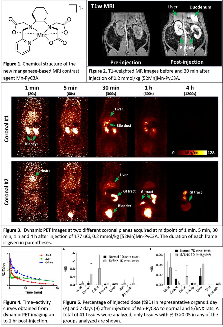
Methods: Adult male and female Wistar rats and 5/6 nephrectomy (5/6 NX) rats were imaged at 4.7 T using a 40 cm-bore MR scanner with a PET insert of 20 cm in diameter with a FOV of 80 x 150 mm (Bruker Biospec). Animals were kept warm by blowing heated air. Temperature and respiration were monitored. Rats were imaged with MRI before contrast agent injection using a multislice 2-dimensional (2D) T1-weighted (T1w) fast low-angle low-shot (FLASH) sequence. An intravenous injection of 0.2 mmol/kg [52Mn]Mn-PyC3A (100-200 uCi) (Figure 1) formulated at 50 mM in sterile water was administered during the PET/MRI acquisition allowing for the biodistribution imaging of the PET tracer and the MRI contrast. Tissues were harvested at the end of the study and biodistribution of residual Mn quantified by gamma counting.

Results: Simultaneous PET/MR imaging in rats with nearly entire torso coverage was successfully achieved. Figure 2 shows the T1w FLASH MRI images before and 30 min after [52Mn]Mn-PyC3A injection showing strong signal enhancement in the kidneys, liver, and bile. Dynamic PET images at 1 min, 5 min, 30 min, 1 h, and 4 h post-injection PET images are shown in Figure 3 showing rapid blood clearance through a predominantly renal pathway but also some liver uptake and clearance into the bile. Time-activity curves for kidney, heart and liver in Figure 4 show renal and hepatic excretion pathways with rapid clearance within 1 hr. Together, the results are consistent with rapid elimination by a mixed renal and hepatobiliary path. Dechelated Mn is known to accumulate in the myocardium, pancreas, and certain glands, but little to no PET signal was detected in these organs over the entire study consistent with high in vivo stability and rapid elimination of Mn-PyC3A in both normal and 5/6 NX rats. Biodistribution analysis of 41 tissues at 24 h showed that 0.32±0.12 %ID and 2.0±1.1 %ID recovered from normal rats and 5/6 NX rats 1 day after injection, respectively, and 0.058±0.051 %ID and 0.14±0.11 %ID recovered from normal and 5/6 NX rats 7 days after injection. Quantities exceeding >0.05 %ID were recovered from the bone, cecum, colon, kidney, liver, skin, and stomach of 5/6NX rats 1 day after injection, but these values decreased to <0.05 %ID by 7 days for all groups analyzed (Figure 5). Conclusion: Dynamic and delayed phase PET/MR results confirm rapid renal and hepatobiliary clearance of Mn-PyC3A in both normal and 5/6 nephrectomized rats with nearly complete elimination within 24 h. This study demonstrates the utility of Mn-52 PET to characterize novel MRI contrast agents. Acknowledgements: Funding from the National Institutes of Health (R44DK113906, S10OD023503, K25HL128899, and R01DK120663) is gratefully acknowledged.

Figure
  • Download figure
  • Open in new tab
  • Download powerpoint
Previous
Back to top

In this issue

Journal of Nuclear Medicine
Vol. 61, Issue supplement 1
May 1, 2020
  • Table of Contents
  • Index by author
Article Alerts
Sign In to Email Alerts with your Email Address
Email Article

Thank you for your interest in spreading the word on Journal of Nuclear Medicine.

NOTE: We only request your email address so that the person you are recommending the page to knows that you wanted them to see it, and that it is not junk mail. We do not capture any email address.

Enter multiple addresses on separate lines or separate them with commas.
Noninvasive Quantification of Whole-Body Distribution and Elimination of [52Mn] Mn-PyC3A Using Simultaneous PET/MRI
(Your Name) has sent you a message from Journal of Nuclear Medicine
(Your Name) thought you would like to see the Journal of Nuclear Medicine web site.
Citation Tools
Noninvasive Quantification of Whole-Body Distribution and Elimination of [52Mn] Mn-PyC3A Using Simultaneous PET/MRI
Iris Zhou, Eric Gale, Nicholas Rotile, Peter Caravan
Journal of Nuclear Medicine May 2020, 61 (supplement 1) 1;

Citation Manager Formats

  • BibTeX
  • Bookends
  • EasyBib
  • EndNote (tagged)
  • EndNote 8 (xml)
  • Medlars
  • Mendeley
  • Papers
  • RefWorks Tagged
  • Ref Manager
  • RIS
  • Zotero
Share
Noninvasive Quantification of Whole-Body Distribution and Elimination of [52Mn] Mn-PyC3A Using Simultaneous PET/MRI
Iris Zhou, Eric Gale, Nicholas Rotile, Peter Caravan
Journal of Nuclear Medicine May 2020, 61 (supplement 1) 1;
Twitter logo Facebook logo LinkedIn logo Mendeley logo
  • Tweet Widget
  • Facebook Like
  • Google Plus One
Bookmark this article

Jump to section

  • Article
  • Figures & Data
  • Info & Metrics

Related Articles

  • No related articles found.
  • Google Scholar

Cited By...

  • No citing articles found.
  • Google Scholar

More in this TOC Section

Molecular Targeting Probes - Radioactive & Nonradioactive

  • A general 11C-carboxylation approach mediated by fluoride-desilylation of organosilanes
  • Relationships between tau, atrophy, regional brain activity and connectivity in Alzheimer's disease: a PET/MRI multimodal study
  • Gray matter structural networks related to 18F-THK5351 retention in cognitively normal older adults and early Alzheimer’s disease patients
Show more Molecular Targeting Probes - Radioactive & Nonradioactive

Center for Molecular Imaging Innovation & Translation (CMIIT) YIA Symposium

  • Imaging activated immune response following therapeutic vaccination in an orthotopic glioma model with 89Zr-DFO-OX40 mAb PET
  • Polyoxometalate nanoclusters act as broad-spectrum antioxidants to alleviate ischemic stroke by intrathecal injection
  • A Pretargeted Strategy for Imaging EGFR Positive Colorectal Carcinoma via the Modulation of Radioligand-Tz Pharmacokinetics
Show more Center for Molecular Imaging Innovation & Translation (CMIIT) YIA Symposium

Similar Articles

SNMMI

© 2025 SNMMI

Powered by HighWire