
Main menu

  • Home
  • Content
    • Current
    • Ahead of print
    • Past Issues
    • JNM Supplement
    • SNMMI Annual Meeting Abstracts
    • Continuing Education
    • JNM Podcasts
  • Subscriptions
    • Subscribers
    • Institutional and Non-member
    • Rates
    • Journal Claims
    • Corporate & Special Sales
  • Authors
    • Submit to JNM
    • Information for Authors
    • Assignment of Copyright
    • AQARA requirements
  • Info
    • Reviewers
    • Permissions
    • Advertisers
  • About
    • About Us
    • Editorial Board
    • Contact Information
  • More
    • Alerts
    • Feedback
    • Help
    • SNMMI Journals
  • SNMMI
    • JNM
    • JNMT
    • SNMMI Journals
    • SNMMI

User menu

  • Subscribe
  • My alerts
  • Log in
  • My Cart

Search

  • Advanced search
Journal of Nuclear Medicine
  • SNMMI
    • JNM
    • JNMT
    • SNMMI Journals
    • SNMMI
  • Subscribe
  • My alerts
  • Log in
  • My Cart
Journal of Nuclear Medicine

Advanced Search

  • Home
  • Content
    • Current
    • Ahead of print
    • Past Issues
    • JNM Supplement
    • SNMMI Annual Meeting Abstracts
    • Continuing Education
    • JNM Podcasts
  • Subscriptions
    • Subscribers
    • Institutional and Non-member
    • Rates
    • Journal Claims
    • Corporate & Special Sales
  • Authors
    • Submit to JNM
    • Information for Authors
    • Assignment of Copyright
    • AQARA requirements
  • Info
    • Reviewers
    • Permissions
    • Advertisers
  • About
    • About Us
    • Editorial Board
    • Contact Information
  • More
    • Alerts
    • Feedback
    • Help
    • SNMMI Journals
  • View or Listen to JNM Podcast
  • Visit JNM on Facebook
  • Join JNM on LinkedIn
  • Follow JNM on Twitter
  • Subscribe to our RSS feeds
Meeting ReportGeneral Clinical Specialties

Sigma-1 receptor PET/MRI for identifying nociceptive sources of radiating low back pain

Daehyun Yoon, Peter Cipriano, Ian Carroll, Catherine Curtin, Eugene Roh, Thomas Wilson and Sandip Biswal
Journal of Nuclear Medicine May 2020, 61 (supplement 1) 178;
Daehyun Yoon
1Radiology Stanford University School of Medicine Stanford CA United States
  • Find this author on Google Scholar
  • Find this author on PubMed
  • Search for this author on this site
Peter Cipriano
1Radiology Stanford University School of Medicine Stanford CA United States
  • Find this author on Google Scholar
  • Find this author on PubMed
  • Search for this author on this site
Ian Carroll
2Anesthesiology Stanford University School of Medicine Stanford CA United States
  • Find this author on Google Scholar
  • Find this author on PubMed
  • Search for this author on this site
Catherine Curtin
3Plastic & Reconstructive Surgery Stanford University School of Medicine Stanford CA United States
  • Find this author on Google Scholar
  • Find this author on PubMed
  • Search for this author on this site
Eugene Roh
4Orthopaedic Surgery Stanford University School of Medicine Stanford CA United States
  • Find this author on Google Scholar
  • Find this author on PubMed
  • Search for this author on this site
Thomas Wilson
5Neurosurgery Stanford University School of Medicine Stanford CA United States
  • Find this author on Google Scholar
  • Find this author on PubMed
  • Search for this author on this site
Sandip Biswal
1Radiology Stanford University School of Medicine Stanford CA United States
  • Find this author on Google Scholar
  • Find this author on PubMed
  • Search for this author on this site
  • Article
  • Figures & Data
  • Info & Metrics
Loading

Abstract

178

Objectives: Accurately identifying nociceptive sources of low back pain has remained to be a great diagnostic challenge despite its pressing need due to the high prevalence and social impact of the condition1. Unfortunately, findings from structural imaging methods, such as MRI, have been often fraught with false positives and non-specific to pain2, which can result in ineffective and unnecessary treatments3. Sigma-1 receptor (S1R) is a very promising alternative biomarker of painful pathologies because it plays a key role in the ion channel modulation for nociceptive processes4. Our group recently demonstrated the feasibility of in vivo PET/MR imaging of S1R using a novel S1R radioligand (S1RR), 18F-FTC-1465,6. The objective of this study is to introduce early results from our PET/MRI of S1R patient study focusing on radiating low back pain. Methods: We recruited seven patients presenting pain radiating from the low back down to buttocks or legs. Our prospective imaging study was approved by the institutional review board. An informed consent form signed by the patient was obtained prior to imaging. We conducted a whole-body PET/MRI 40 minutes after the intravenous injection of 10-mCi dose of S1RR (18F-FTC-146). A GE SIGNA PET/MRI scanner (GE Healthcare, Waukesha, U.S.) was used for collecting the PET/MRI images simultaneously. PET/MRI images of the lumbar spine to the mid-thigh regions were reviewed by two musculoskeletal radiologists. The MRI image review was to find lesions with abnormal signal intensity on T2-weighted contrast or structural changes. The PET image review was mainly to identify focal hotspots of S1RR uptake. The SUVmax of unilateral hotspots in nerves and muscles was compared with that of the contralateral side using the Wilcoxon signed rank test. The identified abnormalities were grouped depending on the source imaging modality: 1) identified in MRI only, 2) identified in PET only, 3) identified in both MRI and PET.

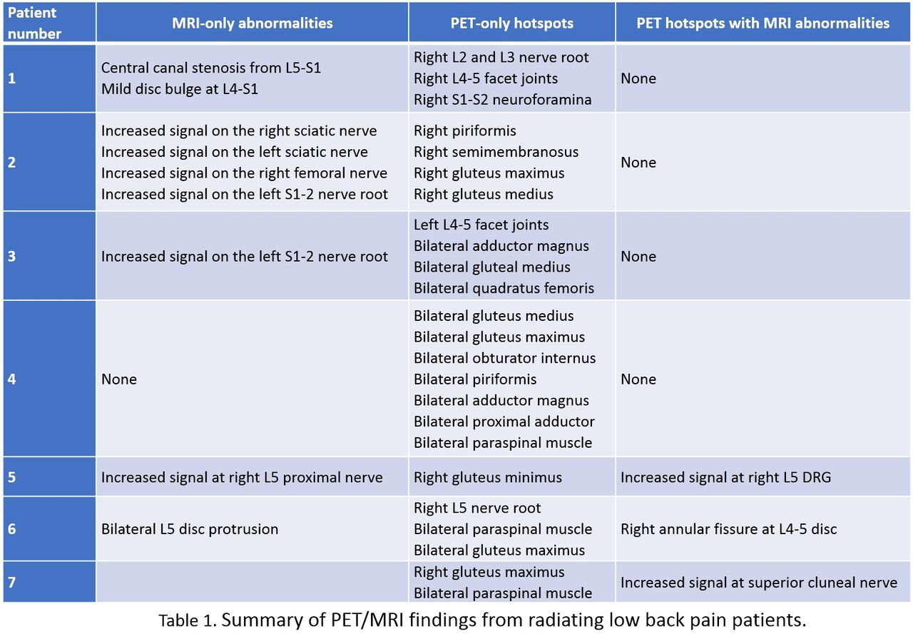
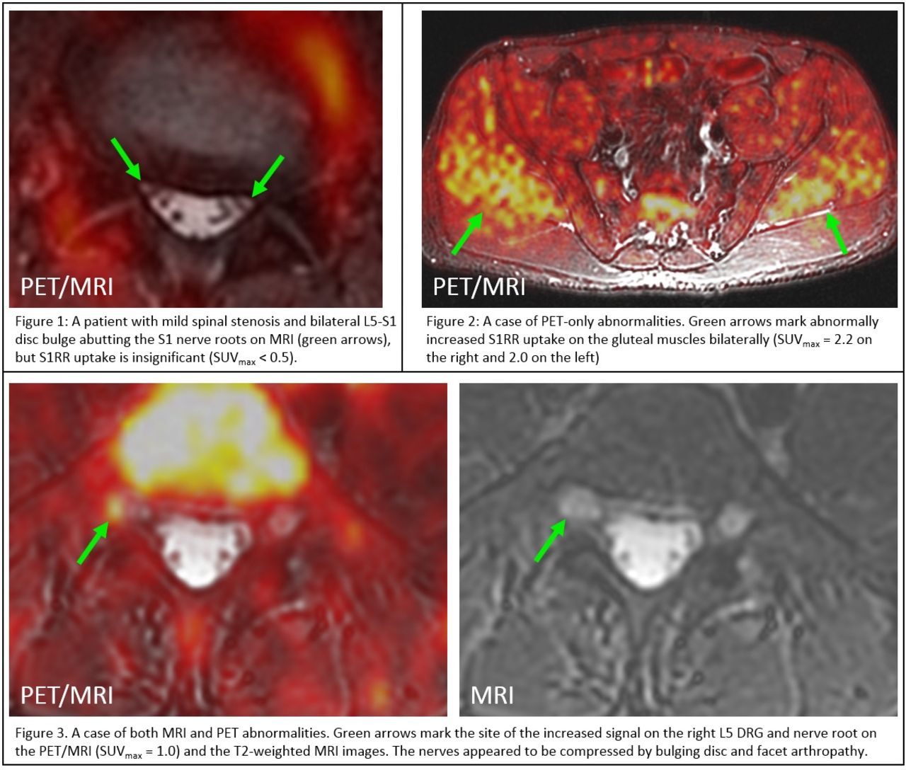
Results: All patients presented abnormalities on either PET (all 7 patients) or MRI (6 patients) while more diverse abnormalities were found on PET images (Table 1). MRI-only abnormalities were mostly increased signal on the peripheral nerves (3 patients) while structural abnormality was found on only two patients as shown in Figure 1 (stenosis and disc abnormalities). On the other hand, S1R PET images showed increased S1RR uptake on a wider variety of tissues such as spinal joints (3 patients), paraspinal/leg muscles (6 patients), and peripheral nerves (4 patients). The SUVmax of unilateral nerve and muscle hotspots was higher than that of the contralateral side with a 5% significance level (Table 2). Except one patient, all other patients presented abnormally increased uptake of S1RR on the muscles in the symptomatic area with any apparent sign of degeneration on MRI (Figure 2). Lesions presenting abnormalities on both S1R PET and MRI were located at either peripheral nerves as shown in Figure 3 or structures close to nerves (patient 6). Conclusion: In summary, we have presented early results of S1R PET/MRI on patients with radiating low back pain. While the MRI abnormalities were mostly found involving the peripheral nerves, the increased S1RR uptake was detected on various tissues including joints, nerves, and muscles around the lumbar spine. The S1RR uptake at the site of the structural abnormalities on MRI can form a stronger evidence in determining the cause of pain, considering the high false-positive rate of the structural abnormalities on MRI in general. Also, the increased S1RR uptake on muscles without the gross structural change may indicate the sensitivity of our S1R PET method to early inflammatory processes. Therefore, our approach demonstrated the promise for the improved confidence and sensitivity in detecting local sources of radiating low back pain. Acknowledgement: GE Healthcare, NIH P41 EB015891.

Figure
  • Download figure
  • Open in new tab
  • Download powerpoint
Figure
  • Download figure
  • Open in new tab
  • Download powerpoint
Figure
  • Download figure
  • Open in new tab
  • Download powerpoint
Previous
Back to top

In this issue

Journal of Nuclear Medicine
Vol. 61, Issue supplement 1
May 1, 2020
  • Table of Contents
  • Index by author
Article Alerts
Sign In to Email Alerts with your Email Address
Email Article

Thank you for your interest in spreading the word on Journal of Nuclear Medicine.

NOTE: We only request your email address so that the person you are recommending the page to knows that you wanted them to see it, and that it is not junk mail. We do not capture any email address.

Enter multiple addresses on separate lines or separate them with commas.
Sigma-1 receptor PET/MRI for identifying nociceptive sources of radiating low back pain
(Your Name) has sent you a message from Journal of Nuclear Medicine
(Your Name) thought you would like to see the Journal of Nuclear Medicine web site.
Citation Tools
Sigma-1 receptor PET/MRI for identifying nociceptive sources of radiating low back pain
Daehyun Yoon, Peter Cipriano, Ian Carroll, Catherine Curtin, Eugene Roh, Thomas Wilson, Sandip Biswal
Journal of Nuclear Medicine May 2020, 61 (supplement 1) 178;

Citation Manager Formats

  • BibTeX
  • Bookends
  • EasyBib
  • EndNote (tagged)
  • EndNote 8 (xml)
  • Medlars
  • Mendeley
  • Papers
  • RefWorks Tagged
  • Ref Manager
  • RIS
  • Zotero
Share
Sigma-1 receptor PET/MRI for identifying nociceptive sources of radiating low back pain
Daehyun Yoon, Peter Cipriano, Ian Carroll, Catherine Curtin, Eugene Roh, Thomas Wilson, Sandip Biswal
Journal of Nuclear Medicine May 2020, 61 (supplement 1) 178;
Twitter logo Facebook logo LinkedIn logo Mendeley logo
  • Tweet Widget
  • Facebook Like
  • Google Plus One
Bookmark this article

Jump to section

  • Article
  • Figures & Data
  • Info & Metrics

Related Articles

  • No related articles found.
  • Google Scholar

Cited By...

  • Development of a PET radioligand for {alpha}2{delta}-1 subunit of calcium channels for imaging neuropathic pain
  • Google Scholar

More in this TOC Section

General Clinical Specialties

  • Exploring variations and utility of FDG- and NaF PET/CT in rheumatoid arthritis and osteoarthritis
  • Application of Lung perfusion only scan combined with SPECT/CT for the post-treatment evaluation of massive and sub-massive pulmonary embolism in the age of covid-19 pandemic: case presentation and literature review
  • Can FDG PET/CT imaging have a role in clinical cases of Dermatomyositis?
Show more General Clinical Specialties

Musculoskeletal

  • Ultra-low-dose total-body 18F-FDG PET/CT in patients with autoimmune inflammatory arthritis: evaluation of image quality with shorter scan time
  • Assessing the effects of age and sex in bone turnover in tibia using NaF-PET/CT
  • Tibial FDG uptake in both females and males increases with age
Show more Musculoskeletal

Similar Articles

SNMMI

© 2025 SNMMI

Powered by HighWire