
Main menu

  • Home
  • Content
    • Current
    • Ahead of print
    • Past Issues
    • JNM Supplement
    • SNMMI Annual Meeting Abstracts
    • Continuing Education
    • JNM Podcasts
  • Subscriptions
    • Subscribers
    • Institutional and Non-member
    • Rates
    • Journal Claims
    • Corporate & Special Sales
  • Authors
    • Submit to JNM
    • Information for Authors
    • Assignment of Copyright
    • AQARA requirements
  • Info
    • Reviewers
    • Permissions
    • Advertisers
  • About
    • About Us
    • Editorial Board
    • Contact Information
  • More
    • Alerts
    • Feedback
    • Help
    • SNMMI Journals
  • SNMMI
    • JNM
    • JNMT
    • SNMMI Journals
    • SNMMI

User menu

  • Subscribe
  • My alerts
  • Log in
  • My Cart

Search

  • Advanced search
Journal of Nuclear Medicine
  • SNMMI
    • JNM
    • JNMT
    • SNMMI Journals
    • SNMMI
  • Subscribe
  • My alerts
  • Log in
  • My Cart
Journal of Nuclear Medicine

Advanced Search

  • Home
  • Content
    • Current
    • Ahead of print
    • Past Issues
    • JNM Supplement
    • SNMMI Annual Meeting Abstracts
    • Continuing Education
    • JNM Podcasts
  • Subscriptions
    • Subscribers
    • Institutional and Non-member
    • Rates
    • Journal Claims
    • Corporate & Special Sales
  • Authors
    • Submit to JNM
    • Information for Authors
    • Assignment of Copyright
    • AQARA requirements
  • Info
    • Reviewers
    • Permissions
    • Advertisers
  • About
    • About Us
    • Editorial Board
    • Contact Information
  • More
    • Alerts
    • Feedback
    • Help
    • SNMMI Journals
  • View or Listen to JNM Podcast
  • Visit JNM on Facebook
  • Join JNM on LinkedIn
  • Follow JNM on Twitter
  • Subscribe to our RSS feeds
Meeting ReportCardiovascular Track

Quantification of myocardial blood flow using dynamic 320-row multi-detector CT with Gauss curve fittingPET

Masahiro Muneta, Yuuki Tomiyama, Masahiko Obara, Masanao Naya, Keiichi Magota, Osamu Manabe, Nagara Tamaki and Chietsugu Katoh
Journal of Nuclear Medicine May 2017, 58 (supplement 1) 589;
Masahiro Muneta
7Hokkaido Universitygraduate School of Medicine Sapporo Japan
  • Find this author on Google Scholar
  • Find this author on PubMed
  • Search for this author on this site
Yuuki Tomiyama
3Hokkaido University Sapporo Japan
  • Find this author on Google Scholar
  • Find this author on PubMed
  • Search for this author on this site
Masahiko Obara
5Hokkaido University Graduate School of Medicine Sapporo, Hokkaido Japan
  • Find this author on Google Scholar
  • Find this author on PubMed
  • Search for this author on this site
Masanao Naya
2Hokkaido University Hokkaido Japan
  • Find this author on Google Scholar
  • Find this author on PubMed
  • Search for this author on this site
Keiichi Magota
4Hokkaido University Graduate School of Medicine Sapporo Japan
  • Find this author on Google Scholar
  • Find this author on PubMed
  • Search for this author on this site
Osamu Manabe
3Hokkaido University Sapporo Japan
  • Find this author on Google Scholar
  • Find this author on PubMed
  • Search for this author on this site
Nagara Tamaki
1Dept. of Nuclear Medicine Sapporo Japan
  • Find this author on Google Scholar
  • Find this author on PubMed
  • Search for this author on this site
Chietsugu Katoh
6Hokkaido University School of Medicine Sapporo Japan
  • Find this author on Google Scholar
  • Find this author on PubMed
  • Search for this author on this site
  • Article
  • Figures & Data
  • Info & Metrics
Loading

Abstract

589

Objectives: Measurements of myocardial blood flow (MBF) and coronary flow reserve (CFR) using multi-detector CT (MDCT) have been reported for evaluating the physiological significance of coronary lesions. However, there was a case in which the beginning of the curve was not stabilized due to deviation of imaging timing or motion. To obtain stable MBF even in such cases, Gaussian curve correction was useful to interpolate the unstable part of the curve. We developed a new algorithm using Gauss curve fitting in the density curve of the myocardial tissue. The aim of this study was to quantify the MBF using dynamic CT with 320-row MDCT with our new algorithm and compared to the conventional method.

Methods: Fifteen patients (64±11 years old) with suspected coronary artery disease were enrolled. All patients refrained from drinking caffeinated beverages for at least 24 hours, eating for more than 6 hours and smoking for at least 4 hours before the scan. Rest dynamic CT was followed by Stress dynamic CT with intravenous ATP infusion (0.016 mg/kg/min) using a second-generation 320-row MDCT system (Aquilion ONE, ViSION Edition, Toshiba Medical Systems). Time attenuation curves (TACs) of Hounsfield (HU) value in the left ventricle (LV) chamber and LV myocardium were derived from the dynamic CT data. HU value of descending aorta was subtracted to estimate the stable dynamic curve from HU value of LV pool. Gauss curves are added to smooth the dynamic curves. Using single-tissue compartment model and Renkin-Crone model analysis, regional MBFs were calculated in the Left anterior descending coronary artery (LAD), Right coronary artery (RCA) and Left circumflex coronary artery (LCX) territories from dynamic CT. These estimates were compared with MBF from 15O-H2O PET.

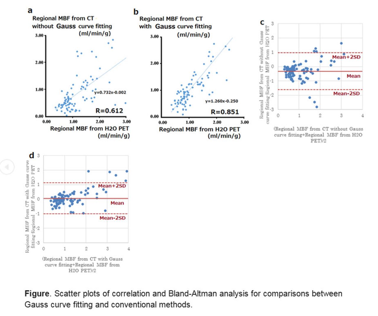
Results: MBF from our new method yielded significant better relationship with MBF from 15O-H2O PET compared to the conventional method. (R=0.851, 0.612, respectively; p < 0.001) (Figure a b). Bland -Altman analysis stated that Gauss curve fitting has less variation in correlation than conventional methods (mean=0.061, -0.323 SD=1.13, 0.960 respectively)(Figure c d).

Conclusion: We developed a new algorithm to estimate MBF with Gauss curve fitting in the TACs of the dynamic CT data. Gauss curve fitting was useful to interpolate MBF from dynamic CT scan.

Figure
  • Download figure
  • Open in new tab
  • Download powerpoint
Previous
Back to top

In this issue

Journal of Nuclear Medicine
Vol. 58, Issue supplement 1
May 1, 2017
  • Table of Contents
  • Index by author
Article Alerts
Sign In to Email Alerts with your Email Address
Email Article

Thank you for your interest in spreading the word on Journal of Nuclear Medicine.

NOTE: We only request your email address so that the person you are recommending the page to knows that you wanted them to see it, and that it is not junk mail. We do not capture any email address.

Enter multiple addresses on separate lines or separate them with commas.
Quantification of myocardial blood flow using dynamic 320-row multi-detector CT with Gauss curve fittingPET
(Your Name) has sent you a message from Journal of Nuclear Medicine
(Your Name) thought you would like to see the Journal of Nuclear Medicine web site.
Citation Tools
Quantification of myocardial blood flow using dynamic 320-row multi-detector CT with Gauss curve fittingPET
Masahiro Muneta, Yuuki Tomiyama, Masahiko Obara, Masanao Naya, Keiichi Magota, Osamu Manabe, Nagara Tamaki, Chietsugu Katoh
Journal of Nuclear Medicine May 2017, 58 (supplement 1) 589;

Citation Manager Formats

  • BibTeX
  • Bookends
  • EasyBib
  • EndNote (tagged)
  • EndNote 8 (xml)
  • Medlars
  • Mendeley
  • Papers
  • RefWorks Tagged
  • Ref Manager
  • RIS
  • Zotero
Share
Quantification of myocardial blood flow using dynamic 320-row multi-detector CT with Gauss curve fittingPET
Masahiro Muneta, Yuuki Tomiyama, Masahiko Obara, Masanao Naya, Keiichi Magota, Osamu Manabe, Nagara Tamaki, Chietsugu Katoh
Journal of Nuclear Medicine May 2017, 58 (supplement 1) 589;
Twitter logo Facebook logo LinkedIn logo Mendeley logo
  • Tweet Widget
  • Facebook Like
  • Google Plus One
Bookmark this article

Jump to section

  • Article
  • Figures & Data
  • Info & Metrics

Related Articles

  • No related articles found.
  • Google Scholar

Cited By...

  • No citing articles found.
  • Google Scholar

More in this TOC Section

Cardiovascular Track

  • To Evaluated the Cardiac Function of Patients with Acute Myocardial Infarction by the Volume and Filling Curve of 99mTc-MIBI SPECT Myocardial Perfusion Imaging
  • Standard versus low-dose rubidium-82 dynamic positron emission tomography imaging with scanner-dependent bias correction for myocardial perfusion imaging and blood flow quantification
  • Evaluation of sympathetic function with PET 11C-hydroxyephedrine (HED) and ammonia (13N-NH3) in a canine pacing model of atrial fibrillation
Show more Cardiovascular Track

Novel Imaging Techniques for Myocardial Phenotyping (Basic Science)

  • Maximum Rubidium-82 activity for accurate myocardial blood flow quantification using digital PET
  • 18F-FDG PET Guided Histological Analysis on Adhesive Molecules in a Rat Model of Acute Myocarditis
  • Combined static-dynamic single-dose cardiac SPECT for quantitative myocardial perfusion imaging
Show more Novel Imaging Techniques for Myocardial Phenotyping (Basic Science)

Similar Articles

SNMMI

© 2025 SNMMI

Powered by HighWire