


In vivo visualization of inflammatory lesions has been revolutionized by PET with 18F-FDG as a tracer and by MRI with gadolinium-labeled contrast media. Apart from other indications, 18F-FDG PET and MRI have substantially improved the diagnosis and monitoring of immune-mediated inflammatory diseases such as arthritis and connective tissue diseases. Although the visualization of active inflammation is well established, the detection of tissue response and tissue remodeling processes, which accompany immune-mediated inflammatory diseases and lead to organ damage, has until recently not been possible. Tissue remodeling processes during inflammation are based on mesenchymal stroma cell activation and expansion in parenchymatous organs or the synovial membrane of inflamed joints. These cells express specific markers, such as fibroblast activation protein (FAP), that can be visualized by radiolabeled compounds (e.g., FAP inhibitors [FAPIs]) using PET. First evidence shows that focal accumulation of FAPI tracers, indicating active tissue remodeling, is observed in patients with immune-mediated inflammatory diseases that are characterized by a combination of chronic inflammation and tissue responses, such as systemic sclerosis, IgG4 syndrome, or spondyloarthritis. Such FAPI-positive remodeling lesions are not always 18F-FDG–positive, indicating that inflammation and tissue responses can be disentangled by such methods. These data suggest that tracers such as FAPI allow visualization of the dynamics of tissue responses in immune-mediated inflammatory diseases in vivo. This development opens new options for recognition of tissue remodeling in the context of chronic inflammation, as expanded herein.
Activated fibroblasts express FAP, a type II transmembrane protease with dipeptidyl peptidase and endopeptidase activity. Resting fibroblasts and most other cell types have only minor or no FAP expression. Recently, radiolabeled quinoline-based tracers suitable for PET that act as FAPIs have been developed (1). The initial goal of this development was to image stromal reactions in tumors and metastases (2). Considerable evidence is emerging on the clinical utility of FAPI PET in oncology (3,4).
Tissue remodeling is also a consequence of chronic inflammation. Activation of fibroblasts is therefore not only confined to tumors but also occurs in immune-mediated inflammatory diseases such as systemic sclerosis and IgG4-related disease and inflammatory arthritis. In these diseases, fibroblast activation may eventually lead to severe organ dysfunction, causing disability or—when parenchymatous organs are involved—even death. Tissue remodeling is the critical step for eliciting damage in immune-mediated inflammatory diseases (5). To date, imaging methods used in immune-mediated inflammatory diseases are mostly confined to detection of inflammation. PET with 18F-FDG or MRI with gadolinium-labeled contrast media has been used to detect and quantify inflammation. However, these methods do not visualize the process of mesenchymal stromal activation and therefore do not allow detection of the process of tissue destruction. Furthermore, techniques such as CT allow quantification of accumulated damage rather than measurement of the dynamic process of tissue change.
Application of FAPI PET in immune-mediated inflammatory diseases has revealed localized tracer accumulation reflecting mesenchymal tissue responses in various diseases such as fibrotic lung and liver diseases, as well as arthritis and colitis (4). A striking example of how mesenchymal stroma activation affects organ function is pulmonary fibrosis, which can arise as an idiopathic disorder or in the context of autoimmune diseases such as systemic sclerosis. Pulmonary fibrosis is often a severe and progressive condition leading to respiratory failure. Diagnosis is established using clinical criteria and high-resolution CT. Two recent publications have shown that FAPIs accumulate in pulmonary fibrosis: Röhrich et al. reported elevated FAPI uptake in 15 patients with different subtypes of fibrotic interstitial lung disease without further specification of their subtypes (6). There was a significant but moderate correlation between CT indices of fibrosis and FAPI uptake measured in the fibrotic areas. The authors hypothesized that FAPI PET/CT might have a role in evaluating the course of pulmonary fibrosis and, in particular, in monitoring the effect of treatment. Bergmann et al. studied a group of 21 patients with systemic sclerosis-associated pulmonary fibrosis using 68Ga-FAPI-04 PET/CT (7). Systemic sclerosis patients also showed an increased FAPI accumulation in the fibrotic areas of the lungs. FAPI uptake was related to parameters of more active disease as measured by higher clinical activity scores. Furthermore, Bergmann et al. could demonstrate that the magnitude of FAPI uptake correlated with progression of disease independently of the extent of involvement on CT scans and lung function at baseline. In 5 patients treated with the tyrosine kinase inhibitor and antifibrotic agent nintedanib, changes in FAPI uptake paralleled the response to treatment as determined by changes in lung function. These latter findings are in contrast to those published for 18F-FDG PET/CT, which were not predictive for treatment response in an article published by Bondue et al. (8), indicating that FAPI PET–based detection of fibrotic tissue responses is more closely related to the pathologic process of systemic sclerosis than the detection of an inflammatory 18F-FDG signal by PET. In any case, larger studies are required to clarify and establish a role for FAPI PET in monitoring the treatment response of interstitial lung disease.
Because 18F-FDG PET detects inflammatory processes, one may ask whether FAPI radioligands provide additional insight into chronic inflammation processes beyond that given by 18F-FDG. In this context, interesting data have been published for IgG4-related disease, a rare prototypical disorder that combines autoimmune inflammation with tumefactive tissue fibrosis affecting the pancreas and biliary tree, the salivary glands, the kidneys, the aorta, and other organs. Immune-targeted therapies effectively inhibit inflammation but may not be suited to tackle fibrotic tissue changes, requiring detection of whether IgG4-related disease is based primarily on inflammatory or fibrotic lesions in an individual patient. Evidence from histopathology indicates that IgG4-related disease can progress from an inflammatory-proliferative to a fibrotic phase, each of which requires different therapeutic approaches. Most patients with IgG4-related disease show 18F-FDG–positive inflammatory lesions; however, FAPI-positive lesions have also been described (9). Schmidkonz et al. used both 18F-FDG and FAPI PET to study a group of 27 patients with IgG4-related disease (10). They demonstrated that 18F-FDG–positive lesions showed dense lymphoplasmacytic infiltrations of IgG4-positive plasma cells in histology, whereas FAPI-positive lesions harbored abundant activated fibroblasts. Moreover, they could also show that FAPI uptake did not correlate with 18F-FDG uptake, suggesting that the two tracers visualize two different aspects of IgG4-related disease. In their patient cohort, the responsiveness of fibrotic lesions to immunotherapy was far less pronounced than that of inflammatory ones, suggesting that FAPI PET might find a role in guiding more specific therapy in this disorder.
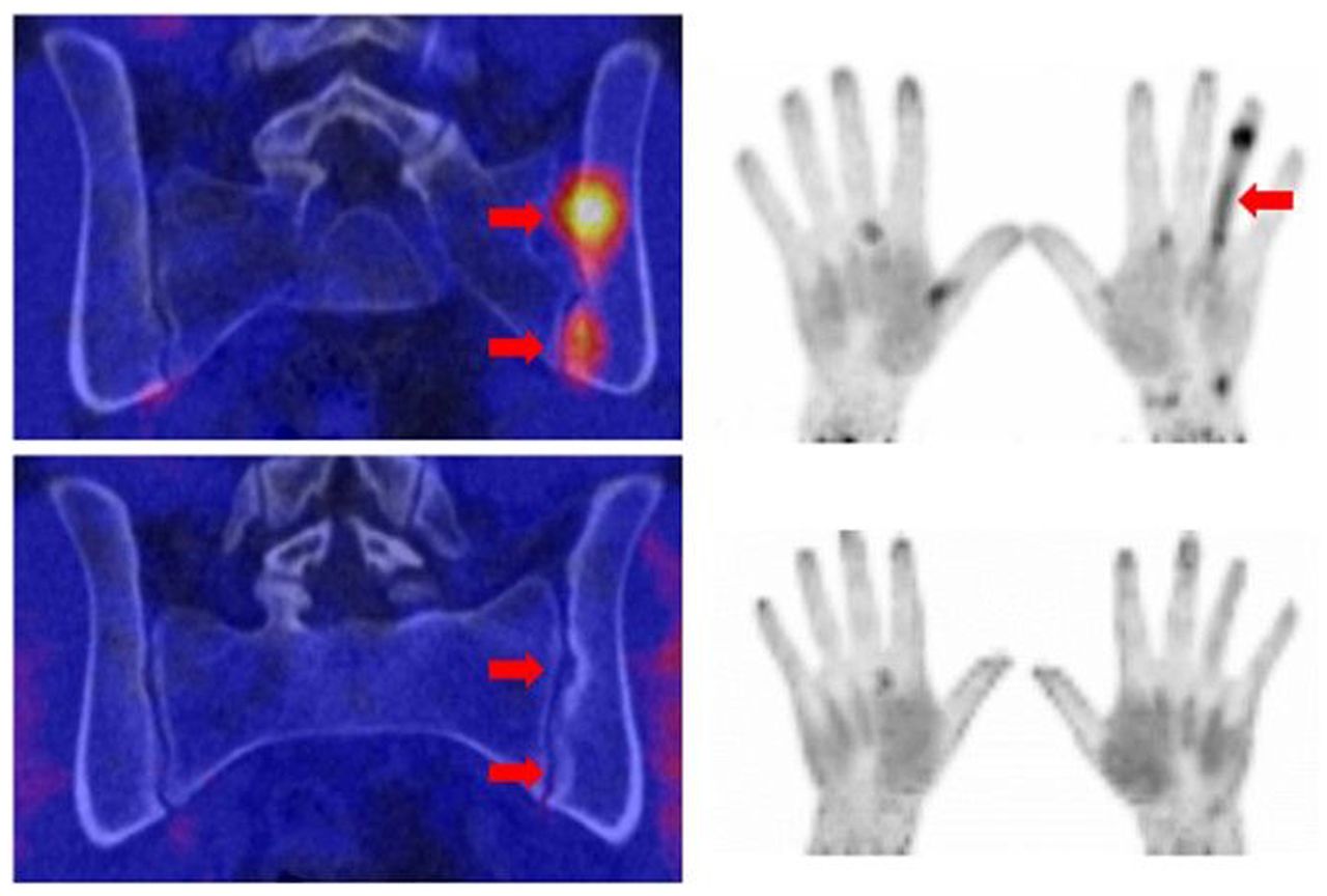
In summary, this paradigmatic evidence is just the start for a wider use of FAPI PET imaging in immune-mediated inflammatory diseases. Other potential indications for FAPI PET are spondyloarthritis, in which tissue responses lead to ankylosis (Fig. 1); rheumatoid arthritis, which is associated with resident tissue responses that manifest in synovial hyperplasia; and colitis, in which tissue responses trigger strictures in the gut. These examples may further extend the clinical role of FAPI PET as it offers a completely new view on tissue remodeling, fibrosis, and damage in chronic inflammatory diseases. These findings also offer new possibilities for early recognition of the tissue remodeling process, prediction of damage, and response to antifibrotic therapies. Hence, nuclear medicine of the future will likely step out of what has been called the “hegemony of 18F-FDG” (11) also in rheumatology.

68Ga-FAPI PET images before treatment (top row) and after treatment (bottom row) in patient with spondyloarthritis manifesting with sacroiliitis (upper left panel) and dactylitis of fourth finger. Left upper and lower images are coronal PET/CT fusion images of the sacroiliac joints, and right upper and lower images 3D–volume-rendered PET datasets of the hands. Arrows indicate tracer accumulation and erosions, respectively.
DISCLOSURE
No potential conflict of interest relevant to this article was reported.
Footnotes
Published online Apr. 7, 2022.
- © 2022 by the Society of Nuclear Medicine and Molecular Imaging.
REFERENCES
- Received for publication February 24, 2022.
- Accepted for publication March 30, 2022.
In this issue


Jump to section
Related Articles
Cited By...
- Collagen Hybridizing Peptide-Based Radiotracers for Molecular Imaging of Collagen Turnover in Pulmonary Fibrosis
- 1,090 Publications and 5 Years Later: Is FAP-Targeted Theranostics Really Happening?
- Anatomical pattern of entheseal and synovial fibroblast activation in patients with psoriasis and its risk of developing psoriatic arthritis
- FAPI PET/CT Immune-Fibrosis Imaging for New Insights into Rheumatologic Disorders
- Molecular Imaging with Fibroblast Activation Protein Tracers depicts Inflammatory Joint Damage and its Transition to Resolution of Inflammation
- Fibroblast Activation Protein Inhibitor Imaging in Nonmalignant Diseases: A New Perspective for Molecular Imaging