


Abstract
Antimicrobial peptides such as ubiquicidin (UBI) are believed to differentiate between mammalian and bacterial or fungal cells. 99mTc-UBI29-41 was previously tested for detecting infection in humans using SPECT. For the present study, the UBI fragment UBI29-41 (TGRAKRRMQYNRR) was conjugated to 1,4,7-triazacyclononane-triacetic acid (NOTA), radiolabeled with 68Ga, and investigated in a rabbit infection model. Methods: 68Ga was obtained from a 1.85-GBq 68Ge/68Ga generator. New Zealand White rabbits were anesthetized with ketamine/medetomidine before tracer administration and placed in a clinical PET/CT scanner. 68Ga-1,4,7-triazacyclononane-1,4,7-triacetic-acid-ubiquicidin29-41 (68Ga-NOTA-UBI29-41) was formulated in saline solution, and 101 ± 41 MBq were administered intravenously. The tracer distribution was studied by PET/CT imaging in animals (a) that were healthy, (b) bearing muscular Staphylococcus aureus infections and turpentine oil-induced muscular inflammations, and (c) bearing ovalbumin-induced lung inflammations. Static PET/CT imaging was performed at different time intervals up to 120 min after injection. For calculation of target-to-nontarget ratios, standardized uptake values were normalized against healthy thigh muscle, representing nontargeted tissue. Results: PET/CT images of healthy animals showed predominant distribution in the kidneys, liver, and bladder; heart and spleen showed moderate, declining uptake, only. The biologic half-life in blood was 29 min. Urinary accumulation of 68Ga-NOTA-UBI29-41 peaked at 3.8 ± 0.91 percentage injected dose per gram (%ID) at 120 min, and 88 ± 5.2 %ID was recovered in total urine. 68Ga-NOTA-UBI29-41 imaging in (b) selectively visualized the muscular infection site and was differentiated from sterile inflammatory processes. Standardized uptake value ratios for muscles (infected/inflamed) were 2.9 ± 0.93, 2.9 ± 0.50, 3.5 ± 0.86, and 3.8 ± 0.90 at 5, 30, 60, and 90 min after injection, respectively. Rabbit lungs with asthma showed insignificant uptake. Conclusion: 68Ga-NOTA-UBI29-41 was strongly localized in bacteria-infected areas and minimally detected in a sterile inflammation area in rabbit muscles. The findings propose this compound to be an excellent first-line PET/CT tracer to allow the distinguishing of infection from inflammation.
Infectious diseases have a major impact on global morbidity and mortality despite advances in both diagnosis and treatment (1). The early identification and correct diagnosis of an active infection is crucial to its effective control or adequate treatment. Million of antibiotic doses are consumed unnecessarily, resulting in bacterial multidrug resistance and cost increases for therapy. Innovative diagnostic radiopharmaceuticals for detecting infection are of paramount importance because nuclear medicine techniques for detecting infections have the advantage of noninvasive whole-body imaging, which might be of great value in cases of occult infection. However, there are limitations, mainly the problem of differentiating between infectious and sterile inflammatory processes (2). To determine the correct course of treatment, clinicians need to be able to discriminate between sites of sterile inflammation and those that are infected by pathogenic microorganisms. Sites that are infected appear, at least superficially, similar to sites that are inflamed due to other reasons, such as trauma, ischemia, or the presence of foreign bodies such as implants or neoplasm (3). Radiolabeled leukocytes and 67Ga-citrate were the most commonly administered tracers for infection imaging (4), but because efficient current good manufacturing practices 68Ge/68Ga generators became commercially available, 68Ga-citrate (5,6) and other novel 68Ga-labeled compounds such as 68Ga-siderophores (7) and 68Ga-apo-transferrin (8) were introduced for specific diagnosis of infected tissues. 68Ga-labeled peptides have also become relevant for diagnostic imaging because of their favorable pharmacokinetics (9). Interestingly, peptides with antimicrobial properties are found in abundance in mammals, amphibians, and plants, as a part of innate immunity against infection (10). Due to differences in electrostatic charges on the cell membrane or cell wall, human cationic antimicrobial peptides such as ubiquicidin (UBI) (11) are believed to differentiate between mammalian and bacterial or fungal cells (12). Fragments of UBI, namely UBI18-35 and UBI29-41 (TGRAKRRMQYNRR), were studied for their visualization of bacterial infections and sterile inflammatory processes (13). 68Ga can be labeled via a chelator-conjugated targeting molecule. One of these chelators is 1,4,7-triazacyclononane-triacetic acid (NOTA), which has a high affinity for 68Ga and therefore conjugates with peptides to functionalize these toward radiolabeling for prospective PET imaging (14). Preclinical publications (13,15,16) reported scintigraphic detection of bacterial and fungal infections using 99mTc-UBI29-41. Clinical studies were performed by Melendez-Alafort et al. in 2004 and Akhtar et al. in 2005 administering 99mTc-UBI29-41 for detecting infection using SPECT imaging (17,18). Key to this approach is that this peptide accumulates at infection sites but not in sterile inflammatory lesions. Studies in humans performed recently have applied 99mTc-UBI29-41 to detect infection associated with fever of unknown origin (19) or used it for antibiotic therapy monitoring (20). For the present study, the UBI fragment UBI29-41 was conjugated with NOTA, subsequently radiolabeled with 68Ga and investigated in a preclinical rabbit infection model. The functional radiolabeling with 68Ga will allow image acquisition with PET/CT.
MATERIALS AND METHODS
General
All animal experiments were conducted according to the South African code of practice for the care and use of animals, after approval from the Medical Research Council ethics committee. Health surveillance was performed weekly for the duration of the experiments. Rabbits were randomly assigned to 1 of 3 groups (NZR-1, healthy condition [n = 8]; NZR-2, muscular infection/inflammation [n = 7]; NZR-3, pulmonary inflammation [asthma] [n = 3]). Animals underwent 14-d antibiosis before the study to preclude random infections. Pharmaceutical grade chemicals were purchased from Merck. NOTA-UBI29-41 was obtained from GL Biochem. C18-SepPak cartridges were obtained from Waters Corp. Instant thin-layer chromatography silica gel (ITLC-SG) was purchased from PALL Life Science. Sterile Millex GV filters were obtained from Millipore. High-performance liquid chromatography–grade water was produced in-house using a Simplicity 185 Millipore system.
Imaging of Staphylococcus Aureus Infection and Turpentine Inflammation in Muscles and Asthma in Lungs of Rabbits
Staphylococcus aureus 25923 was purchased from American Type Culture Collection. Bacilli stock was incubated overnight in nutrient broth at 37°C and agitation at 110 rpm. Sterile frozen aliquots (2 × 10⁸ colony-forming units/cm3) were kept at −70°C. Saline solution (1 cm3) containing 2 × 10⁸ colony-forming units was injected into the right thigh muscle, and turpentine oil (1 cm3) was injected into the left hind thigh muscle of 7 rabbits (group NZR-2). PET/CT imaging was commenced 72 h after induction. Three rabbits (group NZR-3) were actively sensitized weekly for 4 wk with intraperitoneal injections of 1 cm3 of an ovalbumin-containing solution (2.5 mg of ovalbumin in 1 part physiologic saline and 1 part of Alum [Pierce]). These rabbits were challenged by aerosol inhalation of 5% ovalbumin solution 1 wk after the last sensitization injection and again 24 h before PET/CT imaging. Rabbits were euthanized with a lethal dose of sodium pentobarbitone after imaging. For postmortem bacteriologic testing, sections of infected or inflamed sites of thighs were homogenized for cultivation in nutrient broth at 37°C. Bacterial identification was confirmed by biochemical testing. For histopathologic examination, sections of the sterile inflammation site, the infection site, and particular lung sections were fixed in 10% phosphate-buffered saline/buffered formalin and paraffin-embedded. Sections of 0.5 μm were stained with hematoxylin and eosin and examined by light microscopy.
Radiolabeling of UBI-NOTA29-41
Radiolabeling of NOTA-UBI-29-41 was based on a 68Ga-DOTA-TATE labeling procedure (21). Briefly, 68Ga(III)chloride was eluted from a SnO2-based 68Ge/68Ga generator purchased from iThemba LABS using 0.6N HCl. Up to 95% activity yield was harvested in 2 cm3 eluate. The sodium acetate–buffered 68Ga (pH 3.5–4) was incubated in the presence of 50 nM NOTA-UBI29-41 at 90°C for 15 min. In preparation for preclinical use, 68Ga-NOTA-UBI29-41 was purified from free 68Ge-, 68Ga-, and 68Ga-colloids using a C18-SepPak cartridge. The radiolabeled product was filtered through a Millex GV 0.22-μm sterile filter and formulated in sterile saline solution.
PET/CT Imaging
Subcutaneous injection of ketamine (15 mg/kg)/medetomidine (0.5 mg/kg) was given to induce sedation. Anesthesia was maintained by propofol/thiopental infusion. Whole-body PET/CT imaging with the patient supine was performed after the intravenous administration of 29 ± 13 MBq of 68Ga-NOTA-UBI29-41 per kilogram. Images were acquired in 3-dimensional mode and reconstructed with and without attenuation correction (CT-based) using ordered-subset expectation maximization to yield axial, sagittal, and coronal slices (CT parameters: 120 kV; 120 mAs; slice thickness, 5 mm; pitch, 0.8 mm; matrix size, 512 × 512). A minimum of three 68Ga-NOTA-UBI29-41–treated animals were scanned to achieve statistically significant data. Semiquantitative analysis was performed using Siemens eSoft analytic software; 3-dimensional volumes of interest were drawn manually over the liver, kidneys, lung, heart, brain, spleen, bladder, brain, front and hind leg muscles, and infected and inflamed target tissues. When the half-life–corrected activity concentration of the organ volume of interest divided by the animal body weight and the injected dose (ID) was considered, the final values were expressed as organ SUV (standardized uptake value), as target organ–to–muscle ratios, or as percentage ID (%ID).
Distribution of 68Ga-NOTA-UBI29-41 in Rabbit Blood and Urine
Samples were collected from healthy rabbits (group NZR-1). About 1 cm3 of blood was collected in heparinized tubes after intravenous administration of the radiopharmaceutical at 0, 1, 3, 5, 8, 12, 15, 30, 45, 60, and 90 min after injection. Urine (available volume ≤ 5.5 cm3) was harvested from catheterized bladders and collected after intravenous administration of the radiopharmaceutical at 0, 5, 15, 30, 45, 60, 90, and 120 min after injection. Samples were weighed, and radioactivity was measured using a CRC 25 ionization chamber (Capintec Inc.) and corrected for radioactivity decay. After 120 min, the bladder was drained to calculate the total urine recovery. Information gained from the blood and urine samples was used to draw time–activity curves to determine the biologic half-life and the elimination rate of the radiopharmaceutical.
Analysis of 68Ga Activity in Urine
Urine samples collected at 30, 60, and 120 min were analyzed using ITLC-SG (mobile phase, 0.1 M citric acid, pH 5) to calculate the percentage of free 68Ga activity per injected dose.
Serum Integrity of 68Ga-NOTA-UBI29-41
The tracer serum integrity was tested up to 180 min as described by Welling et al. (22) and analyzed by ITLC-SG and expressed as percentage recurrence of free 68Ga.
Statistical Analysis
If systematic errors were nonexistent, outliers were determined by the Grubbs Test. Analytic data were expressed as mean value ± SD. The significance of 2 mean values was calculated by a Student t test (paired and unpaired comparison). The level of significance was set at a P value of less than 0.05.
RESULTS
Radiolabeling and Tracer Injection
The complete labeling procedure including quality control was achieved in 36 ± 8 min. The product was stable in solution for up to 4 h. The product radioactivity achieved was 337 ± 91 MBq (n = 15), sufficient to inject 2 animals within 120 min. The product was sterile, pure (99.2% ± 0.46%, n = 13), and free of 68Ge. The tracer was successfully injected with an activity concentration over the 3 groups of 27 ± 11 MBq (n = 8), 29 ± 15 MBq (n = 7), and 29 ± 13 MBq (n = 3) per kilogram for NZR-1, NZR-2, and NZR-3, respectively.
PET/CT Imaging of Healthy Rabbits and Organ Biodistribution
Static images were obtained 30, 60, and 120 min after tracer injection, and radioactivity in target organs and tissues was quantified as %ID (Table 1) and SUV (Table 2). In Figure 1A, a representative PET image, taken at 60 min, shows highest activity accumulation in the urinary bladder, liver, and kidneys, representing renal excretion. The spleen, heart, and lung showed low uptake with subsequent declining organ activity concentration. Minimal uptake of activity was observed in the musculoskeletal tissue; hence, the quantifications from the frontal leg triceps muscle was used as reference (nontargeted) tissue to calculate the target-to-nontarget (T/NT) ratios (Table 3). There was also no significant difference in the %ID values of frontal leg triceps and hind leg muscle at 30, 60, and 120 min.
Organ and Tissue Biodistribution of 68Ga-NOTA-UBI29-41 in Healthy Rabbits
Organ and Tissue Concentration of 68Ga-NOTA-UBI29-41 in Healthy Rabbits

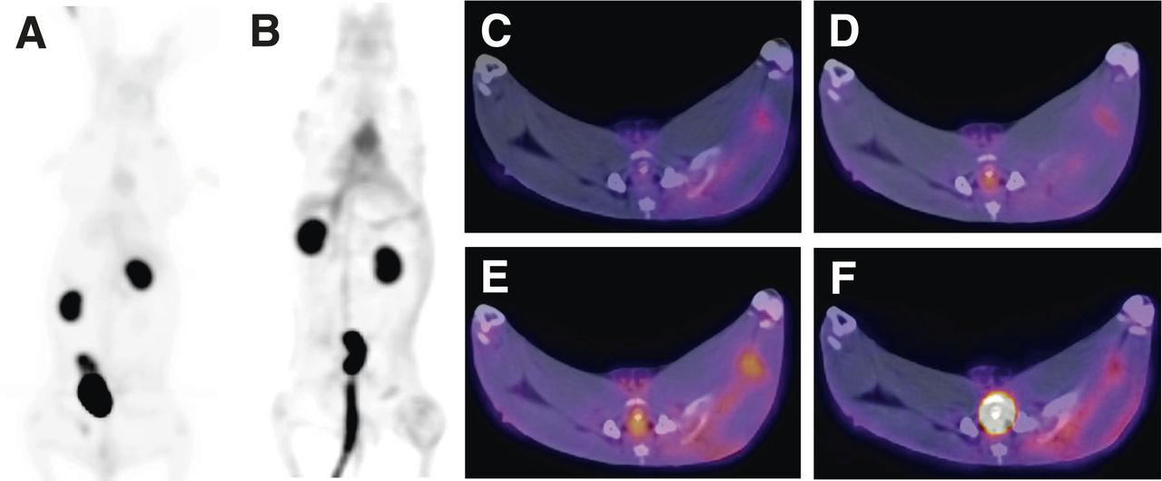
Coronal PET images acquired at 60 min after intravenous injection of 48–155 MBq of 68Ga-NOTA-UBI29-41 into healthy rabbits (NZR-1) (A) and 37–174 MBq of 68Ga-NOTA-UBI29-41 into rabbits with muscular infection (right thigh) and with sterile muscular inflammation muscle (left thigh) (NZR-2) (B). Axial PET/CT images displaying hind thighs 5 (C), 30 (D), 60 (E), and 90 (F) min after injection.
Accumulation of 68Ga-NOTA-UBI29-41 in Staphylococcus Aureus–Infected Rabbits
68Ga-NOTA-UBI29-41 Concentration in Blood and Urine of Healthy Rabbits
Results of the fluid analysis of 68Ga-NOTA-UBI29-41 shows that the peak blood activity concentration was obtained 1 min after injection (0.68 ± 0.07 %ID/g) (Fig. 2A, r2 = 0.886). A fast release of blood-bound activity is indicated by an exponential decline over 60 min, with an elimination rate of 1.44 %ID/h and a biologic blood half-life of 29 min. Between 30 and 60 min, an 18% decline of 68Ga-NOTA-UBI29-41 activity concentration was noted. At approximately 90 min, no measurable activity was found in the obtained blood samples. Urine samples showed accumulating activity levels of 68Ga-NOTA-UBI29-41 following a linear incline peaking at 3.8 ± 0.91 %ID/g at 120 min, with an accumulation rate of 1.72 %ID/h (Fig. 2B, r2 = 0.969). After 120 min, the total urine recovery amounted to 88 ± 5.2 %ID (n = 3).

Activity concentration of 68Ga-NOTA-UBI29-41 in blood (A) and urine (B) obtained from healthy rabbits (group NZR-1). Animals were injected intravenously with 96 ± 35 MBq/kg.
ITLC Analysis of 68Ga Activity in Rabbit Urine
Urine samples were analyzed using ITLC at 30, 60, and 120 min. The peak activity for 68Ga3+ was detected with a retention factor of 0.05. Disintegration of the compound (free 68Ga activity percentage) was found for the 3 groups as 0.88 ± 0.35, 1.6 ± 0.31, and 4.2 ± 0.55 %ID, respectively.
Serum Integrity Analysis
68Ga-NOTA-UBI29-41 appeared stable in serum-challenged samples, with minimal recurrence of free 68Ga up to 120 min; the free 68Ga activity amounted to 3.6 ± 0.41 %ID after 180-min incubation duration.
68Ga-NOTA-UBI29-41 Imaging of Infection
Noninvasive preclinical imaging was performed 72 h after induction to compare healthy animals (NZR-1) with bacteria-infected animals (NZR-2). The biodistribution of 68Ga-NOTA-UBI29-41 and its accumulation in both the muscular infection site and the inflammation site (Figs. 1A and 1B) was followed up to 90 min after injection of the radiopharmaceutical. The Staphylococcus aureus–bearing right thigh muscle was clearly visible in the PET/CT images, showing elevated uptake of 68Ga-NOTA-UBI29-41, compared with the contralateral thigh muscle (bearing sterile inflammation), at 5, 30, 60, and 90 min after injection (Figs. 1C–1F), and the infection site showed gradual accumulation of the tracer over the time. The T/NT ratios representing muscular inflammation (Table 3) ranged from 0.98 to 1.25, with no significant difference, compared with normal muscular tissue (P > 0.349). The maximum 68Ga-NOTA-UBI29-41 accumulation in an infected muscle was found 5.2 times higher than in normal muscle tissue at 60 min after injection. Additionally, the ratio of infected muscle to liver was moderate, with a maximum of 1.35 at 60 min.
68Ga-NOTA-UBI29-41 Uptake and Detection of Pulmonary Inflammation
The ability of 68Ga-NOTA-UBI29-41 to detect pulmonary inflammation was investigated by calculating tracer uptake and distribution in lungs. All acquired images were analyzed by calculating average whole-lung tissue SUV, which was related to the front triceps muscle SUV, yielding lung-to-muscle (L/M) ratios (Table 4). Images obtained at 30 and 60 min showed similar L/M ratios among the 3 groups. The (group-independent) L/M ratios followed a gradual, consistent incline (r2 = 0.923), amounting to 1.95 ± 0.44 (n = 4), 2.46 ± 0.53 (n = 12), 2.51 ± 0.48 (n = 10), 2.73 ± 1.01 (n = 3), and 3.04 ± 0.55 (n = 3) at 5, 30, 60, 90, and 120 min, respectively.
Pulmonary Distribution of 68Ga-NOTA-UBI29-41 in Rabbits: Quantification of Lung Tissue
DISCUSSION
The present study was undertaken with NOTA-UBI29-41 to investigate it as a potential PET/CT imaging infection agent. Moreover, the molecule was tested for its feasibility to differentiate infection from pulmonary and extrapulmonary inflammation in rabbits.
Numerous studies were published evaluating 99mTc-UBI29-41 in infected mice and rabbits (13,16,22,23) for SPECT only. One of the objectives has been to transfer the findings and therefore label the peptide fragment with the PET radionuclide 68Ga instead of 99mTc, without compromising the infection-imaging properties of UBI29-41. Whereas 99mTc can be directly introduced to UBI29-41, because the Arg and Lys residues within the fragment support the complexation of 99mTc (24), the complexation of 68Ga3+ to a peptide requires a chelating molecule. Such bifunctional chelators, of which DOTA is the most commonly used, are macrocyclic compounds designed as carriers for radiometals (25). We have selected NOTA as a chelator; with its 3 carboxyl and 3 tertiary amine residues it has been previously demonstrated to robustly complex 68Ga (26). Contrary to DOTA, the NOTA structure provides better selectivity for 68Ga3+; thus, it will form a conjugate, providing in vivo stability, and voids de-metalation and subsequent nonspecific accumulation of radiogallium.
NOTA-UBI29-41 was labeled using 68Ga, which was eluted from a tin dioxide–based generator, by fractionated elution, a simple but reliable step to reduce coelution of metal impurities such as Ge, Fe, or Zn (27). Labeling and purification was successfully performed for Ga-NOTA-UBI29-41, showing a labeling efficiency comparable to DOTA-conjugated peptides (21).
Healthy rabbits were scanned after 68Ga-NOTA-UBI29-41 administration to reveal generic tracer biodistribution and to obtain pharmacokinetic parameters in vivo. High uptake levels in the urinary bladder and kidneys dominated the 68Ga-NOTA-UBI29-41 biodistribution; however, it’s acceptable for a PET agent, because it can be easily kept under a hazardous radiation level and will seldom disturb image analysis. A 99mTc-UBI29-41 study in healthy rabbits revealed gradual excretion for the kidneys, gradual decline in liver activity, and bladder accumulation up to 76 %ID at 120 min (28). Like Akhtar et al., our study confirmed the %ID accumulation in liver and bladder. Additionally, all organ or tissues (except kidneys) showed peak tracer concentration at 30 min, declining more or less gradually until 120 min. These findings indicate that NOTA conjugation to UBI29-41 may not interfere with its natural biodistribution. The present study also successfully determined the activity levels in blood and urine samples and subsequent calculation of a moderate biologic half-life of 29 min for 68Ga-NOTA-UBI29-41 in blood. Moreover, 88% of the injected activity dose was recovered in total urine, 120 min after injection; a similar investigation was conducted in murine blood and urine by Welling et al. (22). The half-life of 99mTc-labeled UBI29-41 (16% ± 0.85%) was compared with its scrambled UBI29-41 version (21% ± 1.2%), revealing a significant difference, when the amino acid sequence was compromised. The authors also reported the percentage distribution to blood plasma fraction and blood cells; most of the activity amount was associated with the plasma-to-platelet fraction (86 %ID) and less than 15 %ID within erythrocytes and leukocytes. We have positively tested the serum stability of 68Ga-NOTA-UBI29-41 up to 3 h. The urine analysis conducted by Welling et al. showed results similar to our findings, recovering 90 %ID activity in total urine. The 2 clinical studies performed with 99mTc-UBI29-41 also support the hereby published findings of gradual blood-pool-activity decline and similar accumulation in urine (17,18). On the basis of our findings, we have reason to believe that uptake, distribution, and excretion of UBI29-41 were not significantly compromised by the conjugation with NOTA. It is noted, however, that alterations at the complexing moiety can cause a different metabolism of 99mTc-UBI29-41 (i.e., the hydrazinonicotinamide-conjugation showed similar renal clearance and warranted the infection imaging in mice but the N(2)S(2)-conjugated 99mTc-UBI29-41 showed hepatobiliar excretion and was subsequently not recommended for detection of abdominal infection (29)). It is postulated that antimicrobial peptides bind selectively to bacterial cytoplasmic membranes, because the cationic peptide residues are capable of interacting electrostatically with those anionic regions of bacterial membranes (30,31). The rabbits of group NZR-2 were scanned at 5, 30, 60, and 90 min after injection to find the ideal, earliest and most selective time duration for the detection and differentiation of the muscular infection. We found higher 68Ga-NOTA-UBI29-41 uptake in infectious muscle than in normalized muscle in 7 of 7 rabbits and for all time durations up to 90 min, indicating sensitive binding by 68Ga-NOTA-UBI29-41. The muscular inflammation showed significantly lower accumulation for all time durations (P < 0.01); 0 of 7 accumulations were significantly higher than in nontargeted muscle. T/NT ratios could differentiate between infection and inflammation from 20 min and increased gradually until 90 min after injection. Postmortem histopathologic analysis diagnosed a moderate, chronic, nonsuppurative myositis in the left thigh muscle. Distinct slides were also observed in the infected thigh; however, Staphylococcus aureus bacilli were exclusively detected in the right thigh muscle sample, confirming that the NOTA moiety did not compromise the ability of UBI29-41 to selectively visualize bacterial infection.
As the study scope is thought to be extended to noninvasive detection of airway infection, a required property of NOTA-UBI29-41 is an insensitivity to pulmonary inflammatory processes such as asthma. Rabbits sensitized with ovalbumin (NZR-3) will develop an early onset airway response that resembles human asthma with histopathologic changes, thus they can serve as a noninfectious model of inflammation (32). Airway and lung inflammations have been studied noninvasively, using mainly 18F-FDG in both animals and humans (33) and other imaging techniques such as functional MRI (fMRI) (34) and fluorescence-mediated tomography (FMT) (35). We found no significant increases in the L/M ratios in asthmatic inflamed lungs (P = 0.662) and rabbit lungs of animals with an extrapulmonary infection and inflammation (P = 0.820), compared with rabbit lungs in healthy condition. The postmortem histopathologic examination of the ovalbumin-challenged lungs positively confirmed a phenotype of asthmatic inflammation (diffuse moderate interstitial pneumonia was diagnosed with scattered heterophils, metaplasia, areas of emphysema, and areas of congestion). On the basis of this latter approach, we can preclude 68Ga-NOTA-UBI29-41 from accumulating in ovalbumin-challenged lungs and thereof it will not falsely detect an airway inflammation in rabbits.
CONCLUSION
The results support that 68Ga NOTA-UBI29-41 is an efficient and sensitive tracer of in vivo imaging of infection. 68Ga-NOTA-UBI29-41 exhibited significant uptake ratios between muscular infection and inflammation. Further clinical evaluation of this novel metabolic tracer is warranted to investigate its potential use as a first-line PET/CT infection imaging agent.
DISCLOSURE
The costs of publication of this article were defrayed in part by the payment of page charges. Therefore, and solely to indicate this fact, this article is hereby marked “advertisement” in accordance with 18 USC section 1734. We gratefully acknowledge funding provided by the Nuclear Technologies in Medicine and the Biosciences Initiative (NTeMBI). No other potential conflict of interest relevant to this article was reported.
Acknowledgments
We thank Delene van Wyk, Cindy Davis, Roleen Celliers, and Brenda Mokaleng from Steve Biko Academic Hospital for assistance during PET/CT imaging and Dr. Daniel Goosen from Disease Control Africa for his veterinary expertise and for assistance on the animal monitoring.
Footnotes
Published online Jan. 16, 2014.
- © 2014 by the Society of Nuclear Medicine and Molecular Imaging, Inc.
REFERENCES
- Received for publication June 28, 2013.
- Accepted for publication October 22, 2013.


Jump to section
Related Articles
Cited By...
- Molecular Imaging of Cardiovascular Device Infection: Targeting the Bacteria or the Host-Pathogen Immune Response?
- Molecular imaging of bacterial infections: Overcoming the barriers to clinical translation
- 68Ga-NOTA-Functionalized Ubiquicidin: Cytotoxicity, Biodistribution, Radiation Dosimetry, and First-in-Human PET/CT Imaging of Infections
- Metabolic Imaging of Infection
- Specific Imaging of Bacterial Infection Using 6''-18F-Fluoromaltotriose: A Second-Generation PET Tracer Targeting the Maltodextrin Transporter in Bacteria
- A Systematic Approach for Developing Bacteria-Specific Imaging Tracers
- New diagnostic approaches in infective endocarditis
- 68Ga-NOTA-UBI-29-41 as a PET Tracer for Detection of Bacterial Infection