


Abstract
The aim of this study was to investigate joint scintigraphy in rabbits with 99mTc-N-[3-(triethylammonio)propyl]-15ane-N5 (NTP 15-5), a new radiopharmaceutical that specifically localizes in cartilaginous tissues. Methods: Scans obtained after intravenous injection of the 99mTc-labeled compound in normal and arthropathy-induced rabbits were compared with those of the bone-imaging agent 99mTc–methylene diphosphonate (99mTc–MDP). Results: The radioactive uptake of 99mTc–NTP 15-5 was detected in cartilaginous tissues 5 min after injection and was stable for 2 h. The uptake intensity was related to age and joint disease severity, and cartilage alterations not revealed by radiography induced a significant decrease of radiotracer uptake. On the other hand, imaging performed with 99mTc–MDP did not reveal the early changes in arthrosis but was more specific for bone remodeling in advanced stages of diseases or in inflammatory processes. Conclusion: Our results indicate that 99mTc–NTP 15-5 could be a good tracer for human arthrosic and arthritic cartilage detection, especially for the early diagnosis of joint diseases.
To date, detection of arthrosis (a joint disease involving cartilage destruction that is later followed by synovial changes and bone resorption) uses several techniques, each with advantages and drawbacks. Radiography is commonly used in the diagnosis of arthritis, but it does not directly reveal articular cartilage damage at the early stage of disease. This method is valuable to confirm morphologic changes that occur in the advanced stage of joint disease (space narrowing, bone sclerosis, osteophyte formation). MRI is a highly sensitive technique, but its usefulness is limited because of the high cost of the examination and because deeper joints such as the hip are detected only with difficulty. Joint scintigraphic imaging uses mainly the bone imaging agent 99mTc–methylene diphosphonate (99mTc–MDP) and the inflammation imaging agent 67Ga citrate. The former identifies only joint diseases with bone lesions, and the specificity of the latter toward joint tissues is poor.
Use of a radiopharmaceutical that localizes directly in the articular cartilage would greatly enhance the usefulness of joint scintigraphy. 75Se-labeled compounds exhibiting affinity for cartilage have been synthesized (1), but the high toxicity of these products and the long half-life (120 d) of the radionuclide do not permit their clinical use. Another report describes radioimmunodetection in rabbit cartilage using an 123I-labeled monoclonal antibody against a protein extracted from cartilage proteoglycans (2), but the immunoscintigraphic assays performed between 6 and 24 h detected only healthy cartilage tissues and could not be actually applied to arthrosis imaging.
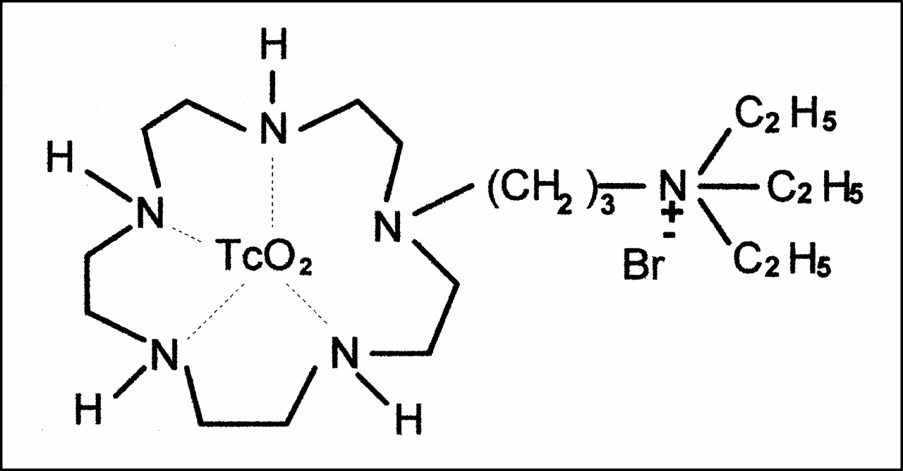
The distribution of some compounds with high affinity for cartilaginous tissues has been shown in rats (3,4); this selectivity is associated with an ionic binding between the basic quaternary ammonium group of the drugs and the polyanionic glycosaminoglycans present in cartilage (5–8). These results allowed us to synthesize molecules containing a quaternary ammonium group that is able to bind anionic proteoglycans in the target tissue and to complex 99mTc with the goal of obtaining new radiopharmaceuticals for cartilage imaging. Several products with this structure have been synthesized (9,10). Pharmacokinetic studies revealed the high affinity of these compounds for cartilage (11). Among them, 99mTc-N-[3-(triethylammonio)propyl]-15ane-N5 (NTP 15-5) (Fig. 1) exhibited the highest affinity for cartilage and was used for scintigraphic studies of rabbits, both healthy and those bearing experimental arthropathies.

Chemical structure of 99mTc–NTP 15-5.
MATERIALS AND METHODS
Tracer Preparation
99mTc–NTP 15–5 was prepared as described (10) by reducing 99mTcO4- with stannous chloride in saline at 85°C for 30 min. The yield was about 100%, and the pH was equivalent to 7. The specific radioactivity was 23 MBq/μmol, and the 99mTc-labeled compound was stable at room temperature for >24 h at pH 7.
99mTc–MDP was prepared using the MEDROCIS kit (CIS-Biointernational, Gif sur Yvette, France). This labeled compound was prepared in saline with a specific radioactivity of 13 MBq/μmol. Radiochemical purity was >98%.
Papain-Induced Arthrosis
Papain, a proteolytic enzyme, mimics the early changes in arthrosis by releasing chondroitin sulfate from the proteoglycans of the articular cartilage matrix when injected intra-articularly into a rabbit’s knee (12). Degenerative arthrosis in the knee joint was induced by a single intra-articular injection of 0.1 mL 1% sterile papain solution, activated with 0.03 mol/L cysteine, into the right knee of twelve 3- to 4-mo-old rabbits (Fauve de Bourgogne, Elevage Scientifique des Dombes, L’Arbresle, France) (same litter and same weight) after local anesthesia by subcutaneous injection of xylocaine. The left knee was used as a control. Scintigraphy was performed 0, 8, 15, 30, and 60 d after induction of arthropathy on animals anesthetized as follows (13): A single dose of 20 mg/kg flunitrazepam (Narcozep; Roche Diagnostic, Meylan, France) was administered by intramuscular injection; a 40 mg/kg dose of 6% pentobarbital diluted in isotonic saline was administered 30 min later by subcutaneous injection in 10 sites on the back of each animal. Radiography of the whole-body rabbit and knee joint was performed immediately after scintigraphy. For the histologic studies of joints, animals were killed 60 d after induction, the treated and control knee joints were removed, and histologic studies were performed using hematoxylin, eosin, and safran or toluidine blue and safranin for staining.
Zymosan-Induced Arthritis
To induce aseptic inflammatory arthritis, we used zymosan, a glycan derived from yeast cell walls. This agent is known to activate complement by the alternative pathway, to induce enzyme secretion from macrophages, and to elicit an inflammatory reaction with synovial hypertrophy, formation of pannus, and cartilage degradation within 7 d of intra-articular injection (14). Zymosan, weighed and sterilized by autoclaving, was suspended in isotonic saline (40 mg/mL) and stirred before use to obtain a homogeneous suspension. The right knee joint of each of ten 3- to 4-mo-old rabbits was injected intra-articularly with 100 μL zymosan suspension. Scintigraphy was performed 7 d after injection on animals anesthetized as described. Immediately after imaging, animals were killed and joints were removed for histologic studies.
Influence of Aging
For this study, five healthy 1-mo-old and five healthy 1-y-old rabbits (Fauve de Bourgogne), each group of the same weight and the same litter, were used. Scintigraphy was performed as described.
Imaging Protocols
Scintigraphy was performed in parallel with 99mTc–NTP 15-5 and 99mTc–MDP on rabbits separated in two groups (six animals per group for arthrosis, five animals per group for arthritis, and five animals per group for the aging study). After anesthetization, one group received by intravenous injection in the auricular vein 37 MBq/kg 99mTc–NTP 15-5, and the other group received 8.6 MBq/kg 99mTc–MDP dissolved in isotonic saline solution. Imaging was performed between 5 min and 2 h after injection for animals given 99mTc–NTP 15-5 and between 1 and 3 h for animals given 99mTc–MDP. Scans were obtained with a gamma camera (Gammatome II; Sopha Camera, France) fitted with a parallel-hole collimator for whole-body views and for joint scintigraphy with a pinhole collimator of 5.36-mm aperture (sensitivity, 3.2 × 10⁻⁴) set at 70 mm from the knee joint. In addition to analog images, static views were acquired on a computer system (Sopha Medical, Paris, France) and stored on a disk for later analysis and display. Scintigrams were obtained before induction of arthropathy (corresponding to healthy animals) and then 8, 15, 30, and 60 d after papain injection and 7 d after zymosan injection.
RESULTS
Whole-Body Scintigraphy on Healthy Animals
Whole-body scintigraphy (Fig. 2A) performed on healthy animals with 99mTc–NTP 15-5 showed a very fast uptake of the radiopharmaceutical in cartilage such as the two epiphyses of the knee articulation, the intervertebral disks, and the humeral head. Maximal concentration was obtained 5 min after injection, and good contrast toward bone and muscle was obtained up to 2 h after dosing. Kidney uptake was observed immediately after injection and a high level of radioactivity concentrated in the bladder after 15 min, in accordance with the urinary elimination of this molecule. A high concentration in the liver was observed 15 min after dosing, and this level persisted up to 2 h after injection of the labeled compound.

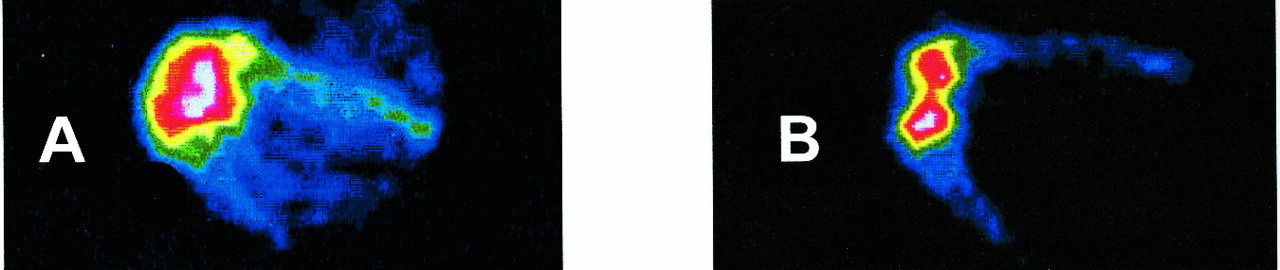
Scintigrams obtained on healthy rabbits. (A) Whole-body scintigram 10 min after administration of 37-MBq/kg dose of 99mTc–NTP 15-5. K = knee; D = intervertebral disk; H = humeral head. (B) Scintigram of right knee (pinhole collimator) 10 min after administration of 37-MBq/kg dose of 99mTc–NTP 15-5. (C) Scintigram of right knee (pinhole collimator) 90 min after administration of 8.6-MBq/kg dose of 99mTc–MDP.
Pinhole Scintigraphy of Healthy Knees
With a pinhole collimator pointed at the joint knee, an image of the tibial plate and femoral head cartilage was easily visible 10 min after injection (Fig. 2B); this pattern persisted up to 90 min after injection. The radiopharmaceutical did not label bone and muscle tissues, and the contrast between cartilage and adjacent tissues was high. In contrast, 99mTc–MDP specifically labeled bone tissues, and tibial plate and femoral head were distinct (Fig. 2C). The images of the two radiopharmaceuticals appeared similar, with a fixing zone that was slightly larger with NTP 15-5 than with MDP.
Papain-Induced Arthrosis
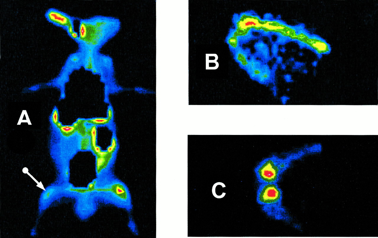
Scintigrams obtained on papain-injected rabbits on days 0, 8, 15, 30, and 60 showed a marked decrease in incorporation of labeled 99mTc–NTP 15-5. The six rabbits tested showed an important hypofixation of the radiopharmaceutical in the arthrosic joint compared with the image of the control knee (Fig. 3A). This hypofixation became significant 15 d after papain injection and increased with time after induction (Fig. 4). Histologic studies performed 60 d after induction showed that papain was responsible for important arthrosic lesions: disruption of cartilage structure, suggesting collagen network destruction clefts; and empty chondrocyte lacunas, suggesting cell death. Nevertheless, radiography performed on the same animals did not reveal any morphological damage to articular cartilage, even though the arthrosis induced by papain was severe. Comparative experiments performed with 99mTc–MDP showed no significant modifications of the radiopharmaceutical uptake before and after treatment (Fig. 3C).

Scintigrams obtained on rabbits bearing papain-induced arthrosis 30 d after induction. (A) Whole-body scintigram 10 min after administration of 37-MBq/kg dose of 99mTc-NTP 15-5. Arrow indicates treated knee. (B) Scintigram of knee with arthrosis (pinhole collimator) 10 min after administration of 37-MBq/kg dose of 99mTc-NTP 15-5. (C) Scintigram of knee with arthrosis (pinhole collimator) 90 min after administration of 8.6-MBq/kg dose of 99mTc-MDP.

Ratio of 99mTc-NTP-15-5 uptake by knee with arthrosis to 99mTc-NTP-15-5 uptake by control knee as function of time after induction of arthrosis. Results are expressed as mean ± SD (n = 6 animals).
Zymosan-Induced Arthritis
Scintigraphy of the treated joint showed an increase in 99mTc–NTP 15-5 uptake, with a significant growth of the labeled area. It was not possible to distinguish tibial plate and femoral head, and the image was fuzzy and intense (Fig. 5A). On the contrary, 99mTc–MDP incorporation in the arthritic joint was only slightly higher after induction of arthritis, and no modification of the area of uptake was observed, the tibial and femoral epiphysis remaining still distinct (Fig. 5B). In contrast to the arthrosic joint, zymosan-induced arthritis showed an inflamed and hypertrophied synovial membrane. Histologic examination of this tissue revealed an intense hypercellularity and a marked inflammatory cell infiltrate, with a predominant lymphocyte presence in the germinative center and a small number of polymorphonuclear leukocytes, histiocytes, and fibroblasts. The synovial pannus of granulation tissue extended over the articular cartilage, which was partially and slightly indented at the upper surface, and with a partial loss of chondrocytes within the matrix, suggesting secondary changes occurring in cartilage under the influence of the inflammatory process. These histopathologic findings confirmed inflammatory arthritis induced by zymosan.
Scintigrams obtained on rabbits bearing zymosan-induced arthritis 7 d after induction. (A) Scintigram of arthritic knee (pinhole collimator) 10 min after administration of 37-MBq/kg dose of 99mTc–NTP 15–5. (B) Scintigram of arthritic knee (pinhole collimator) 90 min after administration of 8.6-MBq/kg dose of 99mTc–MDP.
Influence of Aging
Scintigraphy performed on healthy rabbits between 1 mo and 1 y old showed a high uptake of 99mTc–NTP 15-5 in the young animals with distinct labeled zones for tibial plate and femoral head for knee articulation. The intensity of radiopharmaceutical incorporation decreased with age, and it was difficult to appreciate distinctly labeled epiphyses on 1-y-old rabbits. The 99mTc–MDP uptake decreased with age, as for 99mTc–NTP 15-5, but the epiphysis zones always appeared to be well separated (scintigrams not shown).
DISCUSSION
Arthrosis is a degenerative joint disease with progressive destruction of cartilage followed by changes in bones and synovium at advanced stages of the disease (15). For some years, the bone tracer 99mTc–MDP has been used as an arthrosis-imaging agent to evaluate the extent of this arthropathy. This radiopharmaceutical is known to concentrate in the mineralization zone, especially in calcification areas, such as provisional calcification of the epiphyseal growth plate. The uptake intensity is related to the osteoblastic activity and the vascularization of the bony tissue and reveals functional modification in bone that often precedes morphological alterations that are visible on radiography. At advanced stages of arthrosis, changes occur in the bony tissue of the articulation (osteophyte formation, subchondral bone sclerosis). 99mTc–MDP can visualize these modifications, but this radiopharmaceutical cannot reveal alterations of cartilage that occur at early stages of osteoarthrosic disease.
Therefore, it would be of interest to use an agent that localizes directly in articular cartilage with a view to enhance the usefulness of joint scintigraphy. The results obtained with 99mTc–NTP 15-5 suggest that this radiopharmaceutical could be more specific than 99mTc–MDP for joint diseases involving cartilage degradation. Indeed, 99mTc–MDP concentrates in the bony tissue, whereas 99mTc–NTP 15-5 distributes in cartilage at the surface of the epiphysis and intervertebral disks (11). The image intensity is correlated with the metabolic activity of the osteoblasts and the level of hydroxyapatite in skeletal tissue for 99mTc–MDP (16) and with the level of proteoglycan content of cartilage for 99mTc–NTP 15-5 (5,11). These incorporation mechanisms can explain the differences observed between the two radiopharmaceuticals. In degenerated cartilage such as arthrosis and arthritis experimentally induced by papain or zymosan, respectively, the results differ greatly between the two radiopharmaceuticals. For 99mTc–NTP 15-5, the decrease in uptake observed up to 8 d after papain injection suggests a loss of anionic proteoglycans within the tissue. The uptake intensity of the radiopharmaceutical may be able to measure the proteoglycan content and the metabolic state of the cartilage (17–19).
In early stages of arthropathy, no morphological lesions of the joint appear and no increase in osteoblastic activity is observed. In these conditions, radiography and scintigraphy using the bone-imaging radiopharmaceutical 99mTc–MDP are unable to identify these lesions. Our results obtained on animal models confirm that only a specific radiopharmaceutical concentrating in cartilage is able to identify arthrosis at this early stage of the disease.
In contrast to arthrosis scintigraphy with 99mTc–NTP 15-5, which revealed a decreased uptake in cartilaginous tissue (related to a depletion of proteoglycans), the increased uptake of the tracer in joints injected with zymosan is related to inflammatory reaction. The fuzzy and intense image of the knee joint probably corresponds to a superimposition of two images, one correlated with cartilage uptake and the other with a possible synovial uptake. It is well known that synovial cells synthesize hyaluronic acid, a main glycosaminoglycan of synovial tissue and synovial fluid that is responsible for lubrication of the articular cartilage bound to a nutritive function (16). Inflammatory reactions induced by intra-articular injection of zymosan produce a synovial tissue hypertrophy and perhaps an increased level of synovial fluid with carboxyl and sulfate groups able to bind the quaternary ammonium functions of the radiopharmaceutical by ionic interactions. The number of synoviocytes increases in arthritis, and these cells synthesize more glycosaminoglycans than do the healthy cells (Maurizis JC, Ollier M, March 1998, unpublished data). In arthritic patients, the synovial fluid is more abundant and contains more hyaluronic acid bearing carboxyl residues that are able to increase the uptake of the radiopharmaceutical in the arthritic joint. It is highly likely that the fuzzy, intense, and enlarged images obtained with 99mTc–NTP 15-5 are associated with ionic interactions between the quaternary ammonium function of the radiopharmaceutical and proteoglycans of cartilage and the glycosaminoglycans such as hyaluronic acid present in excess in fluid and synovial tissue. Consequently, the presence of an important and indistinct radiolabeled zone in articulation will be evidence of severe inflammation, with synovial tissue hypertrophy and with the possible presence of synovial fluid in excess, which can be of interest for examination of a deep articulation such as the hip. Scans of arthritic joints obtained with 99mTc–MDP revealed only a slight increase with a distinct epiphysis of the knee articulation, which showed the absence of bone remodeling during this aseptic inflammation induced by zymosan.
CONCLUSION
Our data show that the pattern of scintigraphic imaging performed with 99mTc–NTP 15-5 on healthy joints is identical to that of 99mTc–MDP in normal articulation and that the intensity of uptake decreases with age for both radiopharmaceuticals. On the other hand, the bone tracer 99mTc–MDP is unable to identify the early stages of arthropathy; its uptake is related to the alterations in bone remodeling, suggesting osteoblast activity or calcification, within or outside of articulations. On the contrary, the uptake of 99mTc–NTP 15-5 decreases in the early stages of arthropathy, except under some severe inflammatory processes with induced synovial hypertrophy, which hinders the visualization of the epiphysis. Nevertheless, our results indicate that 99mTc–NTP 15-5 can be used as a cartilage-imaging agent associated with bone-tracer scanning in joint diseases.
Footnotes
Received Feb. 23, 2000; revision accepted Jun. 30, 2000.
For correspondence or reprints contact: Jean-Claude Maurizis, PhD, Institut National de la Santé et de la Recherche Médicale U484, Rue Montalembert, BP184, Clermont-Ferrand Cedex, 63005 France.


