
Main menu

  • Home
  • Content
    • Current
    • Ahead of print
    • Past Issues
    • JNM Supplement
    • SNMMI Annual Meeting Abstracts
    • Continuing Education
    • JNM Podcasts
  • Subscriptions
    • Subscribers
    • Institutional and Non-member
    • Rates
    • Journal Claims
    • Corporate & Special Sales
  • Authors
    • Submit to JNM
    • Information for Authors
    • Assignment of Copyright
    • AQARA requirements
  • Info
    • Reviewers
    • Permissions
    • Advertisers
  • About
    • About Us
    • Editorial Board
    • Contact Information
  • More
    • Alerts
    • Feedback
    • Help
    • SNMMI Journals
  • SNMMI
    • JNM
    • JNMT
    • SNMMI Journals
    • SNMMI

User menu

  • Subscribe
  • My alerts
  • Log in
  • Log out
  • My Cart

Search

  • Advanced search
Journal of Nuclear Medicine
  • SNMMI
    • JNM
    • JNMT
    • SNMMI Journals
    • SNMMI
  • Subscribe
  • My alerts
  • Log in
  • Log out
  • My Cart
Journal of Nuclear Medicine

Advanced Search

  • Home
  • Content
    • Current
    • Ahead of print
    • Past Issues
    • JNM Supplement
    • SNMMI Annual Meeting Abstracts
    • Continuing Education
    • JNM Podcasts
  • Subscriptions
    • Subscribers
    • Institutional and Non-member
    • Rates
    • Journal Claims
    • Corporate & Special Sales
  • Authors
    • Submit to JNM
    • Information for Authors
    • Assignment of Copyright
    • AQARA requirements
  • Info
    • Reviewers
    • Permissions
    • Advertisers
  • About
    • About Us
    • Editorial Board
    • Contact Information
  • More
    • Alerts
    • Feedback
    • Help
    • SNMMI Journals
  • View or Listen to JNM Podcast
  • Visit JNM on Facebook
  • Join JNM on LinkedIn
  • Follow JNM on Twitter
  • Subscribe to our RSS feeds
Research ArticleThe Illustrated Post

CD38-Targeted 89Zr-DFO-Daratumumab PET of Myeloma: Immuno-PET Impacting Clinical Care

Gary A. Ulaner, Jason Lewis and Ola Landgren
Journal of Nuclear Medicine January 2025, jnumed.124.269393; DOI: https://doi.org/10.2967/jnumed.124.269393
Gary A. Ulaner
1Molecular Imaging and Therapy, Hoag Family Cancer Institute, Irvine, California;
2Radiology and Translational Genomics, University of Southern California, Los Angeles, California;
  • Find this author on Google Scholar
  • Find this author on PubMed
  • Search for this author on this site
Jason Lewis
3Radiology, Memorial Sloan Kettering Cancer Center, New York, New York; and
  • Find this author on Google Scholar
  • Find this author on PubMed
  • Search for this author on this site
Ola Landgren
4Division of Myeloma, University of Miami, Miami, Florida
  • Find this author on Google Scholar
  • Find this author on PubMed
  • Search for this author on this site

Currently, the determination of disease burden in multiple myeloma is suboptimal. Myeloma cells may not secrete abnormal immunoglobulins or free light chains, limiting blood and urinary analysis (1). Imaging by radiography and MRI is limited, and approximately 30% of myeloma lesions are not appreciable by 18F-FDG PET (2). Bone marrow biopsies are also limited by the limited sites that can be sampled (3). Thus, better methods of detecting, localizing, and quantitating myeloma cells are needed.

Here, we report on a 76-y-old man with multiple myeloma who experienced new biochemical progression after stem cell transplantation. A masked bone marrow biopsy showed no evidence of myeloma cells. 18F-FDG PET/CT was ordered to measure tumor burden and determine whether systemic therapy was appropriate; however, no lesions were visualized by 18F-FDG PET (Fig. 1).

FIGURE 1.
  • Download figure
  • Open in new tab
  • Download powerpoint
FIGURE 1.

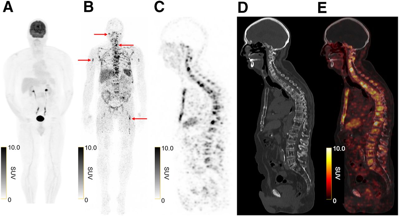
(A) 18F-FDG maximum-intensity projection of 76-y-old patient with biochemically relapsed/refractory multiple myeloma is without abnormal foci of disease. (B) In contrast, 89Zr-DFO-daratumumab maximum-intensity projection, 7 d after tracer administration, demonstrates more than 100 abnormal foci in head, neck, chest, abdomen, pelvis, and extremities (arrows). (C–E) Sagittal 89Zr-DFO-daratumumab PET (C), CT (D), and 89Zr-DFO-daratumumab PET/CT (E) demonstrate abnormal foci corresponding to osseous structures, representing non–18F-FDG–avid osseous myeloma lesions.

He was accrued into a phase II trial (NCT04814615) of CD38-targeted immuno-PET with 89Zr-deferoxamine (DFO)-daratumumab. CD38 is a transmembrane glycoprotein that is expressed on virtually all myeloma cells, as well as at lower levels on normal lymphoid and myeloid cells (4). 89Zr-DFO-daratumumab comprises the CD38-targeting antibody daratumumab, chelated through DFO to the positron-emitting radiometal 89Zr, whose half-life of 78.4 h allows for imaging up to at least 9 d after administration (5). As antibodies such as daratumumab may take 6–7 d to optimally biodistribute onto tumor, the long half-life of 89Zr allows optimal timing of imaging. 89Zr-DFO-daratumumab was able to demonstrate widespread foci of disease, with subsequent image-guided biopsy providing proof of myeloma. The identification, localization, and quantification of disease led to initiation of systemic therapy with daratumumab, lenalidomide, and dexamethasone. This case suggests that 89Zr-DFO-daratumumab immuno-PET has the potential to have an important clinical impact on patients diagnosed with multiple myeloma.

DISCLOSURE

This research was funded in part through NIH R01 CA248398 (Gary Ulaner and Ola Landgren) and the James & Pamela Muzzy Endowed Chair (Gary Ulaner). Ola Landgren is supported by a Sylvester Comprehensive Cancer Center NCI core grant (P30 CA 240139) and by the Rising Tide Foundation, Leukemia & Lymphoma Society, Multiple Myeloma Research Foundation (MMRF), International Myeloma Foundation (IMF), Paula and Rodger Riney Foundation, Tow Foundation, Perelman Family Foundation, Myeloma Solutions Fund, and Cannon Guzy Family Fund. No other potential conflict of interest relevant to this article was reported.

Footnotes

  • Published online Jan. 30, 2025.

  • © 2025 by the Society of Nuclear Medicine and Molecular Imaging.

REFERENCES

  1. 1.
    1. Chawla SS,
    2. Kumar SK,
    3. Dispenzieri A,
    4. et al
    . Clinical course and prognosis of non-secretory multiple myeloma. Eur J Haematol. 2015;95:57–64.
  2. 2.
    1. Hillengass J,
    2. Landgren O
    . Challenges and opportunities of novel imaging techniques in monoclonal plasma cell disorders: imaging “early myeloma.” Leuk Lymphoma. 2013;54:1355–1363.
  3. 3.
    1. Mailankody S,
    2. Korde N,
    3. Lesokhin AM,
    4. et al
    . Minimal residual disease in multiple myeloma: bringing the bench to the bedside. Nat Rev Clin Oncol. 2015;12:286–295.
  4. 4.
    1. Nooka AK,
    2. Kaufman JL,
    3. Hofmeister CC,
    4. et al
    . Daratumumab in multiple myeloma. Cancer. 2019;125:2364–2382.
  5. 5.
    1. Ulaner GA,
    2. Sobol NB,
    3. O’Donoghue JA,
    4. et al
    . CD38-targeted immuno-PET of multiple myeloma: from xenograft models to first-in-human imaging. Radiology. 2020;295:606–615.
  • Received for publication December 18, 2024.
  • Accepted for publication January 6, 2025.
SNMMI

© 2025 SNMMI

Powered by HighWire