


Abstract
P914
Introduction: There is evidence to suggest Total Tumor Volume (TTV) and Whole Body SUVmean on quantitative 177Lu-PSMA SPECT/CT (Lu-SPECT) predicts outcomes in patients with metastatic castration resistant prostate cancer (mCRPC) receiving 177Lu-PSMA therapy (Pathmanandavel S et al. JNM 2022; John N et al. JNM 2022). Generating TTV is a time-consuming process. This study evaluated the accuracy of an algorithm to automatically generate TTV on Lu-SPECT.
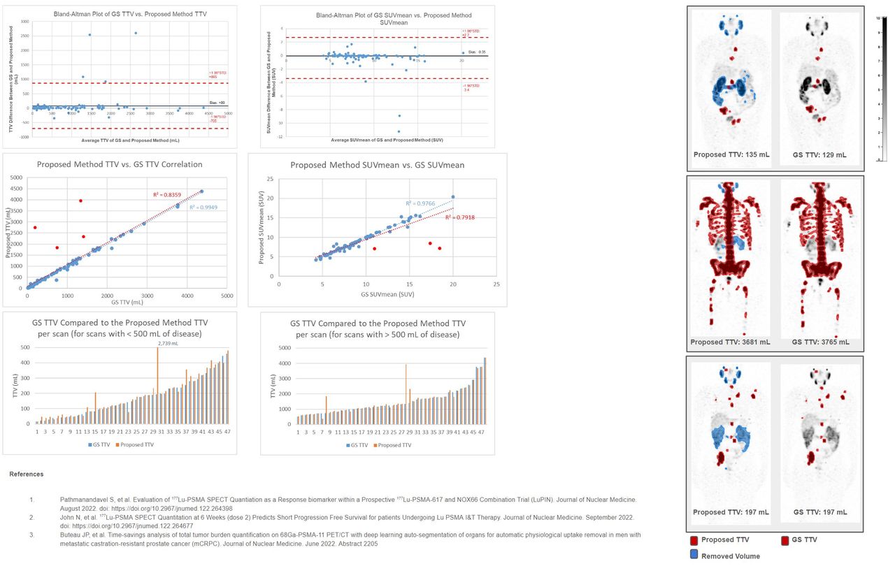
Methods: 95 quantitative 24-hour post 177Lu-PSMA therapy SPECT/CT scans from 36 patients with mCRPC were used from the LuPIN Trial (Pathmanandavel S et al. JNM 2022). Patients received between 1 and 6 cycles of therapy with each cycle occurring every six weeks. The Gold Standard (GS) VOIs were segmented by a nuclear medicine physician using a 3 SUV threshold followed by manual edits. The proposed algorithm segmented all VOIs in the image with a 3 SUV threshold. Then, utilizing normal structures created by a CT-based deep learning convolutional neural network, VOIs contained within organs with physiological uptake were removed from the proposed segmentation. Organ contours used in the algorithm included: liver, kidneys, parotids, submandibular glands, lacrimal glands, bone, bowel, pelvic lymph nodes, prostate, and bladder (Buteau JP et al. JNM 2022). A paired, two-tailed t-test was used to determine statistical significance, with an α of 0.05 indicating a significant difference.
Results: The average TTV for the proposed method was 927 mL (土 988) compared to the average GS TTV of 847 mL (土 910), which was not significantly different (p=0.054). The average absolute difference in TTV between the proposed method and the GS was 110 mL (土 393). Note that with 4 outliers removed, the difference reduces to 34.3 mL (土 55.1) (p=0.46). The correlation coefficient between the proposed method TTV and the GS was 0.84. The proposed method resulted in an average of 25.5 mL (土 28.4) of uptake that was falsely included and an average of 3.42 mL (土 0.43) of uptake that was either falsely removed or not captured. The average SUVmean of the GS was 8.96 SUV (土 3.34), while the average SUVmean of the proposed method was 8.60 SUV (土 3.11). This difference in SUVmean between the proposed method and the GS was significantly different (p=0.03). Although significantly different, the average relative error in SUVmean was 4.7% (土 9.7%), and the median relative error in SUVmean was 1.4%.
Conclusions: The proposed method utilized CT-based, deep learning generated normal organs for automatic removal of physiologic uptake on Lu-SPECT. This method produced comparable TTV and Whole Body SUVmean to that produced by an expert. A small but statistically significant difference in SUVmean was identified between the proposed automated method and the GS, however the clinical significance of this is unclear, and little manual editing would likely be needed to match the GS. A tool using the proposed method could increase efficiency and reproducibility when generating TTV and Whole Body SUVmean for the assessment of 177Lu-PSMA therapy, but additional investigation is needed to assess the actual amount of time saved when utilizing this method of automatic physiological uptake removal.


In this issue


Jump to section
Related Articles
Cited By...
- No citing articles found.