


Abstract
S-11C-methyl-L-cysteine (11C-MCYS), an analog of S-11C-methyl-L-methionine (11C-MET), can potentially serve as an amino acid PET tracer for tumor imaging. The aim of this study was to investigate the radiosynthesis and perform a biologic evaluation of 11C-MCYS as a tumor imaging tracer. The results of the first human PET study are reported. Methods: 11C-MCYS was prepared by 11C-methylation of the precursor L-cysteine with 11CH3I and purification on commercial C18 cartridges. In vitro competitive inhibition experiments were performed with Hepa1–6 hepatoma cell lines, and biodistribution of 11C-MCYS was determined in normal mice. The incorporation of 11C-MCYS into tissue proteins was investigated. In vivo 11C-MCYS uptake studies were performed on hepatocellular carcinoma–bearing nude mice and inflammation models and compared with 11C-MET PET and 18F-FDG PET. In a human PET study, a patient with a recurrence of glioma after surgery was examined with 11C-MCYS PET and 18F-FDG PET. Results: The uncorrected radiochemical yield of 11C-MCYS from 11CH3I was more than 50% with a synthesis time of 2 min, the radiochemical purity of 11C-MCYS was more than 99%, and the enantiomeric purity was more than 90%. In vitro studies showed that 11C-MCYS transport was mediated through transport system L. Biodistribution studies demonstrated high uptake of 11C-MCYS in the liver, stomach wall, and heart and low uptake of 11C-MCYS in the brain. There was higher accumulation of 11C-MCYS in the tumor than in the muscles. The tumor-to-muscle and inflammatory lesion–to–muscle ratios were 7.27 and 1.62, respectively, for 11C-MCYS, 5.08 and 3.88, respectively, for 18F-FDG, and 4.26 and 2.28, respectively, for 11C-MET at 60 min after injection. Almost no 11C-MCYS was incorporated into proteins. For the patient PET study, high uptake of 11C-MCYS with true-positive results, but low uptake of 18F-FDG with false-negative results, was found in the recurrent glioma. Conclusion: Automated synthesis of 11C-MCYS is easy to perform. 11C-MCYS is superior to 11C-MET and 18F-FDG in the differentiation of tumor from inflammation and seems to have potential as an oncologic PET tracer for the diagnosis of solid tumors.
As a glucose metabolism tracer, 18F-FDG has been widely used in PET imaging for oncology and treatment evaluation ( 1). However, clinical 18F-FDG PET studies have demonstrated several limitations, such as difficulty in differentiating tumor from inflammation because of high uptake of 18F-FDG in tumors and in nonmalignant inflammatory tissue ( 2), thus motivating efforts to develop new oncologic PET tracers. Positron-labeled amino acids have proven to be useful for imaging tumors, especially brain tumors and peripheral tumors such as those in the lung, breast, and liver and lymphomas. Currently, S-11C-methyl-L-methionine (11C-MET) is the most commonly used amino acid tracer for tumor imaging with PET ( 3). However, some investigations, including many clinical PET studies, have shown that increased 11C-MET uptake is not specific to malignant tumors ( 4– 6). 11C-MET is also taken up in some inflammatory lesions ( 7). It has been reported that 11C-MET is incorporated not only into protein fractions via the conversion into amino-acyl-transfer RNA but also into nonprotein materials, such as phospholipids and RNA, by the transmethylation process via S-adenosyl-L-methionine ( 8), making correct quantitative investigation difficult. Because of the short half-life of 11C, many 18F-labeled amino acids have been developed. Among them, O-2-18F-fluoroethyl-L-tyrosine (18F-FET) is another commonly used amino acid tracer for the detection of tumors and has been superior to 18F-FDG in distinguishing tumors from inflammation ( 9). However, the automated radiosynthesis of 18F-FET with complex high-performance liquid chromatography purification is cumbersome and time-consuming ( 10), compared with the production of 11C-MET by on-column methylation ( 11). Thus, the easy availability of 11C-methyl–containing tracers in the PET center with an in-house cyclotron gives them potential for use in clinical PET.
S-11C-methyl-L-cysteine (11C-MCYS), an analog of 11C-MET, has not been evaluated as a tumor imaging agent. In this work, we report the easy-to-automate radiosynthesis and perform a biologic evaluation of 11C-MCYS as a malignant tumor tracer more specific than 18F-FDG and 11C-MET. In addition, the first 11C-MCYS PET study on a patient is reported.
MATERIALS AND METHODS
Chemicals
All reagents used in the synthesis were commercial products applied without further purification unless otherwise indicated. Acetonitrile, dimethylsulfoxide, N,N-dimethylformamide, tetrahydrofuran, lithium aluminum hydride (1.0 M in tetrahydrofuran), hydroiodic acid, and L-cysteine were purchased from Sigma-Aldrich.
Cell Culture
A hepatocellular carcinoma cell line (Hepa1–6) was obtained from the Laboratory Animal Center of Sun Yat-Sen University, Guangzhou, China. The cells were cultivated in RPMI 1640 medium with a physiologic glucose concentration (1.0 g/L) containing 5% fetal calf serum at 37°C in a humidified atmosphere of 5% CO2 and 95% air. The medium was routinely renewed 3 times a week. Two days before the in vitro experiments, the cells were trypsinized and 2 × 10⁵ cells per well were seeded into 24-well plates.
Animals and Tumors
Female and male C57BL/6J mice (6 wk old) were obtained from the Laboratory Animal Center of Sun Yat-Sen University. Mice and nude mice were housed 5 animals per cage under standard laboratory conditions at 25°C and 50% humidity. They were allowed free access to food and water. We prepared 3 groups of mice, namely normal mice (n = 30), tumor nude mice (n = 20), and inflammation mice (n = 20). Tumor nude mice were inoculated subcutaneously in the femoral region with 1.0 × 10⁷ Hepa1–6 cells. Inflammation mice were inoculated subcutaneously with 0.2 mL of turpentine oil (Wako Chemical) ( 12). These turpentine-induced masses showed acute inflammation histologically as described in an earlier report ( 13). All animals were kept in cages under standardized conditions. At the time of the experiments, the mice were 9–10 wk old and weighed 18–25 g. The mass of the tumors and inflammatory tissue grew to 10–25 mm during the experiments. The study was performed according to the guidelines and recommendations of the Committee on Animal and Human Research at the First Affiliated Hospital, Sun Yat-Sen University. The protocol was fully approved by the local institutional review committee on animal care.
Radiosynthesis Procedures
11CO2 was produced by 14N(p,α)11C nuclear reactions using a Cyclone 10/5 cyclotron (IBA) and was delivered to the radiochemical laboratory. 11CO2 was trapped in a loop ring cooled at –160°C with liquid nitrogen. After a flush with helium, the ring was warmed and 11CO2 released into the reactor module under a helium flow (20 mL/min), through a dehydrating agent, P2O5. 11CH3I was produced through reduction of 11CO2 with LiAlH4, hydrolysis of the intermediately formed organometallic complex, and subsequent iodination of 11C-methanol with hydrogen iodide. 11C-MCYS were synthesized according to the solid-phase 11C-methylation of the precursor L-cysteine loaded into a C18 column with 11CH3I ( Fig. 1). Briefly, 11CH3I produced as described above was delivered under a helium flow (20 mL/min) to a Sep-Pak Plus C18 cartridge (Waters) previously loaded with a solution of L-cysteine (∼2 to 3 mg) dissolved in NaOH 0.5 mol/L in 50:50 ethanol:water (v/v, 0.210 mL). 11C-MCYS was eluted with NaH2PO4 0.05 M buffer (5 mL, pH ∼3 to 4) and collected in a vented sterile vial. The solution was finally sterilized by a 0.22-mm filter to give the final formulation. The fully automated synthesis of 11C-MCYS was performed on a simple, remotely controlled commercial 11C-methylation module (PET-CS-I-IT-I 11C-methylation synthesizer; PET (Beijing) Technology Co., Ltd.) as shown in Supplemental Figure 1 (supplemental materials are available online only at http://jnm.snmjournals.org).
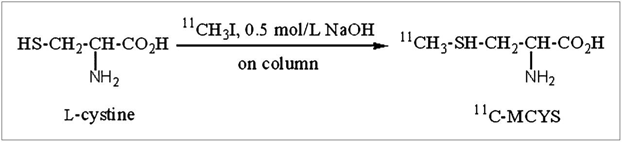
Radiosynthesis of 11C-MCYS by on-column 11C-methylation of L-cysteine.
11C-MET and 18F-FDG were synthesized as published previously by Tang et al. ( 14, 15).
Quality Control Procedures
Radiochemical purity and chemical purity of 11C-MCYS were measured by analytic high-performance liquid chromatography with an Eclipse XDB-C18 analytic column (4.6 × 150 mm, 5 μm; Agilent) using 55:45 CH3OH:3 mM NaH2PO4 (v/v) as the mobile phase at a flow rate of 1 mL/min. The elution was monitored by an ultraviolet detector (254 nm) and radiodetector. The pH was measured by a standard pH meter. Absence of pyrogen in the injectable solutions was checked by limulus amebocyte lysate testing using a gel clotting assay, and samples were tested for sterility.
Transport Assays
Transport assays with Hep1–6 cells were performed at 2 d after seeding during the exponential growth phase. The method of measuring transport was similar to a method reported previously ( 2, 16, 17). The competitive inhibition experiments to characterize the transport system were performed in the presence of the specific competitive inhibitors: for system L, 2′-aminobicyclo( 2, 2, 1)-heptane-2′-carboxylic acid (BCH); for system A, N-methylamino-isobutyric acid (MeAIB); and for system ASC, MeAIB plus serine. The concentration of the inhibitors used was 15 mmol/L. Parallel experiments with increasing concentrations of MCYS were performed to investigate the capacity of the transport system. All experiments were performed in the presence and absence of Na+. In Na+-free experiments, sodium salts were replaced by choline chloride. Each experiment was done in triplicate and averaged and was repeated 5 times on different days. After preincubation of the cells in 200 μL of the medium for 30 min, 200 μL of 11C-MCYS (1.85 MBq/mL) and 200 μL of one of the inhibitors or MCYS (50, 100, 200, 300, or 350 μmol/L) were added and samples were incubated at 37°C for 4 min. After tracer uptake had been stopped with 1 mL of ice-cold 1× phosphate-buffered saline, the cells were washed 3 times with phosphate-buffered saline at 4°C and dissolved in 1.5 mL of 0.1 M NaOH plus 2% Triton X (Sigma Chemical Co.) and the activity measured by a γ-counter.
In Vivo Biodistribution Studies
The animals were anesthetized with pentobarbital (75 mg/kg) before injection of radiotracer and remained anesthetized through the study. They were injected with 0.74–1.48 MBq (20–40 μCi) of radiotracer in 100–200 μL of saline through the tail vein (11C-MCYS and 11C-MET in phosphate-buffered saline and 18F-FDG in normal saline). A prescribed duration was allowed before procurement of organs and tissues. Blood was obtained through the eyeball, tissue samples of interest (blood, brain, heart, lung, liver, kidney, and small intestine in normal mice) were rapidly dissected and weighed, and 11C radioactivity was counted with an automatic γ-counter. All measurements were background-subtracted and decay-corrected to the time of killing and then averaged ( 2). Data were expressed as a percentage of the injected dose per gram of tissue (%ID/g).
Incorporation of 11C-MCYS into Proteins
To determine the extent of protein incorporation of 11C-MCYS, protein-bound activity in the brain, pancreas, tumor, and blood samples was determined at 30 min after injection. For this purpose, tumor, pancreatic, and cerebral tissue (∼50 to 100 mg in wet weight) was homogenized by the addition of 1 mL of distilled water and by ultrasonication at 0°C. Blood samples were immediately centrifuged at 4,000g, and 250 μL of 50% trichloroacetic acid were added to 750 μL of plasma and stirred in a vortex mixer for 1 min. After centrifugation, the supernatants were removed from the pellets. The radioactivity in the acid solution and acid-precipitable fraction was measured with a γ-counter. Sampling of the 11C-activity was performed using the supernatant solution after spiking with a small amount of MCYS by high-performance liquid chromatography (55:45 CH3OH:3 mM NaH2PO4 (v/v)), and the elution profile was detected with an ultraviolet detector at 254 nm. Incorporation of 11C-MCYS into tumor, pancreatic, and cerebral tissue was measured as described. Radioactivities contained in the acid solution (supernatant solution) and acid-precipitable fraction (protein pellet) were also determined.
PET Studies
PET was performed using a Gemini GXL scanner (Philips). Before undergoing 11C-MCYS, 11C-MET, and 18F-FDG PET studies, the animals were kept fasting for at least 4 h. The tumor-bearing mice and inflammation model mice were anesthetized with pentobarbital (75 mg/kg) before injection of the radiotracer and remained anesthetized throughout the study. 11C-MCYS, 11C-MET, or 18F-FDG (10 MBq) was injected in 100–200 μL of phosphate-buffered saline or normal saline through the tail vein. Then, image acquisition was performed at 60 min after intravenous injection in 3-dimensional mode, with emission scans of 4 min per bed position. Imaging started with a low-dose CT scan (30 mAs), immediately followed by a PET scan. The CT scan was used for attenuation correction and localization of the lesion site. In addition, a 45-y-old patient with a history of attempted resection of grade IV glioma 1 y previously was also examined with 18F-FDG and 11C-MCYS PET. The coronal, transaxial, and sagittal PET views of tumor-bearing mice, inflammation model mice, and the patient were obtained after image reconstruction with a slice thickness of 2.0 mm. A region of interest was placed on each tumor, each inflammatory lesion, and a region of femoral muscle. The human study was approved by the Institutional Review Board of the First Affiliated Hospital, Sun Yat-Sen University. Informed consent was obtained from the patient before injection.
Statistical Analysis
Statistical analysis was performed with SPSS software, version 15.0 (SPSS Inc.), for Windows (Microsoft). All data were expressed as mean ± SD. Comparisons between conditions were performed using the unpaired, 2-tailed Student t test. A P value of less than 0.05 was considered to indicate statistical significance.
RESULTS
Radiosynthesis Results
The uncorrected radiochemical yield of 11CH3I was 60%–70% from 11CO2, and the synthesis time was 8–10 min after release of 11CO2. The uncorrected radiochemical yield of 11C-MCYS from 11CH3I was more than 50% with a synthesis time of 2 min. The radiochemical purity of 11C-MCYS was more than 99% ( Fig. 2), the enantiomeric purity was more than 90%, and the total synthesis time from 11CO2 was about 12 min. The retention time was approximately 1.8–2.2 min for 11C-MCYS, less than 1.5 min for cysteine, and approximately 5.8–6.5 min for 11CH3I. 11C-MET was synthesized with high chemical and radiochemical purity ( 13).
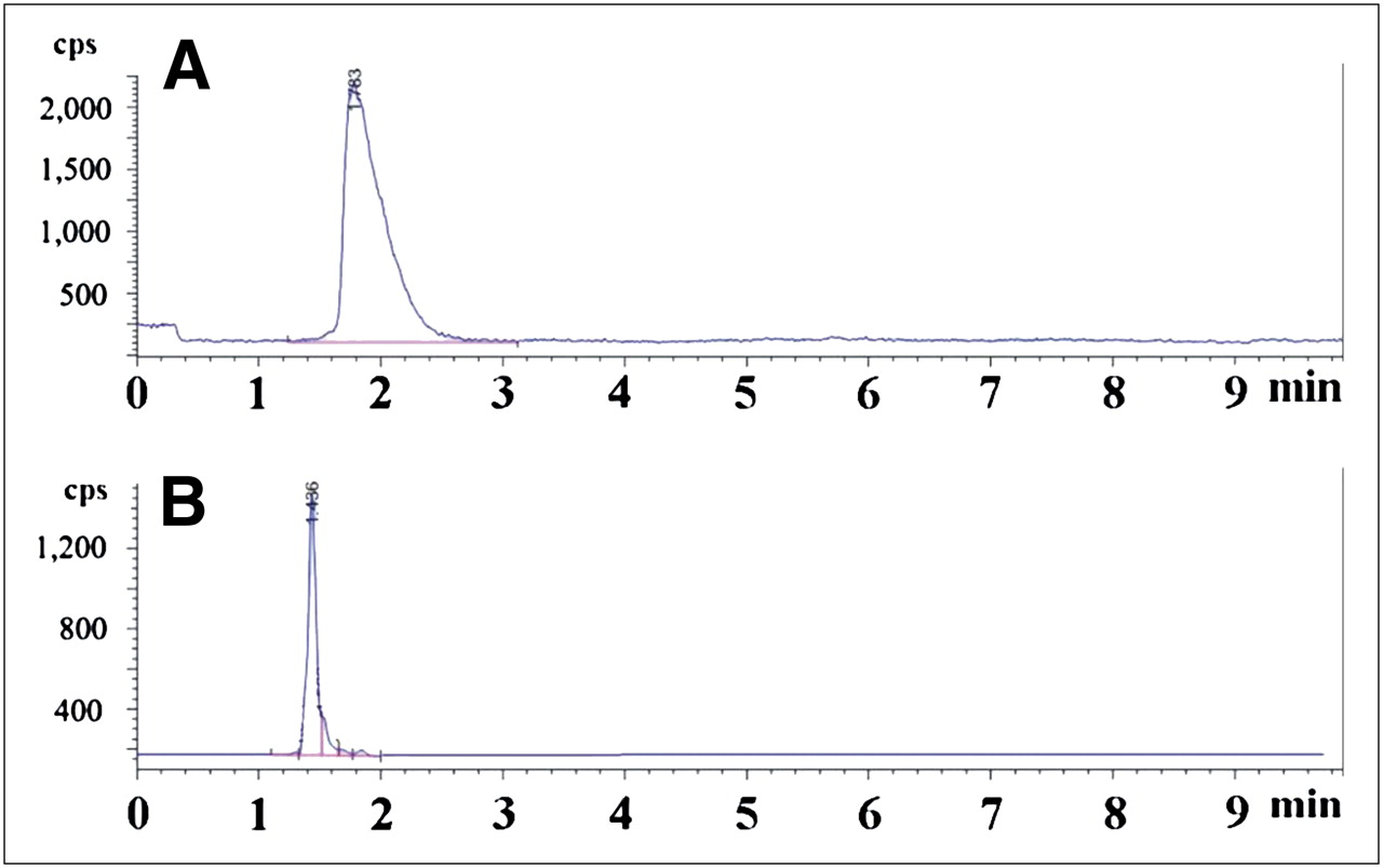
Typical high-performance liquid chromatograms of 11C-MCYS and standard coinjection: radioactive chromatogram of 11C-MCYS (A) and ultraviolet chromatogram of standard MCYS (B).
Transport Mechanism of 11C-MCYS In Vitro
In the competitive inhibition experiments, the specific transport inhibitors BCH, MeAIB, and MeAIB plus serine were used to inhibit the 3 main transport systems of 11C-MCYS. The addition of BCH, specific for system L, resulted in an approximately 80% reduction of 11C-MCYS uptake, whereas the addition of MeAIB and MeAIB plus serine, specific for systems A and ASC, caused no significant change (P = 0.19) in tracer uptake ( Fig. 3A). Therefore, a specific transport of 11C-MCYS in Hep1–6 cells was mediated mainly by system L, and almost no involvement of systems A and ASC could be demonstrated. To clarify the character of the transport system, we examined the Na+ dependency of 11C-MCYS uptake. Uptake of 11C-MCYS was not affected by the presence of Na+ ions. Figure 3 shows the inhibition of Na+-independent amino acid transporters involved in the uptake of 11C-MCYS. The experiments with increasing concentrations of MCYS (0–400 μmol/L) showed no significant change in the radioactivity concentration in the cells ( Fig. 3B). No saturation of transport capacity was found until a concentration of 350 μmol/L had been reached.

(A) Uptake of 11C-MCYS per 106 cells after coincubation with specific transport inhibitors (15 mmol/L) for 4 min: BCH as competitive inhibitor of system L, MeAIB as competitive inhibitor of system A, and MeAIB plus serine as competitive inhibitor of system ASC. (B) Uptake of 11C-MCYS per 106 cells at different concentrations (μmol/L) of MCYS in incubation medium.
Tracer Uptake in Mice
The biodistribution data of 11C-MCYS in normal mice are summarized in Table 1. The highest uptake of 11C-activity in all tissues was found to exceed 0.6 %ID/g at 5 min after injection. The liver had the highest uptake of 11C-MCYS, next to the stomach wall and heart, and the brain had relatively low uptake of 11C-MCYS. Low uptake levels of 11C-MCYS in normal tissues were observed at 30 min after injection The radioactivity in the blood was rapidly decreased from 0.94 ± 0.12 %ID/g at 5 min after injection to 0.17 ± 0.02 %ID/g at 60 min after injection. No elevated organ uptake of 11C-MCYS in the organs sampled was found at 30 min after injection. At 60 min after injection, brain and muscle showed the lowest uptake. Uptake of 11C-MCYS in the brain did not exceed 0.60 %ID/g at any time during the observation except 5 min after injection. Uptake of 11C-MCYS into the brain was slow and decreased slightly at 30 min after injection.
Biodistribution of 11C-MCYS in Normal Mice After Intravenous Injection
Incorporation of 11C-MCYS into Proteins
Protein-bound activity of 11C-MCYS in brain, pancreas, tumor, and blood samples demonstrated less than 1% of the radioactivity to be in the acid-precipitable fraction and almost all the radioactivity to be in the acid solution fraction ( Fig. 4). Thus, almost no incorporation of 11C-MCYS into proteins was observed. Also, high-performance liquid chromatography analysis of plasma samples, brain, pancreas, and tumor tissue at 30 min after injection showed that the activity was completely eluted in the low-molecular-weight fraction. All activity extracted from tissue homogenates, spiked with MCYS, and analyzed on high-performance liquid chromatography was coeluted with the cold standard. In all cases, the major radioactive component, corresponding to parent 11C-MCYS, eluted at a retention time of 2.0 min. In the acid solution fractions from brain, pancreas, tumor, and blood samples, parent 11C-MCYS comprised more than 90% of the total radioactivity. These results showed that 11C-MCYS was metabolically stable in vivo.
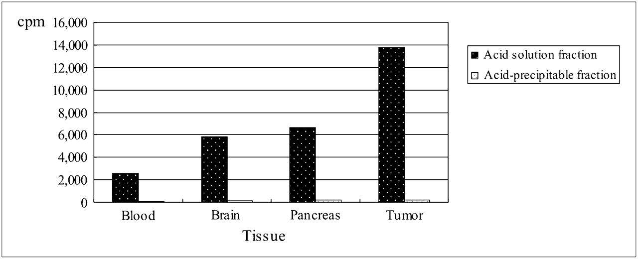
Radioactivities contained in acid solution and acid-precipitable fractions at 30 min after injection.
PET Studies
Whole-body imaging provided the consistent distribution data (Suppl. Fig. 2) obtained from the tissue dissection assays. As for uptake into the inoculated tumors, high 18F-FDG and 11C-MCYS uptake (Suppl. Figs. 2A and 2C) and relatively low 11C-MET uptake (Suppl. Fig. 2B) were found, and tumor-to-muscle ratios were highest for 11C-MCYS uptake. Similarly, high 18F-FDG and 11C-MET uptake was observed in the inflammation group. In contrast, low 11C-MCYS uptake was observed in inflammatory tissue. The uptake percentages of 11C-MCYS, 18F-FDG, and 11C-MET in tumor, inflammatory lesions, and muscle at 60 min after injection are shown in Table 2.
Uptake of 11C-MCYS, 18F-FDG, and 11C-MET in Tumor, Inflammatory Lesion, and Muscle at 60 Minutes After Injection in Tumor-Model Mice and Inflammation-Model Mice
As for the patient with postoperative grade IV glioma, MRI showed a new abnormal enhancing lesion ( Fig. 5), and 18F-FDG PET showed heterogeneous hypometabolism in the right temporal lobe ( Fig. 5B). However, 11C-MCYS PET showed markedly high uptake in the lesion ( Fig. 5A), and histopathologic examination confirmed tumor recurrence.
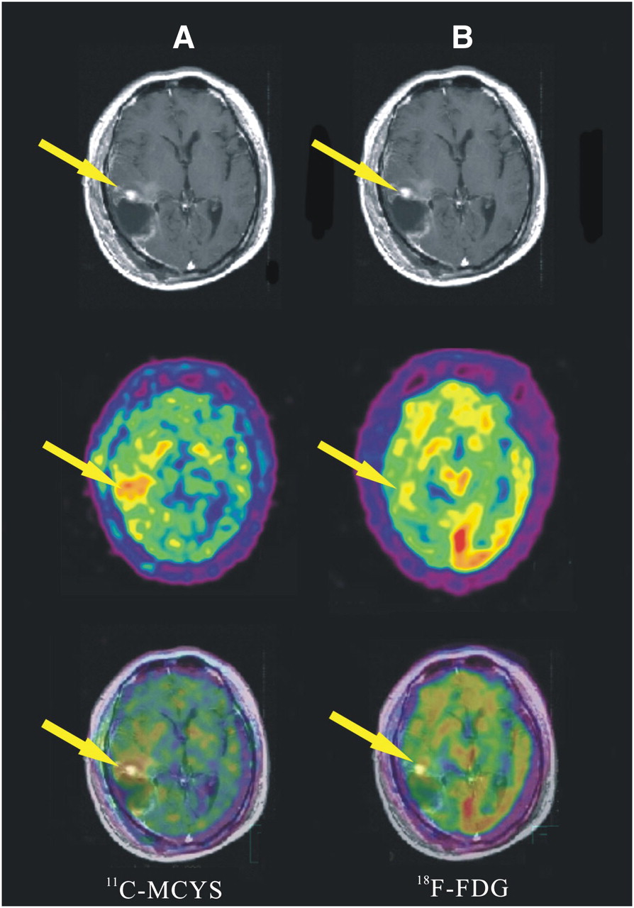
MRI, PET, and coregistered PET/MRI images from top to bottom, respectively, in 45-year-old man with history of attempted resection of grade IV glioma. (A) 11C-MCYS PET illustrates obvious hypermetabolic lesion as depicted by gadolinium-enhanced MRI (arrow), suggesting predominant high-grade tumor recurrence, which was confirmed on histopathology. (B) 18F-FDG PET shows slightly low uptake in patch-shaped heterogeneous hypometabolic area in right temporal lobe (arrow).
DISCUSSION
11C-MCYS, as an analog of 11C-MET, can efficiently be produced by a method similar to that for 11C-MET and exhibits similar biologic activity ( 14, 18). MCYS is a naturally occurring inexpensive water-soluble compound found abundantly in cabbage, garlic, and turnips that has antioxidative and antiinflammatory properties and no adverse effects ( 19– 21). Therefore, evaluation of 11C-MCYS and its automated synthesis is important.
Tumor uptake is thought to reflect increased active transport and protein synthesis ( 3, 22). Most amino acids are taken up by tumor cells through the sodium-independent L-type amino acid transporter system and the sodium-dependent transporter systems A and ASC ( 16). Several positron-labeled naturally occurring amino acids show protein incorporation (e.g., L-1-11C-tyrosine and, partly, 11C-MET) ( 3, 22), and labeled non–protein-composition amino acids show amino acid transport (e.g., 18F-FET and, partly, 2-18F-fluoro-L-phenylalanine) ( 3, 22). For example, 18F-FET is not incorporated into proteins with relatively stable metabolism in vivo, and 18F-FET uptake is mediated through system L ( 23). Like 18F-FET, 11C-MCYS is a non–protein-composition amino acid tracer with relative metabolic stability in vivo and not incorporated into proteins, confirmed by 11C-MCYS incorporation and metabolism experiments in this work. In addition, we found that a specific transport of 11C-MCYS in Hep1–6 cells was mediated mainly by system L, with almost no involvement of systems A and ASC, and that 11C-MCYS uptake was not affected by the presence of Na+. These findings are consistent with the fact that system L has no requirement for sodium and energy sources, in contrast to systems A and ASC ( 24). However, 11C-MET is an 11C-labeled naturally occurring amino acid that can be used to estimate muscle protein synthetic rates ( 25) and also generates substantial amounts of nonprotein metabolites in vivo ( 22). Interestingly, the rapid initial uptake of 11C-MET in tissues mainly reflects amino acid transport, rather than protein synthesis ( 3). Like the uptake mechanism of 11C-MCYS, 11C-MET transport is usually mediated through system L, with minor contributions from systems A and ASC ( 22).
Currently, 4 subtypes of the sodium-independent L-type amino acid transporter (LAT) have been reported: LAT1, LAT2, LAT3, and LAT4 ( 26, 27). The well-known LAT1 seems to be an excellent and promising target for molecular imaging and shows increased transport activity in many cancer cells via transporting neutral amino acids, including methionine ( 27). Higher LAT1 protein expression levels in high-grade glioma cells than in low-grade gliomas and normal brain tissue have been found ( 28), as has LAT1 expression in hepatocarcinoma cells ( 29). 11C-MET transport may be increased by an increased number of microvessels combined with a higher density or activity of LAT1 in the tumor endothelial cells in high-grade gliomas ( 28). 18F-FET can be selectively transported by LAT2 and can differentiate tumor from inflammation, possibly contributing to increased amino acid transport and no expression of LAT2 in inflammatory tissue ( 23). In this work, high uptake of 11C-MCYS in hepatoma tissue and low 11C-MCYS uptake in inflammatory tissue are possibly associated with the increased amino acid transport of 11C-MCYS and its lack of incorporation into protein. Thus, we can assume that 11C-MCYS uptake is transported mainly by LAT1, LAT2, or both. PET of the glioma patient showed high 11C-MCYS uptake in tumor tissue and low tracer uptake in normal brain tissue, further supporting this assumption.
The biodistribution demonstrated a continually increasing accumulation of 11C-MCYS in tumor up to 30 min after injection, relatively low uptake of 11C-MCYS in brain and muscle during the entire observation time, and rapid radioactivity clearance from most tissues. Thus, fast tumor uptake kinetics and low accumulation of 11C-MCYS in the brain and most tissues showed that 11C-MCYS is a potential amino acid PET tracer for imaging of cerebral and peripheral tumors. The good scan time was within 30 min after injection of 11C-MCYS.
Several investigators have shown high accumulation of 18F-FDG in turpentine-induced inflammatory tissue ( 12) and Staphylococcus aureus–inoculated inflammation models ( 30). This study found high uptake of 18F-FDG and 11C-MET and low uptake of 11C-MCYS in the turpentine-induced inflammatory tissues, whereas high uptake of 18F-FDG and 11C-MCYS and relatively low uptake of 11C-MET were found in Hepa1–6 tumors. These results show that 11C-MCYS has advantages over 18F-FDG and 11C-MET in the differentiation of tumor from inflammation. Furthermore, in the patient with brain glioma, the high uptake of 11C-MCYS in contrast to the very low uptake of 18F-FDG indicated that 11C-MCYS is superior to 18F-FDG for malignant glioma imaging. However, all these results require further confirmation before clinical application is possible.
CONCLUSION
The automated synthesis of 11C-MCYS is easy to perform. Our initial study results show that 11C-MCYS is transported by the specific amino acid transport system L and is not incorporated into proteins. 11C-MCYS is superior to 18F-FDG and 11C-MET in the differentiation of tumor from inflammation. 11C-MCYS PET is more sensitive and specific than 18F-FDG PET in the detection of cancer. Therefore, 11C-MCYS may be potentially better than 11C-MET as an amino acid PET tracer for tumors, but further confirmation is needed in clinical studies.
Acknowledgments
We thank Xiangsong Zhang and Zhenhua Gao for assistance with this work. This work was supported by the National Natural Science Foundation (grant 30970856), the National High Technology Research and Development Program of China (program 863, grant 2008AA02Z430), and Sun Yat-Sen University (grant 18901205).
- © 2011 by Society of Nuclear Medicine
REFERENCES
- Received for publication July 16, 2010.
- Accepted for publication November 18, 2010.


Jump to section
Related Articles
Cited By...
- No citing articles found.