
Main menu

  • Home
  • Content
    • Current
    • Ahead of print
    • Past Issues
    • JNM Supplement
    • SNMMI Annual Meeting Abstracts
    • Continuing Education
    • JNM Podcasts
  • Subscriptions
    • Subscribers
    • Institutional and Non-member
    • Rates
    • Journal Claims
    • Corporate & Special Sales
  • Authors
    • Submit to JNM
    • Information for Authors
    • Assignment of Copyright
    • AQARA requirements
  • Info
    • Reviewers
    • Permissions
    • Advertisers
  • About
    • About Us
    • Editorial Board
    • Contact Information
  • More
    • Alerts
    • Feedback
    • Help
    • SNMMI Journals
  • SNMMI
    • JNM
    • JNMT
    • SNMMI Journals
    • SNMMI

User menu

  • Subscribe
  • My alerts
  • Log in
  • My Cart

Search

  • Advanced search
Journal of Nuclear Medicine
  • SNMMI
    • JNM
    • JNMT
    • SNMMI Journals
    • SNMMI
  • Subscribe
  • My alerts
  • Log in
  • My Cart
Journal of Nuclear Medicine

Advanced Search

  • Home
  • Content
    • Current
    • Ahead of print
    • Past Issues
    • JNM Supplement
    • SNMMI Annual Meeting Abstracts
    • Continuing Education
    • JNM Podcasts
  • Subscriptions
    • Subscribers
    • Institutional and Non-member
    • Rates
    • Journal Claims
    • Corporate & Special Sales
  • Authors
    • Submit to JNM
    • Information for Authors
    • Assignment of Copyright
    • AQARA requirements
  • Info
    • Reviewers
    • Permissions
    • Advertisers
  • About
    • About Us
    • Editorial Board
    • Contact Information
  • More
    • Alerts
    • Feedback
    • Help
    • SNMMI Journals
  • View or Listen to JNM Podcast
  • Visit JNM on Facebook
  • Join JNM on LinkedIn
  • Follow JNM on Twitter
  • Subscribe to our RSS feeds
Meeting ReportPhysics, Instrumentation & Data Sciences

Objective evaluation of voxelwise partial volume correction methods in brain PET imaging

Murat Bilgel, Yansong Zhu, Olivier Rousset, Dean Wong, Susan Resnick and Arman Rahmim
Journal of Nuclear Medicine May 2020, 61 (supplement 1) 72;
Murat Bilgel
1Laboratory of Behavioral Neuroscience National Institute on Aging Baltimore MD United States
  • Find this author on Google Scholar
  • Find this author on PubMed
  • Search for this author on this site
Yansong Zhu
2Department of Radiology and Radiological Science Johns Hopkins University Baltimore MD United States
3Departments of Radiology and Physics University of British Columbia Vancouver BC Canada
  • Find this author on Google Scholar
  • Find this author on PubMed
  • Search for this author on this site
Olivier Rousset
2Department of Radiology and Radiological Science Johns Hopkins University Baltimore MD United States
  • Find this author on Google Scholar
  • Find this author on PubMed
  • Search for this author on this site
Dean Wong
2Department of Radiology and Radiological Science Johns Hopkins University Baltimore MD United States
  • Find this author on Google Scholar
  • Find this author on PubMed
  • Search for this author on this site
Susan Resnick
1Laboratory of Behavioral Neuroscience National Institute on Aging Baltimore MD United States
  • Find this author on Google Scholar
  • Find this author on PubMed
  • Search for this author on this site
Arman Rahmim
3Departments of Radiology and Physics University of British Columbia Vancouver BC Canada
2Department of Radiology and Radiological Science Johns Hopkins University Baltimore MD United States
  • Find this author on Google Scholar
  • Find this author on PubMed
  • Search for this author on this site
  • Article
  • Figures & Data
  • Info & Metrics
Loading

Abstract

72

Introduction: Partial volume effects impair the accurate quantification of PET scans, and are important consideration in voxelwise statistical analyses. A number of methods have been proposed for voxelwise partial volume correction (PVC); however, comparison of PVC methods in real data sets is difficult due to the lack of objective criteria.

Objectives: develop objective criteria to assess the performance of voxelwise PVC methods and apply these criteria to compare three PVC methods using 15O-H2O brain PET scans.

Methods: We used cross-sectional 15O-H2O brain PET data acquired on a GE Advance scanner for 175 participants from the Baltimore Longitudinal Study of Aging to assess the performance of the following voxelwise PVC methods: reblurred Van Cittert (VC) [1], Region-Based Voxelwise (RBV) [2], and Parallel Level Set (PLS) with a non-smooth optimization technique (split Bregman) [3]. Images were reconstructed as a single time frame using filtered backprojection, yielding ~7.5 mm FWHM resolution at the center of the field of view [4]. For each participant, we coregistered their inhomogeneity-corrected and skull-stripped structural MRI with their PET. Anatomical regions were defined on the MRI using a multi-atlas labeling approach [5] and mapped onto the PET using the coregistration result. We used the cerebellar gray matter as the reference region to compute standardized uptake value ratios (SUVR). To aid in the evaluation of the performance of voxelwise PVC methods, we calculated the error in SUVR estimates. Assuming that voxelwise intensities are independent, and that within a given region, they are identically distributed as a normal with mean μ and variance σ2 , a first-order Taylor series expansion of the definition of SUVR yields Var(SUVR)≍(μT/μR)2[σT2/(n μT2) + σR2 / (m μR2)], where n and m are the number of voxels within the target (T) and reference (R) regions, respectively. A lower variance is desirable, since it indicates a smaller error in the SUVR estimate. To assess the performance of voxelwise PVC methods, we used the following criteria: the lowest uncertainty in regional SUVR estimates, given by Var(SUVR), and the greatest negative age association at baseline (after adjusting for sex), given by the number of voxels with statistically significant differences within the cortical gray matter.

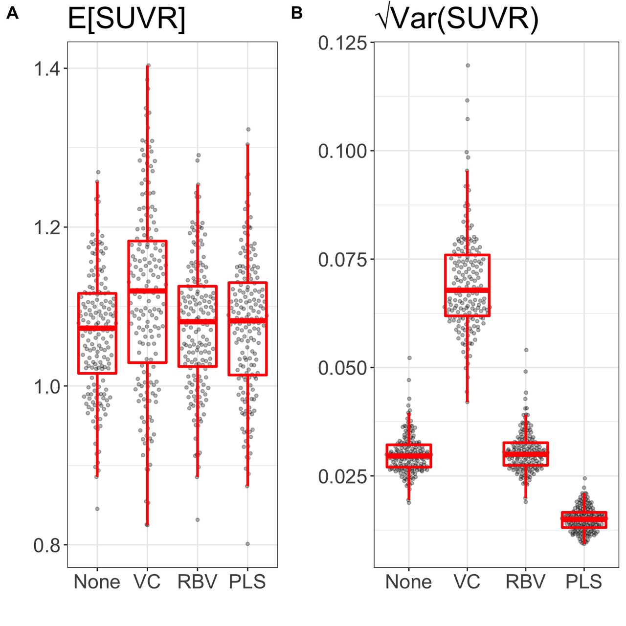
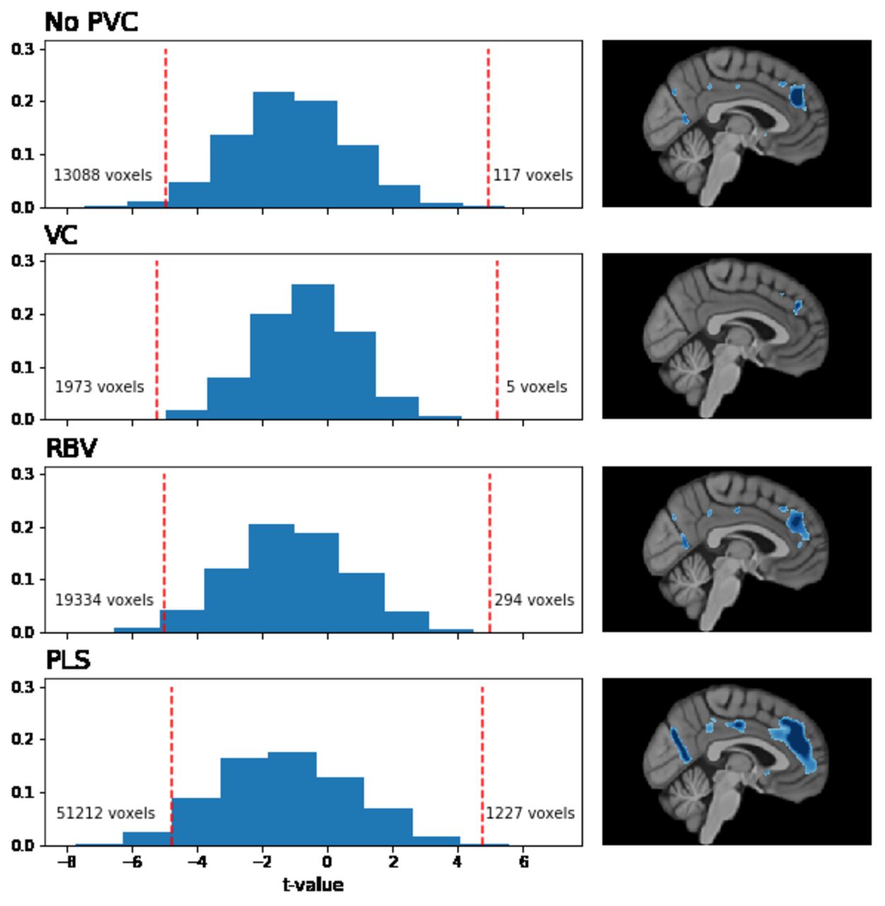
Results: PLS yielded the lowest Var(SUVR), resulting in a lower uncertainty in regional SUVR compared to not performing PVC, while not affecting the overall SUVR levels (Figure 1). In the voxelwise analysis, PLS yielded the greatest number of statistically significant voxels where older individuals had lower SUVR (Figure 2). Conclusion: PLS can improve statistical analysis by reducing the uncertainty around regional SUVR estimates, and may enable the discovery of effects that would not be considered statistically significant without PVC or other PVC methods. Acknowledgments: This research was supported in part by the Intramural Research Program of the National Institute on Aging, National Institutes of Health, and R21 Grant No. AG056142. Figure 1. Swarm and box plots of (A) the expected value of SUVR and (B) square root of the SUVR variance for the right posterior cingulate gyrus. Results were similar in other cortical gray matter regions. None=No PVC, VC=reblurred Van Cittert, RBV=Region-Based Voxelwise, PLS=Parallel Level Set. Figure 2. Normalized histograms of two-sided t-values for the age term in the linear regression model with voxelwise SUVR as outcome (adjusted for sex). Each row corresponds to a different PVC method. Red vertical dashed lines indicate the t-values that correspond to a multiple comparison-corrected (via permutation tests) p<0.05 threshold. Number of statistically significant cortical gray matter voxels with a negative and positive association are indicated to the left and right of the dashed lines, respectively. Sagittal slices illustrate the extent of statistically significant clusters on the left medial cortical surface.

Figure
  • Download figure
  • Open in new tab
  • Download powerpoint
Figure
  • Download figure
  • Open in new tab
  • Download powerpoint
Previous
Back to top

In this issue

Journal of Nuclear Medicine
Vol. 61, Issue supplement 1
May 1, 2020
  • Table of Contents
  • Index by author
Article Alerts
Sign In to Email Alerts with your Email Address
Email Article

Thank you for your interest in spreading the word on Journal of Nuclear Medicine.

NOTE: We only request your email address so that the person you are recommending the page to knows that you wanted them to see it, and that it is not junk mail. We do not capture any email address.

Enter multiple addresses on separate lines or separate them with commas.
Objective evaluation of voxelwise partial volume correction methods in brain PET imaging
(Your Name) has sent you a message from Journal of Nuclear Medicine
(Your Name) thought you would like to see the Journal of Nuclear Medicine web site.
Citation Tools
Objective evaluation of voxelwise partial volume correction methods in brain PET imaging
Murat Bilgel, Yansong Zhu, Olivier Rousset, Dean Wong, Susan Resnick, Arman Rahmim
Journal of Nuclear Medicine May 2020, 61 (supplement 1) 72;

Citation Manager Formats

  • BibTeX
  • Bookends
  • EasyBib
  • EndNote (tagged)
  • EndNote 8 (xml)
  • Medlars
  • Mendeley
  • Papers
  • RefWorks Tagged
  • Ref Manager
  • RIS
  • Zotero
Share
Objective evaluation of voxelwise partial volume correction methods in brain PET imaging
Murat Bilgel, Yansong Zhu, Olivier Rousset, Dean Wong, Susan Resnick, Arman Rahmim
Journal of Nuclear Medicine May 2020, 61 (supplement 1) 72;
Twitter logo Facebook logo LinkedIn logo Mendeley logo
  • Tweet Widget
  • Facebook Like
  • Google Plus One
Bookmark this article

Jump to section

  • Article
  • Figures & Data
  • Info & Metrics

Related Articles

  • No related articles found.
  • Google Scholar

Cited By...

  • No citing articles found.
  • Google Scholar

More in this TOC Section

Physics, Instrumentation & Data Sciences

  • Keel-Edge Height Selection for Improved Multi-Pinhole 123I Brain SPECT Imaging
  • Ultra-Fast Reconstruction of Short List-Mode PET Data Frames for Real-Time Visualization and Processing
  • Deep Learning Based 3D Dose Estimation from Prompt Gamma Distribution for Proton Therapy Monitoring
Show more Physics, Instrumentation & Data Sciences

Brain (Data Analysis & Management)

  • Monte-Carlo Simulations for Evaluating Sensitivity of Tau Detection in [F-18]MK-6240 PET Scans of Early Stage Alzheimer’s Disease
  • SV2A synaptic density imaging in the neonate rhesus macaque brain: Analysis with partial volume correction
  • Measurement of cerebral circulation and oxygen metabolism using integrated PET/MRI scanner with oxygen-15 labeled gases: Effects of attenuation correction and real-time motion correction
Show more Brain (Data Analysis & Management)

Similar Articles

SNMMI

© 2025 SNMMI

Powered by HighWire