
Main menu

  • Home
  • Content
    • Current
    • Ahead of print
    • Past Issues
    • JNM Supplement
    • SNMMI Annual Meeting Abstracts
    • Continuing Education
    • JNM Podcasts
  • Subscriptions
    • Subscribers
    • Institutional and Non-member
    • Rates
    • Journal Claims
    • Corporate & Special Sales
  • Authors
    • Submit to JNM
    • Information for Authors
    • Assignment of Copyright
    • AQARA requirements
  • Info
    • Reviewers
    • Permissions
    • Advertisers
  • About
    • About Us
    • Editorial Board
    • Contact Information
  • More
    • Alerts
    • Feedback
    • Help
    • SNMMI Journals
  • SNMMI
    • JNM
    • JNMT
    • SNMMI Journals
    • SNMMI

User menu

  • Subscribe
  • My alerts
  • Log in
  • My Cart

Search

  • Advanced search
Journal of Nuclear Medicine
  • SNMMI
    • JNM
    • JNMT
    • SNMMI Journals
    • SNMMI
  • Subscribe
  • My alerts
  • Log in
  • My Cart
Journal of Nuclear Medicine

Advanced Search

  • Home
  • Content
    • Current
    • Ahead of print
    • Past Issues
    • JNM Supplement
    • SNMMI Annual Meeting Abstracts
    • Continuing Education
    • JNM Podcasts
  • Subscriptions
    • Subscribers
    • Institutional and Non-member
    • Rates
    • Journal Claims
    • Corporate & Special Sales
  • Authors
    • Submit to JNM
    • Information for Authors
    • Assignment of Copyright
    • AQARA requirements
  • Info
    • Reviewers
    • Permissions
    • Advertisers
  • About
    • About Us
    • Editorial Board
    • Contact Information
  • More
    • Alerts
    • Feedback
    • Help
    • SNMMI Journals
  • View or Listen to JNM Podcast
  • Visit JNM on Facebook
  • Join JNM on LinkedIn
  • Follow JNM on Twitter
  • Subscribe to our RSS feeds
Meeting ReportOncology: Clinical Therapy and Diagnosis

Characterization of Segmented 18F-DCFPyL PET/CT Lesions in the Context of PSMA-RADS Structured Reporting

Saeed Ashrafinia, Mohammad Salehi Sadaghiani, Pejman Dalaie, Rima Tulbah, Yafu Yin, Kevin Leung, Ryan VanDenBerg, Jeffrey Leal, Michael Gorin, Martin Pomper, Arman Rahmim and Steven Rowe
Journal of Nuclear Medicine May 2019, 60 (supplement 1) 1565;
Saeed Ashrafinia
4Electrical and Computer Engineering Johns Hopkins University Baltimore MD United States
5The Russell H. Morgan Department of Radiology and Radiological Sciences Johns Hopkins University Baltimore MD United States
  • Find this author on Google Scholar
  • Find this author on PubMed
  • Search for this author on this site
Mohammad Salehi Sadaghiani
5The Russell H. Morgan Department of Radiology and Radiological Sciences Johns Hopkins University Baltimore MD United States
  • Find this author on Google Scholar
  • Find this author on PubMed
  • Search for this author on this site
Pejman Dalaie
5The Russell H. Morgan Department of Radiology and Radiological Sciences Johns Hopkins University Baltimore MD United States
  • Find this author on Google Scholar
  • Find this author on PubMed
  • Search for this author on this site
Rima Tulbah
5The Russell H. Morgan Department of Radiology and Radiological Sciences Johns Hopkins University Baltimore MD United States
  • Find this author on Google Scholar
  • Find this author on PubMed
  • Search for this author on this site
Yafu Yin
6The first hospital of China Medical University Shenyang China
  • Find this author on Google Scholar
  • Find this author on PubMed
  • Search for this author on this site
Kevin Leung
3Biomedical Engineering Johns Hopkins University Baltimore MD United States
  • Find this author on Google Scholar
  • Find this author on PubMed
  • Search for this author on this site
Ryan VanDenBerg
5The Russell H. Morgan Department of Radiology and Radiological Sciences Johns Hopkins University Baltimore MD United States
  • Find this author on Google Scholar
  • Find this author on PubMed
  • Search for this author on this site
Jeffrey Leal
5The Russell H. Morgan Department of Radiology and Radiological Sciences Johns Hopkins University Baltimore MD United States
  • Find this author on Google Scholar
  • Find this author on PubMed
  • Search for this author on this site
Michael Gorin
1Baltimore MD United States
  • Find this author on Google Scholar
  • Find this author on PubMed
  • Search for this author on this site
Martin Pomper
3Biomedical Engineering Johns Hopkins University Baltimore MD United States
5The Russell H. Morgan Department of Radiology and Radiological Sciences Johns Hopkins University Baltimore MD United States
  • Find this author on Google Scholar
  • Find this author on PubMed
  • Search for this author on this site
Arman Rahmim
2Integrative Oncology BC Cancer Research Centre Vancouver BC Canada
7Radiology University of British Columbia Vancouver BC Canada
5The Russell H. Morgan Department of Radiology and Radiological Sciences Johns Hopkins University Baltimore MD United States
  • Find this author on Google Scholar
  • Find this author on PubMed
  • Search for this author on this site
Steven Rowe
5The Russell H. Morgan Department of Radiology and Radiological Sciences Johns Hopkins University Baltimore MD United States
  • Find this author on Google Scholar
  • Find this author on PubMed
  • Search for this author on this site
  • Article
  • Figures & Data
  • Info & Metrics
Loading

Abstract

1565

Objectives: A prostate-specific membrane antigen (PSMA) reporting and data system (PSMA-RADS version 1.0) was recently introduced for PSMA-targeted PET imaging studies, in order to classify individual findings into categories that reflect the likelihood of the presence of prostate cancer (PCa). This structured reporting framework is based on imaging findings including anatomic location and intensity of radiotracer uptake. We aim to report on imaging features and segmentation characteristics of 18F-DCFPyL (PyL) lesions as categorized based on the PSMA-RADS scoring scheme. Our work also provides more insight into segmentation of PyL PET images in general.

Methods: The dataset contains 277 PCa patients imaged with PyL PET/CT at 60 min post-injection. Four trained nuclear medicine physicians manually segmented 3,794 lesions regions of interest (ROI), in addition to a spherical ROI (30 mm diameter) in the liver for reference, yielding a total of 4,071 ROIs. They subsequently performed PSMA-RADS scoring for every lesion utilizing both PET and CT images. Detailed underlying anatomic location was also recorded (572 sites), categorized into 9 broad anatomic categories. SUVmax, tumor volume and effective thresholding level for each manually segmented ROI was also calculated. The effective threshold was computed as min{ SUVminROI, (SUVminROI+SUVmaxring)/2} (expressed as percentage of SUVmaxROI), where SUVmaxring was calculated outside of the ROI on the immediately adjacent 1 to 2 rings of voxels.

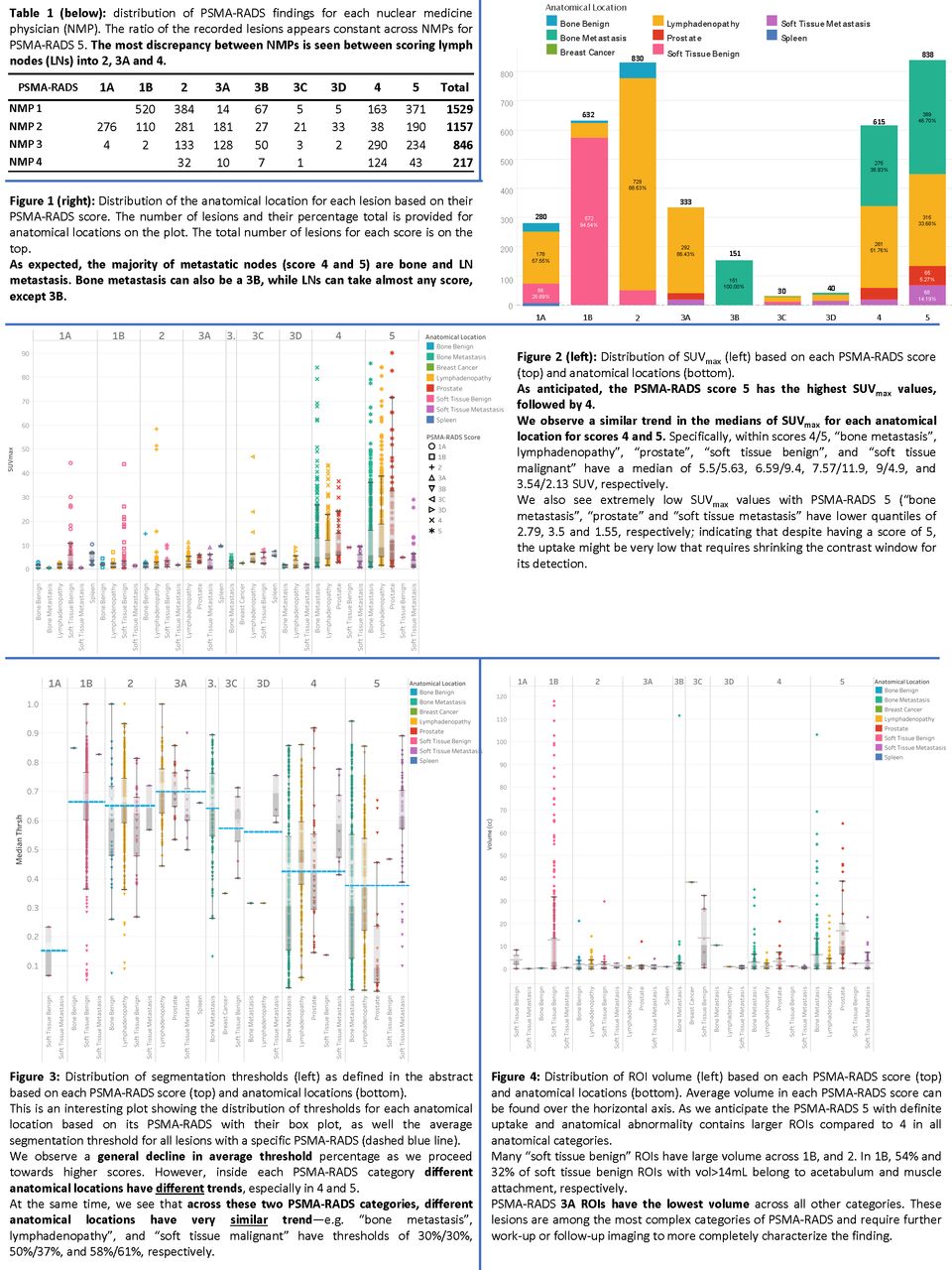
Results: The distribution of lesions was as follows: 280 PSMA-RADS 1As, 632 1Bs, 830 2s, 337 3As, 147 3Bs, 30 3Cs, 40 3Ds, 615 4s, and 838 5s, showing higher prevalence of intermediate-to-high uptake lesions with scores 4 and 5, followed by lower uptake lesions such as 3A. Axillary and inguinal lymph nodes together accounted for 599 lesions, and were generally considered PSMA-RADS-2 as they were not a common site of PCa. While the internal ROI segmentation thresholds varied highly within each PSMA-RADS score, the thresholding was less variable within the same location. Generally, lesions with intermediate uptake in 1B, 2, 3A, 3B, 3C and 3D had low SUVmax (average 2.2, 1.7, 2.8, 1.8, 6.6, and 2.1, for each score, respectively) compared to 4 and 5 (average SUVmax of 9.8 and 14.1, respectively). Linked to this, the former group of lesions had a higher effective segmentation threshold compared to the latter, as their uptake was closer to the background. In nearly half the ROIs, the immediate neighborhoods contained voxel(s) with value >SUVminROI indicating that more sophisticated thresholding techniques are required for automatic/semiautomatic segmentation of PyL PET images. This was observed even for scores 4 and 5 with high uptakes. Proximity to normal areas with intermediate uptake (vessels, bowel loops) for scores 1B, 2, 3A, and 3B, or high uptake (kidney, bladder) for 4 and 5 contributed to that effect. PSMA-RADS scores for lesions with highest volumes (mean/median in mL) were 1B (14.2/3.1), 3C (16.4/13.4), and 5(5.6/2.1). The median volume across all lesions was 1.14 mL with only 7.6% of the lesions larger than 10 mL.

Conclusions: Median effective segmentation thresholds were low (43%, 35%) for PSMA-RADS scores 4 and 5 but higher (61.4% ~ 70%) across 2 and all 3 subcategories. The challenging segmentation task for PyL PET images requires supervision by an expert or use of an advanced method such as deep learning. The relatively small sizes of the PyL lesions demonstrate its high focal concentration and specificity; yet, it also indicates that a radiomics analysis of these lesions may not provide new, significant information. Nonetheless, the differences in parameters related to each of the PSMA-RADS categories suggests that a sufficiently large dataset of lesions could be used with artificial intelligence applications to automate lesion categorization.

Figure
  • Download figure
  • Open in new tab
  • Download powerpoint
View this table:
  • View inline
  • View popup

Distribution of Lesion Underlying Tissue Sites for Each PSMA-RADS Score

Previous
Back to top

In this issue

Journal of Nuclear Medicine
Vol. 60, Issue supplement 1
May 1, 2019
  • Table of Contents
  • Index by author
Article Alerts
Sign In to Email Alerts with your Email Address
Email Article

Thank you for your interest in spreading the word on Journal of Nuclear Medicine.

NOTE: We only request your email address so that the person you are recommending the page to knows that you wanted them to see it, and that it is not junk mail. We do not capture any email address.

Enter multiple addresses on separate lines or separate them with commas.
Characterization of Segmented 18F-DCFPyL PET/CT Lesions in the Context of PSMA-RADS Structured Reporting
(Your Name) has sent you a message from Journal of Nuclear Medicine
(Your Name) thought you would like to see the Journal of Nuclear Medicine web site.
Citation Tools
Characterization of Segmented 18F-DCFPyL PET/CT Lesions in the Context of PSMA-RADS Structured Reporting
Saeed Ashrafinia, Mohammad Salehi Sadaghiani, Pejman Dalaie, Rima Tulbah, Yafu Yin, Kevin Leung, Ryan VanDenBerg, Jeffrey Leal, Michael Gorin, Martin Pomper, Arman Rahmim, Steven Rowe
Journal of Nuclear Medicine May 2019, 60 (supplement 1) 1565;

Citation Manager Formats

  • BibTeX
  • Bookends
  • EasyBib
  • EndNote (tagged)
  • EndNote 8 (xml)
  • Medlars
  • Mendeley
  • Papers
  • RefWorks Tagged
  • Ref Manager
  • RIS
  • Zotero
Share
Characterization of Segmented 18F-DCFPyL PET/CT Lesions in the Context of PSMA-RADS Structured Reporting
Saeed Ashrafinia, Mohammad Salehi Sadaghiani, Pejman Dalaie, Rima Tulbah, Yafu Yin, Kevin Leung, Ryan VanDenBerg, Jeffrey Leal, Michael Gorin, Martin Pomper, Arman Rahmim, Steven Rowe
Journal of Nuclear Medicine May 2019, 60 (supplement 1) 1565;
Twitter logo Facebook logo LinkedIn logo Mendeley logo
  • Tweet Widget
  • Facebook Like
  • Google Plus One
Bookmark this article

Jump to section

  • Article
  • Figures & Data
  • Info & Metrics

Related Articles

  • No related articles found.
  • Google Scholar

Cited By...

  • No citing articles found.
  • Google Scholar

More in this TOC Section

Oncology: Clinical Therapy and Diagnosis

  • Added value of single photon emission computed tomography (SPECT) / Computed Tomography (CT) with radioiodine whole body scan in follow up of patients with differentiated thyroid cancer
  • Simultaneous PET/MR reveals significant correlation between the intravoxel incoherent motion parameters and standardized uptake values in nasopharyngeal carcinoma patients
  • Accuracy of 18F-fluoroethyltyrosine PET for detecting high grade glioma recurrence: size matters
Show more Oncology: Clinical Therapy and Diagnosis

Prostate/GU Imaging Posters

  • [18F]-fluciclovine positivity rate is not affected by androgen deprivation therapy (ADT) in recurrent prostate cancer post-prostatectomy.
  • Test-retest study of tumor blood flow quantification with 82Rubidium-PET/CT in prostate cancer
  • Comparison of three interpretation criteria of 68Ga-PSMA PET based on inter- and intra-reader agreement
Show more Prostate/GU Imaging Posters

Similar Articles

SNMMI

© 2025 SNMMI

Powered by HighWire