
Main menu

  • Home
  • Content
    • Current
    • Ahead of print
    • Past Issues
    • JNM Supplement
    • SNMMI Annual Meeting Abstracts
    • Continuing Education
    • JNM Podcasts
  • Subscriptions
    • Subscribers
    • Institutional and Non-member
    • Rates
    • Journal Claims
    • Corporate & Special Sales
  • Authors
    • Submit to JNM
    • Information for Authors
    • Assignment of Copyright
    • AQARA requirements
  • Info
    • Reviewers
    • Permissions
    • Advertisers
  • About
    • About Us
    • Editorial Board
    • Contact Information
  • More
    • Alerts
    • Feedback
    • Help
    • SNMMI Journals
  • SNMMI
    • JNM
    • JNMT
    • SNMMI Journals
    • SNMMI

User menu

  • Subscribe
  • My alerts
  • Log in
  • Log out
  • My Cart

Search

  • Advanced search
Journal of Nuclear Medicine
  • SNMMI
    • JNM
    • JNMT
    • SNMMI Journals
    • SNMMI
  • Subscribe
  • My alerts
  • Log in
  • Log out
  • My Cart
Journal of Nuclear Medicine

Advanced Search

  • Home
  • Content
    • Current
    • Ahead of print
    • Past Issues
    • JNM Supplement
    • SNMMI Annual Meeting Abstracts
    • Continuing Education
    • JNM Podcasts
  • Subscriptions
    • Subscribers
    • Institutional and Non-member
    • Rates
    • Journal Claims
    • Corporate & Special Sales
  • Authors
    • Submit to JNM
    • Information for Authors
    • Assignment of Copyright
    • AQARA requirements
  • Info
    • Reviewers
    • Permissions
    • Advertisers
  • About
    • About Us
    • Editorial Board
    • Contact Information
  • More
    • Alerts
    • Feedback
    • Help
    • SNMMI Journals
  • View or Listen to JNM Podcast
  • Visit JNM on Facebook
  • Join JNM on LinkedIn
  • Follow JNM on Twitter
  • Subscribe to our RSS feeds
Meeting ReportOncology, Clinical Diagnosis Track

Measuring Differences in Y90 Delivery Techniques: A Potential Tool for Physician Training, Optimization of Technique, and Confirmation of Dose Delivery

Dustin Osborne, Shelley Acuff, Cain Green, Rachel Taylor, Stephen Leschak and Christopher Stephens
Journal of Nuclear Medicine May 2018, 59 (supplement 1) 660;
Dustin Osborne
2Radiology University of Tennessee: Graduate School of Medici Knoxville TN United States
  • Find this author on Google Scholar
  • Find this author on PubMed
  • Search for this author on this site
Shelley Acuff
4UT Medical Center Maynardville TN United States
  • Find this author on Google Scholar
  • Find this author on PubMed
  • Search for this author on this site
Cain Green
3UT Medical Center Knoxville TN United States
  • Find this author on Google Scholar
  • Find this author on PubMed
  • Search for this author on this site
Rachel Taylor
1Department of Radiology Knoxville TN United States
  • Find this author on Google Scholar
  • Find this author on PubMed
  • Search for this author on this site
Stephen Leschak
6UTMC Knoxville TN United States
  • Find this author on Google Scholar
  • Find this author on PubMed
  • Search for this author on this site
Christopher Stephens
5UT Radiology Knoxville TN United States
  • Find this author on Google Scholar
  • Find this author on PubMed
  • Search for this author on this site
  • Article
  • Figures & Data
  • Info & Metrics
Loading

Abstract

660

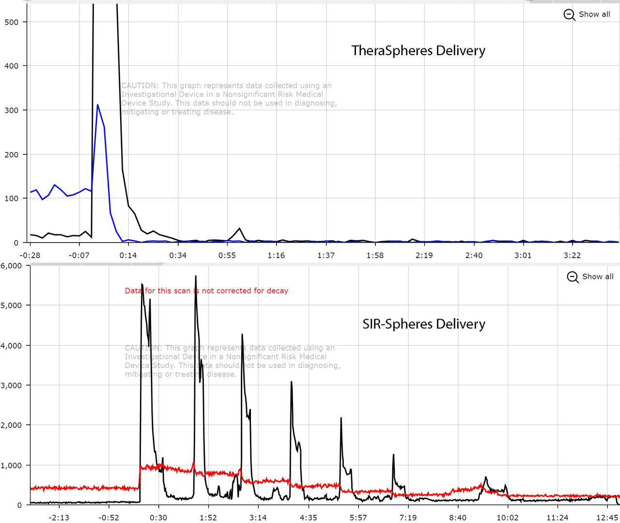
Introduction: Two devices are currently approved for Y90 radioembolization: SIR-Spheres and TheraSpheres. Each have different characteristics with SIR-Spheres being made of resin, ranging in size from 20-60 microns, and having a per-sphere activity of approximately 50 Bq. TheraSpheres beads are made of glass with a size range of 20-30 microns and activities of 2,500 Bq. Each of these spheres require similar but unique manual delivery techniques to properly deliver the radioactive beads to the tumor site. This study examines the feasibility of using modular detectors to measure time activity curves (TACs) related to the Y90 dose infusion and provide visual feedback that can be used to optimize delivery techniques for experienced physicians and provide robust training for radiology residents. This study also uses TACs derived from this system to provide near real-time feedback regarding whether or not the full dose was delivered successfully to the patient. Complete dose delivery is often a concern with Y90 radioembolization procedures as issues can arise, such as stasis, that interfere with the full dose infusion. Materials and Methods: For each radioembolization device, a modular detector (Lucerno Dynamics, LLC) was placed at a location near the primary dose vial as well as near the dose delivery catheter. The detector near the dose vial monitored for baseline activity levels of the total dose to be administered and to determine if the total dose was completely delivered. The detector near the catheter line provided time activity curves (TACs) of the actual dose delivered through the catheter allowing for post-therapy analysis of the technique used by a physician for a given patient and radioembolization device. Results: Time activity curves show the differences between the delivery methods used for each of the radioembolization devices. The characteristic delivery techniques of each is clearly observed. Theraspheres cases readily showed the required three flushes of 60 ml of saline resulting in a TAC with three boluses of decreasing activity with each saline flush. The classic pulsed or puffed injections of SIR-Spheres is seen with multiple puffs observed during the infusion each with decreasing activity. The baseline curves on the attached images show that over the course of the infusion, the primary vial reading drops to zero indicating the dose vial has been completely evacuated.

Conclusions: Analysis of time activity curves from both detectors indicate that use of these detectors can provide specific information about a particular radioactive sphere infusion procedure. Data can be used to optimize techniques for a given Y90 therapy device or used to train radiology residents on proper technique. This method can also provide near real-time confirmation of a successful complete delivery of the desired patient dose which may also maximize therapy efficacy and improve patient outcomes.

Figure
  • Download figure
  • Open in new tab
  • Download powerpoint
Previous
Back to top

In this issue

Journal of Nuclear Medicine
Vol. 59, Issue supplement 1
May 1, 2018
  • Table of Contents
  • Index by author
Article Alerts
Sign In to Email Alerts with your Email Address
Email Article

Thank you for your interest in spreading the word on Journal of Nuclear Medicine.

NOTE: We only request your email address so that the person you are recommending the page to knows that you wanted them to see it, and that it is not junk mail. We do not capture any email address.

Enter multiple addresses on separate lines or separate them with commas.
Measuring Differences in Y90 Delivery Techniques: A Potential Tool for Physician Training, Optimization of Technique, and Confirmation of Dose Delivery
(Your Name) has sent you a message from Journal of Nuclear Medicine
(Your Name) thought you would like to see the Journal of Nuclear Medicine web site.
Citation Tools
Measuring Differences in Y90 Delivery Techniques: A Potential Tool for Physician Training, Optimization of Technique, and Confirmation of Dose Delivery
Dustin Osborne, Shelley Acuff, Cain Green, Rachel Taylor, Stephen Leschak, Christopher Stephens
Journal of Nuclear Medicine May 2018, 59 (supplement 1) 660;

Citation Manager Formats

  • BibTeX
  • Bookends
  • EasyBib
  • EndNote (tagged)
  • EndNote 8 (xml)
  • Medlars
  • Mendeley
  • Papers
  • RefWorks Tagged
  • Ref Manager
  • RIS
  • Zotero
Share
Measuring Differences in Y90 Delivery Techniques: A Potential Tool for Physician Training, Optimization of Technique, and Confirmation of Dose Delivery
Dustin Osborne, Shelley Acuff, Cain Green, Rachel Taylor, Stephen Leschak, Christopher Stephens
Journal of Nuclear Medicine May 2018, 59 (supplement 1) 660;
Twitter logo Facebook logo LinkedIn logo Mendeley logo
  • Tweet Widget
  • Facebook Like
  • Google Plus One
Bookmark this article

Jump to section

  • Article
  • Figures & Data
  • Info & Metrics

Related Articles

  • No related articles found.
  • Google Scholar

Cited By...

  • No citing articles found.
  • Google Scholar

More in this TOC Section

Oncology, Clinical Diagnosis Track

  • Immunohistochemical analysis of Gastrin-Releasing-Peptide receptor (GRPr) and Prostate-Specific- Membrane Antigen (PSMA) in primary prostate cancer: comparison with radiolabeled GRPr antagonist (68Ga-RM2) PET/CT
  • Relationship between FDG PETCT imaging and CA 125 levels in treated patients with Ovarian cancers - Can FDG PETCT define and predict the disease burden in clinically suspected recurrence ?
  • Cancer-associated fibroblasts enhance tumor 18F-FDG uptake and contribute to the intratumor heterogeneity of SUVmax
Show more Oncology, Clinical Diagnosis Track

Image Guided Therapy

  • Intratumoral heterogeneity of radioisotopic FDG and pathology H&E images using earth mover distance and radiomics in TNBC PDX
  • Real-time pharmacodynamic evaluation of a therapeutic system XC- inhibitor using [18F]FSPG-PET
  • CD54-targeted PET for predicting the abscopal effect of local radiotherapy
Show more Image Guided Therapy

Similar Articles

SNMMI

© 2025 SNMMI

Powered by HighWire