
Main menu

  • Home
  • Content
    • Current
    • Ahead of print
    • Past Issues
    • JNM Supplement
    • SNMMI Annual Meeting Abstracts
    • Continuing Education
    • JNM Podcasts
  • Subscriptions
    • Subscribers
    • Institutional and Non-member
    • Rates
    • Journal Claims
    • Corporate & Special Sales
  • Authors
    • Submit to JNM
    • Information for Authors
    • Assignment of Copyright
    • AQARA requirements
  • Info
    • Reviewers
    • Permissions
    • Advertisers
  • About
    • About Us
    • Editorial Board
    • Contact Information
  • More
    • Alerts
    • Feedback
    • Help
    • SNMMI Journals
  • SNMMI
    • JNM
    • JNMT
    • SNMMI Journals
    • SNMMI

User menu

  • Subscribe
  • My alerts
  • Log in
  • My Cart

Search

  • Advanced search
Journal of Nuclear Medicine
  • SNMMI
    • JNM
    • JNMT
    • SNMMI Journals
    • SNMMI
  • Subscribe
  • My alerts
  • Log in
  • My Cart
Journal of Nuclear Medicine

Advanced Search

  • Home
  • Content
    • Current
    • Ahead of print
    • Past Issues
    • JNM Supplement
    • SNMMI Annual Meeting Abstracts
    • Continuing Education
    • JNM Podcasts
  • Subscriptions
    • Subscribers
    • Institutional and Non-member
    • Rates
    • Journal Claims
    • Corporate & Special Sales
  • Authors
    • Submit to JNM
    • Information for Authors
    • Assignment of Copyright
    • AQARA requirements
  • Info
    • Reviewers
    • Permissions
    • Advertisers
  • About
    • About Us
    • Editorial Board
    • Contact Information
  • More
    • Alerts
    • Feedback
    • Help
    • SNMMI Journals
  • View or Listen to JNM Podcast
  • Visit JNM on Facebook
  • Join JNM on LinkedIn
  • Follow JNM on Twitter
  • Subscribe to our RSS feeds
Meeting ReportInstrumentation & Data Analysis Track

Motion-compensated 90Y-PET/MR image reconstruction for enhanced localization and quantification of liver 90Y dose maps

Nicolas Karakatsanis, Mootaz Eldib, David Faul, Matthias Fenchel, Lale Kostakoglu, Karin Knesaurek and Zahi Fayad
Journal of Nuclear Medicine May 2018, 59 (supplement 1) 653;
Nicolas Karakatsanis
4Department of Radiology Weill Cornell Medical College New York NY United States
1Translational and Molecular Imaging Institute Icahn School of Medicine at Mount Sinai New York NY United States
  • Find this author on Google Scholar
  • Find this author on PubMed
  • Search for this author on this site
Mootaz Eldib
1Translational and Molecular Imaging Institute Icahn School of Medicine at Mount Sinai New York NY United States
  • Find this author on Google Scholar
  • Find this author on PubMed
  • Search for this author on this site
David Faul
3Siemens Healthineers New York NY United States
  • Find this author on Google Scholar
  • Find this author on PubMed
  • Search for this author on this site
Matthias Fenchel
3Siemens Healthineers New York NY United States
  • Find this author on Google Scholar
  • Find this author on PubMed
  • Search for this author on this site
Lale Kostakoglu
2Radiology Mount Sinai Medical Center New York NY United States
  • Find this author on Google Scholar
  • Find this author on PubMed
  • Search for this author on this site
Karin Knesaurek
2Radiology Mount Sinai Medical Center New York NY United States
  • Find this author on Google Scholar
  • Find this author on PubMed
  • Search for this author on this site
Zahi Fayad
1Translational and Molecular Imaging Institute Icahn School of Medicine at Mount Sinai New York NY United States
  • Find this author on Google Scholar
  • Find this author on PubMed
  • Search for this author on this site
  • Article
  • Figures & Data
  • Info & Metrics
Loading

Abstract

653

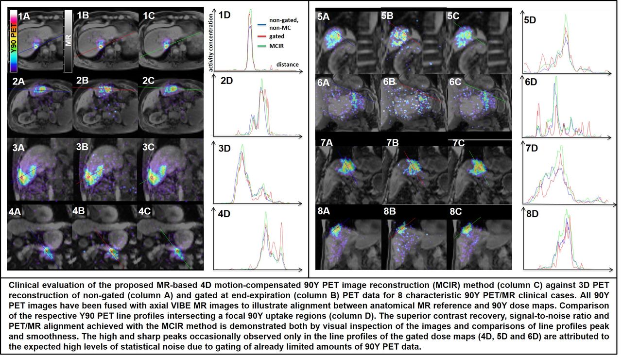
Objectives: 90Y PET has been employed to image and quantify the absorbed 90Y dose distribution in liver tumors and surrounding normal tissue, following 90Y radioembolization [1]. However, PET/CT liver dose maps can suffer from respiratory motion-induced blurring, due to the relatively poor 4D motion tracking capabilities of the CT, and the low 90Y PET signal, due to low positron yield [2]. In fact, clinical PET/CT studies have shown 90Y dose differences of as low as 25% between responding and non-responding lesions [3]. Nevertheless, the advent of simultaneous PET/MR enabled the utilization of the superior soft-tissue contrast of the MR to improve tumors localization and determine a stronger correlation between delivered dose and tumor’s response [4]. In this study, we aim to exploit the superior MR-based motion tracking capabilities of clinical PET/MR and employ a 4D respiratory motion-compensated 90Y PET image reconstruction method to improve the accuracy of the 90Y liver dose maps without further amplifying the statistical noise. Methods: A cohort of 21 patients (66.7 +/- 9.9 y.o., 77.7 +/- 14 kg) underwent a PET/MR scan at a Siemens Biograph mMR scanner, starting 3.3 +/- 0.8 h post injection of a 2 +/- 1 GBq of 90Y dosage. The PET/MR protocol consisted of a 20min list-mode (LM) PET acquisition, in parallel to a 19sec VIBE MR sequence with 2-point Dixon fat/water separation, to support PET attenuation correction with 4-class tissue (air, lungs, fat, water) segmentation, and a 20min 3D radial VIBE MR prototype sequence with self-navigation to track the respiratory motion phase and accordingly sort the MR and the synchronized LM PET data into 5 gates from end-expiration to end-inspiration. The LM PET gates were later histogrammed into 5 sinogram gates, respectively. Hierarchical diffeomorphic image registration was then applied to the 5 MR gates to derive the 3D motion transformations between a reference gate (end-expiration) and each of the other gates (forward model) and vice-versa (reverse model). Subsequently, we developed a sinogram-based 4D motion-compensated image reconstruction (MCIR) method by incorporating the forward and reverse motion components into the forward- and back-projection operators, respectively, of an Ordinary Poisson Ordered Subsets Expectation Maximization (OP-OSEM) algorithm. Finally all the gated PET sinograms were passed to the 4D MCIR algorithm to produce a) a motion-compensated (MC) 90Y dose map and compare it against dose maps reconstructed from b) non-gated non-MC, as well as c) gated (end-expiration) 90Y data. Results: Visual inspection and quantitative evaluation along line profiles intersecting points of suspected focal liver uptake demonstrated that the in-vivo application of the 4D MCIR method on 90Y-PET/MR liver data for all 21 cases resulted in significantly higher signal contrast recovery (12% higher line profile peaks on average) compared to non-gated non-MC dose maps for the same noise levels. In addition, the dose maps derived with the 4D MCIR method were associated with significantly lower noise levels and higher signal-to-noise ratio (smoother line profiles with more concentrated peaks), compared to the gated dose maps at end-expiration position. Finally, the alignment between the 90Y dose maps and the MR-defined liver regions was significantly improved with the 4D MCIR method, compared to non-gated non-MC dose maps, as suggested by the elimination of 90Y dose outside the liver after 4D MCIR in nearly all cases. This is of high importance in clinical practice, as occasionally a percentage of 90Y dose may be observed in lungs due to air embolization.

Conclusions: 90Y-PET/MRI exploiting MR-based respiratory motion models and 4D motion-compensated PET image reconstruction may provide highly accurate 90Y dose liver maps to significantly enhance hepatic tumor response assessments after 90Y radioembolization. Research Support: This work was supported by NIH/NHLBI R01HL071021 grant.

Figure
  • Download figure
  • Open in new tab
  • Download powerpoint
Previous
Back to top

In this issue

Journal of Nuclear Medicine
Vol. 59, Issue supplement 1
May 1, 2018
  • Table of Contents
  • Index by author
Article Alerts
Sign In to Email Alerts with your Email Address
Email Article

Thank you for your interest in spreading the word on Journal of Nuclear Medicine.

NOTE: We only request your email address so that the person you are recommending the page to knows that you wanted them to see it, and that it is not junk mail. We do not capture any email address.

Enter multiple addresses on separate lines or separate them with commas.
Motion-compensated 90Y-PET/MR image reconstruction for enhanced localization and quantification of liver 90Y dose maps
(Your Name) has sent you a message from Journal of Nuclear Medicine
(Your Name) thought you would like to see the Journal of Nuclear Medicine web site.
Citation Tools
Motion-compensated 90Y-PET/MR image reconstruction for enhanced localization and quantification of liver 90Y dose maps
Nicolas Karakatsanis, Mootaz Eldib, David Faul, Matthias Fenchel, Lale Kostakoglu, Karin Knesaurek, Zahi Fayad
Journal of Nuclear Medicine May 2018, 59 (supplement 1) 653;

Citation Manager Formats

  • BibTeX
  • Bookends
  • EasyBib
  • EndNote (tagged)
  • EndNote 8 (xml)
  • Medlars
  • Mendeley
  • Papers
  • RefWorks Tagged
  • Ref Manager
  • RIS
  • Zotero
Share
Motion-compensated 90Y-PET/MR image reconstruction for enhanced localization and quantification of liver 90Y dose maps
Nicolas Karakatsanis, Mootaz Eldib, David Faul, Matthias Fenchel, Lale Kostakoglu, Karin Knesaurek, Zahi Fayad
Journal of Nuclear Medicine May 2018, 59 (supplement 1) 653;
Twitter logo Facebook logo LinkedIn logo Mendeley logo
  • Tweet Widget
  • Facebook Like
  • Google Plus One
Bookmark this article

Jump to section

  • Article
  • Figures & Data
  • Info & Metrics

Related Articles

  • No related articles found.
  • Google Scholar

Cited By...

  • No citing articles found.
  • Google Scholar

More in this TOC Section

Instrumentation & Data Analysis Track

  • Deep Learning Based Kidney Segmentation for Glomerular Filtration Rate Measurement Using Quantitative SPECT/CT
  • The Benefit of Time-of-Flight in Digital Photon Counting PET Imaging: Physics and Clinical Evaluation
  • Preclinical validation of a single-scan rest/stress imaging technique for 13NH3 cardiac perfusion studies
Show more Instrumentation & Data Analysis Track

PET/MR Quantitation (Image Generation)

  • Attenuation Correction of PET/MR Using Deep Neural Network Based on Dixon and ZTE MR Images
  • Impact of attenuation map generation schemes in lesion quantification in prostate cancer patients with PET/MRI
  • PET-MR Attenuation Correction using 3D Multi-Echo Dixon-Based UTE Sequence
Show more PET/MR Quantitation (Image Generation)

Similar Articles

SNMMI

© 2025 SNMMI

Powered by HighWire