
Main menu

  • Home
  • Content
    • Current
    • Ahead of print
    • Past Issues
    • JNM Supplement
    • SNMMI Annual Meeting Abstracts
    • Continuing Education
    • JNM Podcasts
  • Subscriptions
    • Subscribers
    • Institutional and Non-member
    • Rates
    • Journal Claims
    • Corporate & Special Sales
  • Authors
    • Submit to JNM
    • Information for Authors
    • Assignment of Copyright
    • AQARA requirements
  • Info
    • Reviewers
    • Permissions
    • Advertisers
  • About
    • About Us
    • Editorial Board
    • Contact Information
  • More
    • Alerts
    • Feedback
    • Help
    • SNMMI Journals
  • SNMMI
    • JNM
    • JNMT
    • SNMMI Journals
    • SNMMI

User menu

  • Subscribe
  • My alerts
  • Log in
  • My Cart

Search

  • Advanced search
Journal of Nuclear Medicine
  • SNMMI
    • JNM
    • JNMT
    • SNMMI Journals
    • SNMMI
  • Subscribe
  • My alerts
  • Log in
  • My Cart
Journal of Nuclear Medicine

Advanced Search

  • Home
  • Content
    • Current
    • Ahead of print
    • Past Issues
    • JNM Supplement
    • SNMMI Annual Meeting Abstracts
    • Continuing Education
    • JNM Podcasts
  • Subscriptions
    • Subscribers
    • Institutional and Non-member
    • Rates
    • Journal Claims
    • Corporate & Special Sales
  • Authors
    • Submit to JNM
    • Information for Authors
    • Assignment of Copyright
    • AQARA requirements
  • Info
    • Reviewers
    • Permissions
    • Advertisers
  • About
    • About Us
    • Editorial Board
    • Contact Information
  • More
    • Alerts
    • Feedback
    • Help
    • SNMMI Journals
  • View or Listen to JNM Podcast
  • Visit JNM on Facebook
  • Join JNM on LinkedIn
  • Follow JNM on Twitter
  • Subscribe to our RSS feeds
Meeting ReportGeneral Clinical Specialties Track

Quantification of Lymphoma using FDG PET Heterogeneity Features for Improved Prediction of Clinical Outcome

Arman Rahmim, Saeed Ashrafinia, Steven Rowe, Charles Schmidtlein, Mikkel Vendelbo, Tarec El-Galaly, Lars Gormsen and Ole Munk
Journal of Nuclear Medicine May 2018, 59 (supplement 1) 594;
Arman Rahmim
3Department of Radiology Johns Hopkins University Baltimore MD United States
  • Find this author on Google Scholar
  • Find this author on PubMed
  • Search for this author on this site
Saeed Ashrafinia
3Department of Radiology Johns Hopkins University Baltimore MD United States
  • Find this author on Google Scholar
  • Find this author on PubMed
  • Search for this author on this site
Steven Rowe
3Department of Radiology Johns Hopkins University Baltimore MD United States
  • Find this author on Google Scholar
  • Find this author on PubMed
  • Search for this author on this site
Charles Schmidtlein
4Department of Medical Physics Memorial Sloan-Kettering Cancer Center New York NY United States
  • Find this author on Google Scholar
  • Find this author on PubMed
  • Search for this author on this site
Mikkel Vendelbo
2Department of Nuclear Medicine & PET Center Aarhus University Hospital Aarhus Denmark
  • Find this author on Google Scholar
  • Find this author on PubMed
  • Search for this author on this site
Tarec El-Galaly
1Department of Hematology Aalborg University Hospital Aalborg Denmark
  • Find this author on Google Scholar
  • Find this author on PubMed
  • Search for this author on this site
Lars Gormsen
2Department of Nuclear Medicine & PET Center Aarhus University Hospital Aarhus Denmark
  • Find this author on Google Scholar
  • Find this author on PubMed
  • Search for this author on this site
Ole Munk
2Department of Nuclear Medicine & PET Center Aarhus University Hospital Aarhus Denmark
  • Find this author on Google Scholar
  • Find this author on PubMed
  • Search for this author on this site
  • Article
  • Figures & Data
  • Info & Metrics
Loading

Abstract

594

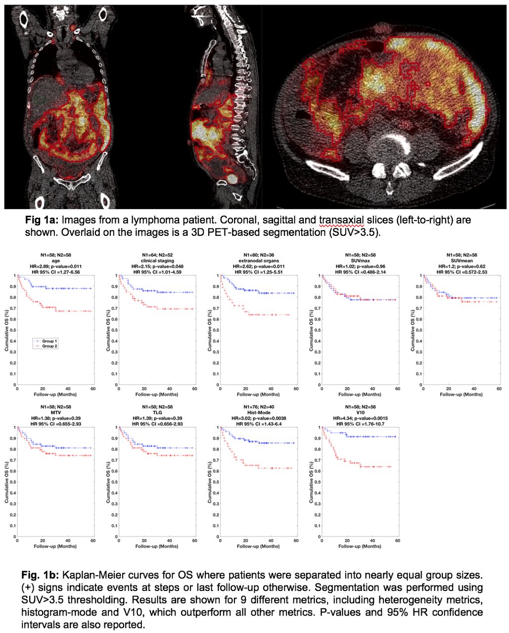
Objectives: Although current visual grading systems for response assessment on follow-up FDG PET scans in lymphomas work well, prediction of patient outcomes and information on selecting treatment derived from baseline scans is lacking. In this work, we aim to improve prediction of clinical outcome, by moving beyond conventional imaging measures, and to perform heterogeneity quantification of FDG uptake. Methods: We analyzed pre-treatment FDG PET images from 116 patients with lymphoma (73 (62.9%) males; mean age 64.1 years [SD 15.3; range 16-88]), and assessed prediction of overall survival (OS) and progression-free survival (PFS). The number of events for OS (death) and PFS (progression) were 28 and 36, respectively. First, volumetric tumor segmentations were performed: thresholding (1) 41% SUVmax, (2) SUV>2.5, and (3) SUV>3.5 (Hermes Hybrid Viewer PDR). A total of 45 features were extracted from each patient. These included (1) sex, (2) age, (3) clinical staging, (4) PET-only staging, and (5) extra-nodal involvement. We also extracted 40 quantitative imaging measures, including SUVmax, SUVmean, metabolic tumor volume (MTV) and total lesion glycolysis (TLG) (n=4), the recently introduced class of generalized effective total uptake (gETU) measures [1] which place varying degrees of emphasis on volumetric vs. uptake information (n=10), intensity histogram (n=19) and intensity-volume histogram (n=7) measures [2, 3]. All metrics were standardized according to the framework of the image biomarker standardization initiative (IBSI) [4]. Feature selection was then performed. Metrics with Pearson correlations (r) > 0.95 were considered relatively redundant, and a total of 26 features were retained. Finally, univariate and multivariate analyses were performed, which included statistical considerations (to discourage false discovery and overfitting), assessing prediction of OS and PFS. Specifically, Kaplan-Meier survival analyses were carried out, where the subjects were divided into high-risk and low-risk groups of nearly equal patients, from which the hazard ratios (HR) were computed via Cox proportional hazards regression. Results: The third segmentation method (SUV>3.5) outperformed other methods and is reported for all subsequent analysis. Table 1 depicts univariate Cox regression analysis for OS and PFS (we report on the performance of SUV, MTV, TLG, and any metric found to be significant). It is seen that even though volumetric measures (MTV and TLG) outperform SUV metrics, all these measures still perform relatively poorly for prediction of OS and PFS. By contrast, age, clinical stage, and extra-nodal involvement are stronger predictors (with respective HR values of 2.89, 2.15 and 2.62 for OS, and 2.05, 2.49 and 2.75 for PFS). By comparison, we found that two PET-based heterogeneity features, namely histogram-mode and V10, resulted in stronger predictions of outcome (HR values of 3.02 and 4.34 for OS, and 2.31 and 2.86 for PFS) [histogram-mode denotes the most common discretized gray level in histogram binning of the segmentation, while V10 quantifies the tumor volume fraction containing at least 10% of the maximum gray level (intensity)]. After correcting for multiple testing (to control for false discovery), V10 was seen to be a significant predictor of outcome for both OS and PFS, unlike all other features. Finally, multivariate analysis (Table 2) revealed that improved HR=3.64 could be obtained for PFS, when combining heterogeneity feature V10 with extra-nodal involvement in the final model. Conclusions: It was demonstrated that FDG PET heterogeneity features, in contrast to commonly invoked imaging features, hold significant potential for improved prediction of clinical outcome in patients with lymphoma.

View this table:
  • View inline
  • View popup

Table 1: Univariate Cox regression ([asterisk] indicates significance after correction for multiple testing)

View this table:
  • View inline
  • View popup

Table 2: Final multivariate Cox regression model for OS and PFS

Figure
  • Download figure
  • Open in new tab
  • Download powerpoint
Previous
Back to top

In this issue

Journal of Nuclear Medicine
Vol. 59, Issue supplement 1
May 1, 2018
  • Table of Contents
  • Index by author
Article Alerts
Sign In to Email Alerts with your Email Address
Email Article

Thank you for your interest in spreading the word on Journal of Nuclear Medicine.

NOTE: We only request your email address so that the person you are recommending the page to knows that you wanted them to see it, and that it is not junk mail. We do not capture any email address.

Enter multiple addresses on separate lines or separate them with commas.
Quantification of Lymphoma using FDG PET Heterogeneity Features for Improved Prediction of Clinical Outcome
(Your Name) has sent you a message from Journal of Nuclear Medicine
(Your Name) thought you would like to see the Journal of Nuclear Medicine web site.
Citation Tools
Quantification of Lymphoma using FDG PET Heterogeneity Features for Improved Prediction of Clinical Outcome
Arman Rahmim, Saeed Ashrafinia, Steven Rowe, Charles Schmidtlein, Mikkel Vendelbo, Tarec El-Galaly, Lars Gormsen, Ole Munk
Journal of Nuclear Medicine May 2018, 59 (supplement 1) 594;

Citation Manager Formats

  • BibTeX
  • Bookends
  • EasyBib
  • EndNote (tagged)
  • EndNote 8 (xml)
  • Medlars
  • Mendeley
  • Papers
  • RefWorks Tagged
  • Ref Manager
  • RIS
  • Zotero
Share
Quantification of Lymphoma using FDG PET Heterogeneity Features for Improved Prediction of Clinical Outcome
Arman Rahmim, Saeed Ashrafinia, Steven Rowe, Charles Schmidtlein, Mikkel Vendelbo, Tarec El-Galaly, Lars Gormsen, Ole Munk
Journal of Nuclear Medicine May 2018, 59 (supplement 1) 594;
Twitter logo Facebook logo LinkedIn logo Mendeley logo
  • Tweet Widget
  • Facebook Like
  • Google Plus One
Bookmark this article

Jump to section

  • Article
  • Figures & Data
  • Info & Metrics

Related Articles

  • No related articles found.
  • Google Scholar

Cited By...

  • No citing articles found.
  • Google Scholar

More in this TOC Section

General Clinical Specialties Track

  • Quantitative Evaluation of Parathyroid Adenoma and Hyperplasia in Reference to Thyroid using Tc-99m MIBI SPECT/CT
  • A primitive study for clinical application of 18F-AlF-NOTA-octreotide PET/CT in combination with 18F-FDG PET/CT for imaging neuroendocrine neoplasms
  • Role of 18F-FDG PET/CT in detection of disease burden and response assessment in patients with myeloid sarcoma
Show more General Clinical Specialties Track

Leukemia/Lymphoma/Myeloma II

  • Predictive Value of Interim FDG-PET/CT Interpreted with Deauville 5-point Score in Diffuse Large B-cell Lymphoma
  • Prognostic value of interim FDG and FLT PET/CT in diffuse large B cell lymphoma
Show more Leukemia/Lymphoma/Myeloma II

Similar Articles

SNMMI

© 2025 SNMMI

Powered by HighWire