
Main menu

  • Home
  • Content
    • Current
    • Ahead of print
    • Past Issues
    • JNM Supplement
    • SNMMI Annual Meeting Abstracts
    • Continuing Education
    • JNM Podcasts
  • Subscriptions
    • Subscribers
    • Institutional and Non-member
    • Rates
    • Journal Claims
    • Corporate & Special Sales
  • Authors
    • Submit to JNM
    • Information for Authors
    • Assignment of Copyright
    • AQARA requirements
  • Info
    • Reviewers
    • Permissions
    • Advertisers
  • About
    • About Us
    • Editorial Board
    • Contact Information
  • More
    • Alerts
    • Feedback
    • Help
    • SNMMI Journals
  • SNMMI
    • JNM
    • JNMT
    • SNMMI Journals
    • SNMMI

User menu

  • Subscribe
  • My alerts
  • Log in
  • My Cart

Search

  • Advanced search
Journal of Nuclear Medicine
  • SNMMI
    • JNM
    • JNMT
    • SNMMI Journals
    • SNMMI
  • Subscribe
  • My alerts
  • Log in
  • My Cart
Journal of Nuclear Medicine

Advanced Search

  • Home
  • Content
    • Current
    • Ahead of print
    • Past Issues
    • JNM Supplement
    • SNMMI Annual Meeting Abstracts
    • Continuing Education
    • JNM Podcasts
  • Subscriptions
    • Subscribers
    • Institutional and Non-member
    • Rates
    • Journal Claims
    • Corporate & Special Sales
  • Authors
    • Submit to JNM
    • Information for Authors
    • Assignment of Copyright
    • AQARA requirements
  • Info
    • Reviewers
    • Permissions
    • Advertisers
  • About
    • About Us
    • Editorial Board
    • Contact Information
  • More
    • Alerts
    • Feedback
    • Help
    • SNMMI Journals
  • View or Listen to JNM Podcast
  • Visit JNM on Facebook
  • Join JNM on LinkedIn
  • Follow JNM on Twitter
  • Subscribe to our RSS feeds
Meeting ReportEducational Exhibits Track

Dynamic whole-body PET imaging: principles, potentials and applications

Arman Rahmim, Martin Lodge, Nicolas Karakatsanis, Vladimir Panin, Yun Zhou, Alan McMillan, Steve Cho, Habib Zaidi, Michael Casey and Richard Wahl
Journal of Nuclear Medicine May 2018, 59 (supplement 1) 1158;
Arman Rahmim
2Department of Radiology Johns Hopkins University Baltimore MD United States
  • Find this author on Google Scholar
  • Find this author on PubMed
  • Search for this author on this site
Martin Lodge
2Department of Radiology Johns Hopkins University Baltimore MD United States
  • Find this author on Google Scholar
  • Find this author on PubMed
  • Search for this author on this site
Nicolas Karakatsanis
6Department of Radiology Weill Cornell Medical College New York NY United States
  • Find this author on Google Scholar
  • Find this author on PubMed
  • Search for this author on this site
Vladimir Panin
3Siemens Healthineers Knoxville TN United States
  • Find this author on Google Scholar
  • Find this author on PubMed
  • Search for this author on this site
Yun Zhou
2Department of Radiology Johns Hopkins University Baltimore MD United States
  • Find this author on Google Scholar
  • Find this author on PubMed
  • Search for this author on this site
Alan McMillan
4Department of Radiology University of Wisconsin Madison WI United States
  • Find this author on Google Scholar
  • Find this author on PubMed
  • Search for this author on this site
Steve Cho
4Department of Radiology University of Wisconsin Madison WI United States
  • Find this author on Google Scholar
  • Find this author on PubMed
  • Search for this author on this site
Habib Zaidi
1Division of Nuclear Medicine and Molecular Imaging Geneva University Hospital Geneva Switzerland
  • Find this author on Google Scholar
  • Find this author on PubMed
  • Search for this author on this site
Michael Casey
3Siemens Healthineers Knoxville TN United States
  • Find this author on Google Scholar
  • Find this author on PubMed
  • Search for this author on this site
Richard Wahl
5Department of Radiology Washington University St. Louis MO United States
  • Find this author on Google Scholar
  • Find this author on PubMed
  • Search for this author on this site
  • Article
  • Figures & Data
  • Info & Metrics
Loading

Abstract

1158

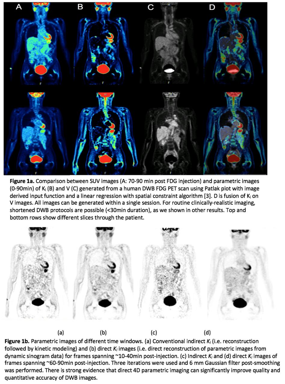
Objectives: To demonstrate that dynamic whole-body (DWB) PET is a clinical imaging tool with significant potential. DWB PET can be performed within reasonable clinical imaging times and enables generation of multiple types of PET images with complementary information in a single imaging session (<30min). Methods: Current clinical PET protocols mirror the pattern established for traditional radiology in that they are optimized for qualitative as opposed to quantitative assessment, with documented limitations [1]. Radiotracer distribution is a dynamic process that varies substantially between organs (particularly tumors) and between patients. The uptake periods used in clinical protocols are somewhat arbitrary and are not expected to be optimal for all clinical cases. We argue that a new paradigm of DWB-PET is both feasible and has significant potential. It is made possible by ongoing technical developments; significant advancements in PET hardware, combined with statistical image reconstruction have made it possible to acquire multi-pass eyes-to-thighs imaging in clinically feasible times, achieving adequate statistical quality in less than 5 min/pass. Patlak analysis [2] is particularly suited for generation of parametric images from DWB PET because it is applicable to FDG and it does not require PET scans to sample the early tracer kinetics. Thus, DWB imaging can be used to produce parametric images of (i) Patlak slope (influx rate Ki) and (ii) intercept (distribution volume V), while also providing (iii) conventional SUV images by summation of dynamic frames. There are also means to reduce noise in the parametric Patlak slope and intercept images, by use of constrained statistical regression [3, 4], and direct 4D reconstruction of parametric images from sinogram data [5]. Results: Parametric images of both Patlak slope Ki and intercept V can be generated, in addition to conventional SUV images by summation of the dynamic frames (Figure 1a). Thus, three distinct images can be obtained from the same imaging session. The Ki image generally shows reduced normal organ uptake, particularly in the liver. Furthermore, direct 4D parametric image reconstruction can significantly improve quality and quantitative accuracy of DWB images (Figure 1b). We also have results demonstrating application of DWB imaging to beyond FDG PET/CT, for instance to PET/MR imaging, as well as Ga-68 DOTATOC PET/CT. Conclusions: DWB-PET has a number of advantages. It can minimize time dependence of SUV activity: SUV uptake changes in time in direct proportion to changes in image uptake. Given variable scan times inherent in a busy clinical practice, this is an issue, and the proposed measures are expected to be less subject to such alterations. Furthermore, it can remove background uptake, allow small and less FDG avid tumors to be identified, and produce more quantitative estimates of tumor uptake. Overall, a new paradigm of DWB PET imaging (applicable to both PET/CT and PET/MRI) generates quantitative measures that may contribute significantly added clinical value to conventional SUV images.

Figure
  • Download figure
  • Open in new tab
  • Download powerpoint
Previous
Back to top

In this issue

Journal of Nuclear Medicine
Vol. 59, Issue supplement 1
May 1, 2018
  • Table of Contents
  • Index by author
Article Alerts
Sign In to Email Alerts with your Email Address
Email Article

Thank you for your interest in spreading the word on Journal of Nuclear Medicine.

NOTE: We only request your email address so that the person you are recommending the page to knows that you wanted them to see it, and that it is not junk mail. We do not capture any email address.

Enter multiple addresses on separate lines or separate them with commas.
Dynamic whole-body PET imaging: principles, potentials and applications
(Your Name) has sent you a message from Journal of Nuclear Medicine
(Your Name) thought you would like to see the Journal of Nuclear Medicine web site.
Citation Tools
Dynamic whole-body PET imaging: principles, potentials and applications
Arman Rahmim, Martin Lodge, Nicolas Karakatsanis, Vladimir Panin, Yun Zhou, Alan McMillan, Steve Cho, Habib Zaidi, Michael Casey, Richard Wahl
Journal of Nuclear Medicine May 2018, 59 (supplement 1) 1158;

Citation Manager Formats

  • BibTeX
  • Bookends
  • EasyBib
  • EndNote (tagged)
  • EndNote 8 (xml)
  • Medlars
  • Mendeley
  • Papers
  • RefWorks Tagged
  • Ref Manager
  • RIS
  • Zotero
Share
Dynamic whole-body PET imaging: principles, potentials and applications
Arman Rahmim, Martin Lodge, Nicolas Karakatsanis, Vladimir Panin, Yun Zhou, Alan McMillan, Steve Cho, Habib Zaidi, Michael Casey, Richard Wahl
Journal of Nuclear Medicine May 2018, 59 (supplement 1) 1158;
Twitter logo Facebook logo LinkedIn logo Mendeley logo
  • Tweet Widget
  • Facebook Like
  • Google Plus One
Bookmark this article

Jump to section

  • Article
  • Figures & Data
  • Info & Metrics

Related Articles

  • No related articles found.
  • Google Scholar

Cited By...

  • No citing articles found.
  • Google Scholar

More in this TOC Section

Educational Exhibits Track

  • Global assessment of PET tracer uptake in joints using CT segmentation: a novel approach to quantify global metabolic activity.
  • Acute leukemia: The wide spectrum of imaging findings on an FDG PET-CT study.
  • PET/MRI imaging as a practical tool in evaluating mild traumatic brain injury (mTBI)
Show more Educational Exhibits Track

Educational Exhibits

  • Do They Work? Utilizing the "Hoshin Kanri" Process in the Nuclear Medicine Department
  • Challenges in F-18 FDG PET/CT: Benign Osseous Hypermetabolic Lesions
  • Early small bowel activity may predict rapid gastric transit on gastric emptying scintigraphy
Show more Educational Exhibits

Similar Articles

SNMMI

© 2025 SNMMI

Powered by HighWire