


Abstract
842
Objectives: Full Monte Carlo simulations using time series of nuclear medicine images are considered the most accurate way to determine the absorbed radiation dose at the voxel level. However, this method is time consuming because of the simulation time involved, mainly because of the electron simulation, and is therefore less suitable for routine planning and evaluation of the radiation absorbed dose on an individual patient basis in the clinic. Recently, a semi Monte Carlo approach was introduced for 177Lu dosimetry that addresses this shortcoming of full Monte Carlo simulations by providing a fast voxel-based method. The purpose of this study is to validate this experimental software package for patient-specific, voxel-based radionuclide dosimetry using both patient SPECT-CT data and another dosimetry software, and using measurements with a 3D printed phantom and thermoluminescent dosimeters (TLD).
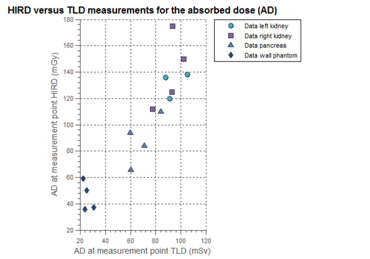
Methods: A program based on semi-Monte Carlo simulations (sMC) (Hermes Medical Solutions, Stockholm, Sweden) was used to estimate the dose distributions from 18 patient 111In-Exendin SPECT-CT scans, each consisting of 3 or 5 acquisitions. The kidney volume was manually delineated using the CT scan. The resulting mean kidney dose estimates were compared to those obtained using OLINDA/EXM 1.1 from the same SPECT-CT acquisitions. For the latter method, the kidney volume was delineated on the first SPECT scan using a 25% of the maximum voxel value in the kidney, to ensure that the spill out was included in the volume of interest (VOI). The kidney mass was scaled in OLINDA/EXM using the kidney volume as obtained from the CT VOI. A previously developed 3D printed phantom of the pancreas and kidneys inside a shell of a NEMA IEC body phantom was filled with aqueous 111In-chloride solutions of different activity concentrations in the background and in the organs. Dose distribution estimates for both the photon and electron contribution were obtained with the sMC software of a time series of 5 SPECT-CT scans of the phantom. The absorbed dose as a result of the photon contribution was compared with local dose measurements performed using TLDs at 16 locations inside the phantom. The absorbed dose was measured in the voxel at the position of the TLD inside the phantom as registered on the CT scan. The electron contribution was not taken into account because of absorption of this radiation in the walls of the organs.
Results: The mean kidney doses in the patient studies obtained using the sMC software correlated strongly with the values from OLINDA/EXM (slope = 0.86, R2 = 0.92). The phantom study resulted in a photon contribution in the sMC software that was 1.52 times higher (SD = 0.37) than the absorbed dose as measured by the TLDs (mean factors of 1.5, 1.3, 1.8, for kidneys, pancreas and background, respectively).
Conclusion: Although good agreement was obtained when comparing software-based estimates, the experimental validation led to differences that could not be fully explained by electron absorption in the walls of the phantom organs. The partial volume effect in the SPECT images at the edge of the organs could explain part of the difference between the software and the TLD measurements. Furthermore, the TLD calibration could be affected by the electrons emitted by 111In. Therefore, further work is needed to quantify these factors.


In this issue


Jump to section
Related Articles
Cited By...
- No citing articles found.