
Main menu

  • Home
  • Content
    • Current
    • Ahead of print
    • Past Issues
    • JNM Supplement
    • SNMMI Annual Meeting Abstracts
    • Continuing Education
    • JNM Podcasts
  • Subscriptions
    • Subscribers
    • Institutional and Non-member
    • Rates
    • Journal Claims
    • Corporate & Special Sales
  • Authors
    • Submit to JNM
    • Information for Authors
    • Assignment of Copyright
    • AQARA requirements
  • Info
    • Reviewers
    • Permissions
    • Advertisers
  • About
    • About Us
    • Editorial Board
    • Contact Information
  • More
    • Alerts
    • Feedback
    • Help
    • SNMMI Journals
  • SNMMI
    • JNM
    • JNMT
    • SNMMI Journals
    • SNMMI

User menu

  • Subscribe
  • My alerts
  • Log in
  • My Cart

Search

  • Advanced search
Journal of Nuclear Medicine
  • SNMMI
    • JNM
    • JNMT
    • SNMMI Journals
    • SNMMI
  • Subscribe
  • My alerts
  • Log in
  • My Cart
Journal of Nuclear Medicine

Advanced Search

  • Home
  • Content
    • Current
    • Ahead of print
    • Past Issues
    • JNM Supplement
    • SNMMI Annual Meeting Abstracts
    • Continuing Education
    • JNM Podcasts
  • Subscriptions
    • Subscribers
    • Institutional and Non-member
    • Rates
    • Journal Claims
    • Corporate & Special Sales
  • Authors
    • Submit to JNM
    • Information for Authors
    • Assignment of Copyright
    • AQARA requirements
  • Info
    • Reviewers
    • Permissions
    • Advertisers
  • About
    • About Us
    • Editorial Board
    • Contact Information
  • More
    • Alerts
    • Feedback
    • Help
    • SNMMI Journals
  • View or Listen to JNM Podcast
  • Visit JNM on Facebook
  • Join JNM on LinkedIn
  • Follow JNM on Twitter
  • Subscribe to our RSS feeds
Meeting ReportInstrumentation & Data Analysis Track

A Viewer for Dynamic Whole Body PET/CT Studies

Jeffrey Leal, Evrim Turkbey, Lilja Solnes, Steven Rowe, Arman Rahmim and Martin Lodge
Journal of Nuclear Medicine May 2017, 58 (supplement 1) 705;
Jeffrey Leal
1Johns Hopkins University Baltimore MD United States
  • Find this author on Google Scholar
  • Find this author on PubMed
  • Search for this author on this site
Evrim Turkbey
1Johns Hopkins University Baltimore MD United States
  • Find this author on Google Scholar
  • Find this author on PubMed
  • Search for this author on this site
Lilja Solnes
1Johns Hopkins University Baltimore MD United States
  • Find this author on Google Scholar
  • Find this author on PubMed
  • Search for this author on this site
Steven Rowe
1Johns Hopkins University Baltimore MD United States
  • Find this author on Google Scholar
  • Find this author on PubMed
  • Search for this author on this site
Arman Rahmim
1Johns Hopkins University Baltimore MD United States
  • Find this author on Google Scholar
  • Find this author on PubMed
  • Search for this author on this site
Martin Lodge
1Johns Hopkins University Baltimore MD United States
  • Find this author on Google Scholar
  • Find this author on PubMed
  • Search for this author on this site
  • Article
  • Figures & Data
  • Info & Metrics
Loading

Abstract

705

Objectives: To provide a system for the review and quantitative analysis of dynamically acquired whole-body PET/CT studies. While single bed position dynamic imaging is standard practice in much of PET-based research, only recently have PET/CT scanners possessed the capability to acquire whole-body dynamic PET images. Even so, few clinical review systems have the ability, or user-interface, to allow the appropriate review and analysis of these new datasets.

Methods: A PET/CT viewer was developed in the Java programming language for the display and quantitative analysis of dynamic whole body PET/CT data. The program supports multi-time point DICOM encoded series data as well as the manual selection of multiple time frames of whole-body DICOM series. In addition, the software supports the display of co-registered CT data, allowing full PET/CT display along all three primary display planes (Axial, Sagittal and Coronal) and time points. Separate lookup tables are provided for functional and anatomical display data with a blending control to adjust the relative contribution of each to the visual display. The software includes basic parametric image calculation, including ‘integral’ images, in which summed images from the initial scan through to each time point are calculated and available for display. Similarly, voxel-by-voxel least-squares linear fitting of the source image data provides ‘slope’, ‘intercept’, and ‘correlation’ datasets for display as well. These basic parametric datasets can be displayed and used to drive volume-of-interest (VOI) placement for quantification of the source SUV data. This functionality is a preview of Patlak capability which is forthcoming. Quantification is provided through the placement of resizable VOIs. The user is able to place VOIs upon any of the source or derived datasets. A variety of quantitative metrics, in particular MAX and PEAK values, are presented in real-time as time-activity-curve (TAC) data which is dynamically presented to the user as ‘live’ plots which auto-recalculate as the user adjusts and relocates the individual spherical VOIs. Tabular data can be reviewed and exported for further analysis outside the software. TAC plots, as well as the myriad display images (SUV, ‘Integral’, ‘slope’, ‘intercept’, etc.) can be exported in a variety of formats, both as static images as well as dynamic AVI movie loops.

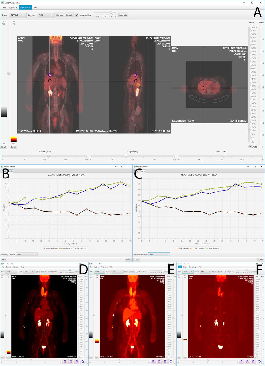
Results: Integrated PET/CT display was provided, allowing user navigation of study time points identically to navigating the data spatially. Figure 1(A) is an example of a dynamic whole-body FDG PET/CT study. Intense FDG uptake in the enlarged thyroid gland due to patient’s known thyroiditis was ignored for the purpose of this demonstration. A VOI placed centrally over the liver for reference measurement, as well as VOIs located over lesions in the liver, can be seen. “Live” TAC plots are shown in Figures 1(B) for MAX voxel and Figure 1(C) for maximum PEAK voxel. Liver lesions were pathologically proven to be metastatic adenocarcinoma. Figures 1(D), 1(E), and 1(F) demonstrate experimental parametric capabilities through dynamic MIP displays of ‘integral’, ‘intercept', and ‘slope' images.

Conclusion: Today’s modern PET/CT scanners are faster and more efficient than ever before, enabling acquisitions once not even considered. Dynamic whole body PET/CT has the potential of bringing an entirely new dimension of capabilities to our clinical practice. While PET/CT scanners of today may be capable of acquiring these new studies, the image display and analysis tools which are typically available to our clinicians have not kept pace with these new datasets. The viewer presented here provides many of the required tools to properly display and analyze these new datasets, and does so in an implementation which is easily navigable by the user and executable on the widest variety of computing platforms. Research Support: National Institutes of Health (P30 CA006973)

Figure
  • Download figure
  • Open in new tab
  • Download powerpoint
Previous
Back to top

In this issue

Journal of Nuclear Medicine
Vol. 58, Issue supplement 1
May 1, 2017
  • Table of Contents
  • Index by author
Article Alerts
Sign In to Email Alerts with your Email Address
Email Article

Thank you for your interest in spreading the word on Journal of Nuclear Medicine.

NOTE: We only request your email address so that the person you are recommending the page to knows that you wanted them to see it, and that it is not junk mail. We do not capture any email address.

Enter multiple addresses on separate lines or separate them with commas.
A Viewer for Dynamic Whole Body PET/CT Studies
(Your Name) has sent you a message from Journal of Nuclear Medicine
(Your Name) thought you would like to see the Journal of Nuclear Medicine web site.
Citation Tools
A Viewer for Dynamic Whole Body PET/CT Studies
Jeffrey Leal, Evrim Turkbey, Lilja Solnes, Steven Rowe, Arman Rahmim, Martin Lodge
Journal of Nuclear Medicine May 2017, 58 (supplement 1) 705;

Citation Manager Formats

  • BibTeX
  • Bookends
  • EasyBib
  • EndNote (tagged)
  • EndNote 8 (xml)
  • Medlars
  • Mendeley
  • Papers
  • RefWorks Tagged
  • Ref Manager
  • RIS
  • Zotero
Share
A Viewer for Dynamic Whole Body PET/CT Studies
Jeffrey Leal, Evrim Turkbey, Lilja Solnes, Steven Rowe, Arman Rahmim, Martin Lodge
Journal of Nuclear Medicine May 2017, 58 (supplement 1) 705;
Twitter logo Facebook logo LinkedIn logo Mendeley logo
  • Tweet Widget
  • Facebook Like
  • Google Plus One
Bookmark this article

Jump to section

  • Article
  • Figures & Data
  • Info & Metrics

Related Articles

  • No related articles found.
  • Google Scholar

Cited By...

  • No citing articles found.
  • Google Scholar

More in this TOC Section

Instrumentation & Data Analysis Track

  • Deep Learning Based Kidney Segmentation for Glomerular Filtration Rate Measurement Using Quantitative SPECT/CT
  • The Benefit of Time-of-Flight in Digital Photon Counting PET Imaging: Physics and Clinical Evaluation
  • Preclinical validation of a single-scan rest/stress imaging technique for 13NH3 cardiac perfusion studies
Show more Instrumentation & Data Analysis Track

Quantification, Image Reconstruction and Methodologies in Nuclear Medicine

  • Automated Motion Correction in Quantitative SPECT Reconstruction: A Feasibility Study of a Method Framework Applied First to Bone Imaging
  • Convolutional neural network (CNN) of MRI and FDG-PET images may predict hypoxia in glioblastoma.
  • A PET cylinder phantom positioned at an oblique angle.
Show more Quantification, Image Reconstruction and Methodologies in Nuclear Medicine

Similar Articles

SNMMI

© 2025 SNMMI

Powered by HighWire