
Main menu

  • Home
  • Content
    • Current
    • Ahead of print
    • Past Issues
    • JNM Supplement
    • SNMMI Annual Meeting Abstracts
    • Continuing Education
    • JNM Podcasts
  • Subscriptions
    • Subscribers
    • Institutional and Non-member
    • Rates
    • Journal Claims
    • Corporate & Special Sales
  • Authors
    • Submit to JNM
    • Information for Authors
    • Assignment of Copyright
    • AQARA requirements
  • Info
    • Reviewers
    • Permissions
    • Advertisers
  • About
    • About Us
    • Editorial Board
    • Contact Information
  • More
    • Alerts
    • Feedback
    • Help
    • SNMMI Journals
  • SNMMI
    • JNM
    • JNMT
    • SNMMI Journals
    • SNMMI

User menu

  • Subscribe
  • My alerts
  • Log in
  • Log out
  • My Cart

Search

  • Advanced search
Journal of Nuclear Medicine
  • SNMMI
    • JNM
    • JNMT
    • SNMMI Journals
    • SNMMI
  • Subscribe
  • My alerts
  • Log in
  • Log out
  • My Cart
Journal of Nuclear Medicine

Advanced Search

  • Home
  • Content
    • Current
    • Ahead of print
    • Past Issues
    • JNM Supplement
    • SNMMI Annual Meeting Abstracts
    • Continuing Education
    • JNM Podcasts
  • Subscriptions
    • Subscribers
    • Institutional and Non-member
    • Rates
    • Journal Claims
    • Corporate & Special Sales
  • Authors
    • Submit to JNM
    • Information for Authors
    • Assignment of Copyright
    • AQARA requirements
  • Info
    • Reviewers
    • Permissions
    • Advertisers
  • About
    • About Us
    • Editorial Board
    • Contact Information
  • More
    • Alerts
    • Feedback
    • Help
    • SNMMI Journals
  • View or Listen to JNM Podcast
  • Visit JNM on Facebook
  • Join JNM on LinkedIn
  • Follow JNM on Twitter
  • Subscribe to our RSS feeds
Meeting ReportInstrumentation & Data Analysis Track

Joint Segmentation of Multi-Channel Images Using Multigraph Cuts

Samadrita Roy Chowdhury, Jiarui Yang, Georges El Fakhri, Quanzheng Li and Joyita Dutta
Journal of Nuclear Medicine May 2017, 58 (supplement 1) 1308;
Samadrita Roy Chowdhury
5UMass Lowell Lowell MA United States
  • Find this author on Google Scholar
  • Find this author on PubMed
  • Search for this author on this site
Jiarui Yang
1Boston University Boston MA United States
  • Find this author on Google Scholar
  • Find this author on PubMed
  • Search for this author on this site
Georges El Fakhri
3Massachusetts General Hospital, Harvard Medical Sc Boston MA United States
  • Find this author on Google Scholar
  • Find this author on PubMed
  • Search for this author on this site
Quanzheng Li
2Massachusetts General Hospital Boston MA United States
  • Find this author on Google Scholar
  • Find this author on PubMed
  • Search for this author on this site
Joyita Dutta
4MGH / Harvard / UMass Lowell Boston MA United States
  • Find this author on Google Scholar
  • Find this author on PubMed
  • Search for this author on this site
  • Article
  • Figures & Data
  • Info & Metrics
Loading

Abstract

1308

Objectives: Diagnostic and therapeutic tasks often rely on multiple images, such as those derived from multimodal, multi-time-frame (e.g. dynamic scanning), or multi-parametric (e.g. different MR pulse sequence based) acquisitions. Quantitative interpretation and analysis of such image groups or “channels” leads to the need for tools to jointly segment such multi-channel images. Our objective is to develop a joint segmentation approach based on multigraph cuts.

Methods: Our joint segmentation approach is based on unified cuts across a multigraph, defined as a composite of graphs generated from different image channels, thereby sharing the same node set but exhibiting different topologies. A composite normalized Laplacian for the multigraph is computed from the normalized Laplacians for each of the individual graphs. The unified cut is obtained by solving an optimization problem that seeks to ensure that the composite cut cost (based on the multigraph Laplacian) is minimized while ensuring similarity with each individual graph’s minimum cut. Minimum cuts for the individual graphs are first computed using a spectral clustering step. The segmentation problem is then posed as an eigenvalue decomposition problem based on the multigraph Laplacian. We solve the eigenvalue decomposition problem for the composite Laplacian, creating an effective cut that is constrained to be similar to each individual cut thus ensuring qualitative similarity between the unified cut and the individual cuts.

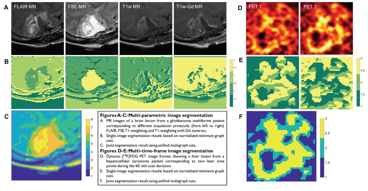
Results: The multigraph segmentation technique was first tested in digital phantom studies and then applied to the following clinical settings: 1) Dynamic imaging: Segmentation of liver lesions in hepatocellar carcinoma patients using dynamic [18F]FDG-PET. 2) Multi-parametric Imaging: Segmentation of glioblastoma multiforme (GBM) using MR images (FLAIR, FSE, T1 weighting, T1 weighting with Gd contrast). The joint segmentation was compared with single-channel segmentation results based on each individual graph’s minimum cut.

Conclusion: Multimodal, multi-time-frame, and multi-parametric imaging accumulates information from multiple structural and/or functional sources thereby facilitating diagnostic and therapeutic decisions. Joint segmentation based on unified multigraph cuts enables us to analyze data by combining disparate information sources. In comparison, single-channel segmentation approaches fail to capture the full-spectrum of anatomical/physiological information available. We have successfully applied the method to segment cancerous lesions in different clinical cases and contrasted the results with single-channel segmentation. Research Support: This work has been supported by NIH grants K01AG050711 and R01EB013293.

Figure
  • Download figure
  • Open in new tab
  • Download powerpoint
Previous
Back to top

In this issue

Journal of Nuclear Medicine
Vol. 58, Issue supplement 1
May 1, 2017
  • Table of Contents
  • Index by author
Article Alerts
Sign In to Email Alerts with your Email Address
Email Article

Thank you for your interest in spreading the word on Journal of Nuclear Medicine.

NOTE: We only request your email address so that the person you are recommending the page to knows that you wanted them to see it, and that it is not junk mail. We do not capture any email address.

Enter multiple addresses on separate lines or separate them with commas.
Joint Segmentation of Multi-Channel Images Using Multigraph Cuts
(Your Name) has sent you a message from Journal of Nuclear Medicine
(Your Name) thought you would like to see the Journal of Nuclear Medicine web site.
Citation Tools
Joint Segmentation of Multi-Channel Images Using Multigraph Cuts
Samadrita Roy Chowdhury, Jiarui Yang, Georges El Fakhri, Quanzheng Li, Joyita Dutta
Journal of Nuclear Medicine May 2017, 58 (supplement 1) 1308;

Citation Manager Formats

  • BibTeX
  • Bookends
  • EasyBib
  • EndNote (tagged)
  • EndNote 8 (xml)
  • Medlars
  • Mendeley
  • Papers
  • RefWorks Tagged
  • Ref Manager
  • RIS
  • Zotero
Share
Joint Segmentation of Multi-Channel Images Using Multigraph Cuts
Samadrita Roy Chowdhury, Jiarui Yang, Georges El Fakhri, Quanzheng Li, Joyita Dutta
Journal of Nuclear Medicine May 2017, 58 (supplement 1) 1308;
Twitter logo Facebook logo LinkedIn logo Mendeley logo
  • Tweet Widget
  • Facebook Like
  • Google Plus One
Bookmark this article

Jump to section

  • Article
  • Figures & Data
  • Info & Metrics

Related Articles

  • No related articles found.
  • Google Scholar

Cited By...

  • No citing articles found.
  • Google Scholar

More in this TOC Section

Instrumentation & Data Analysis Track

  • Deep Learning Based Kidney Segmentation for Glomerular Filtration Rate Measurement Using Quantitative SPECT/CT
  • The Benefit of Time-of-Flight in Digital Photon Counting PET Imaging: Physics and Clinical Evaluation
  • Preclinical validation of a single-scan rest/stress imaging technique for 13NH3 cardiac perfusion studies
Show more Instrumentation & Data Analysis Track

Data Analysis and Management Posters

  • Semi-automated quantification of osseous prostate metastatic burden to the spine and pelvis with sodium fluoride PET/CT
  • Quantification of 99mTc-MDP and 99mTc-HMDP concentration in normal bone with SPECT/CT
  • A texture analysis approach to evaluating SPECT system quality assurance
Show more Data Analysis and Management Posters

Similar Articles

SNMMI

© 2025 SNMMI

Powered by HighWire