
Main menu

  • Home
  • Content
    • Current
    • Ahead of print
    • Past Issues
    • JNM Supplement
    • SNMMI Annual Meeting Abstracts
    • Continuing Education
    • JNM Podcasts
  • Subscriptions
    • Subscribers
    • Institutional and Non-member
    • Rates
    • Journal Claims
    • Corporate & Special Sales
  • Authors
    • Submit to JNM
    • Information for Authors
    • Assignment of Copyright
    • AQARA requirements
  • Info
    • Reviewers
    • Permissions
    • Advertisers
  • About
    • About Us
    • Editorial Board
    • Contact Information
  • More
    • Alerts
    • Feedback
    • Help
    • SNMMI Journals
  • SNMMI
    • JNM
    • JNMT
    • SNMMI Journals
    • SNMMI

User menu

  • Subscribe
  • My alerts
  • Log in
  • My Cart

Search

  • Advanced search
Journal of Nuclear Medicine
  • SNMMI
    • JNM
    • JNMT
    • SNMMI Journals
    • SNMMI
  • Subscribe
  • My alerts
  • Log in
  • My Cart
Journal of Nuclear Medicine

Advanced Search

  • Home
  • Content
    • Current
    • Ahead of print
    • Past Issues
    • JNM Supplement
    • SNMMI Annual Meeting Abstracts
    • Continuing Education
    • JNM Podcasts
  • Subscriptions
    • Subscribers
    • Institutional and Non-member
    • Rates
    • Journal Claims
    • Corporate & Special Sales
  • Authors
    • Submit to JNM
    • Information for Authors
    • Assignment of Copyright
    • AQARA requirements
  • Info
    • Reviewers
    • Permissions
    • Advertisers
  • About
    • About Us
    • Editorial Board
    • Contact Information
  • More
    • Alerts
    • Feedback
    • Help
    • SNMMI Journals
  • View or Listen to JNM Podcast
  • Visit JNM on Facebook
  • Join JNM on LinkedIn
  • Follow JNM on Twitter
  • Subscribe to our RSS feeds
Meeting ReportNeurosciences Track

Regional correlation of subcortical structures with clinical phenotypes in Parkinson’s disease: DAT SPECT imaging approach

Yousef Salimpour, Esmaeil Davoodi-Bojd, Sima Fotouhi, Hamid Soltanian-Zadeh, Sahar Mirpour, Rongkai Yan, Vesna Sossi and Arman Rahmim
Journal of Nuclear Medicine May 2017, 58 (supplement 1) 1277;
Yousef Salimpour
3Neurosurgery Johns Hopkins Baltimore MD United States
  • Find this author on Google Scholar
  • Find this author on PubMed
  • Search for this author on this site
Esmaeil Davoodi-Bojd
1Henry Ford Health System Detroit MI United States
  • Find this author on Google Scholar
  • Find this author on PubMed
  • Search for this author on this site
Sima Fotouhi
2Johns Hopkins Baltimore MD United States
  • Find this author on Google Scholar
  • Find this author on PubMed
  • Search for this author on this site
Hamid Soltanian-Zadeh
1Henry Ford Health System Detroit MI United States
  • Find this author on Google Scholar
  • Find this author on PubMed
  • Search for this author on this site
Sahar Mirpour
2Johns Hopkins Baltimore MD United States
  • Find this author on Google Scholar
  • Find this author on PubMed
  • Search for this author on this site
Rongkai Yan
2Johns Hopkins Baltimore MD United States
  • Find this author on Google Scholar
  • Find this author on PubMed
  • Search for this author on this site
Vesna Sossi
5UBC Vancouver BC Canada
  • Find this author on Google Scholar
  • Find this author on PubMed
  • Search for this author on this site
Arman Rahmim
4Johns Hopkins University Baltimore MD United States
  • Find this author on Google Scholar
  • Find this author on PubMed
  • Search for this author on this site
  • Article
  • Figures & Data
  • Info & Metrics
Loading

Abstract

1277

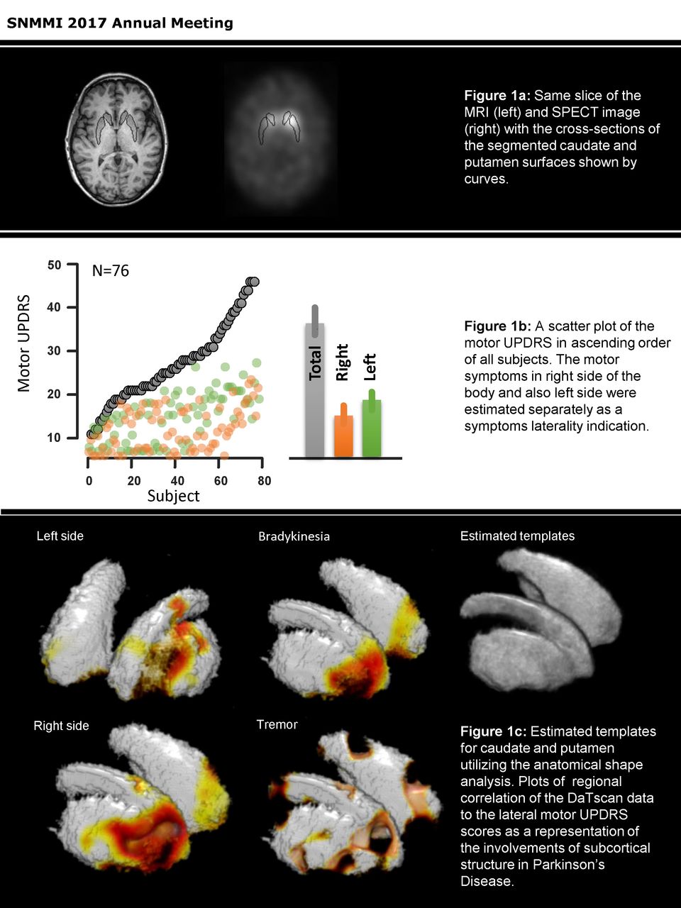
Objectives: To make use of SPECT imaging of Parkinson’s disease (PD), aligned to a common space, to investigate the correlation of functional neuroanatomy and regional contribution of subcortical structures with PD clinical phenotypes.

Methods: We analyzed 76 subjects with mild to severe PD and asymmetric motor symptom manifestation, based on the unified PD rating scale (UPDRS). A common coordinate system for the population was defined based on MR images and DAT SPECT intensities across subjects were compared. A multi stage surface registration algorithm was employed to arrive at a common standard space for all subjects. Image analysis included segmentation of the structures of interest in MRI, registration of SPECT images to MRI, and then overlaying of the structures segmented in MRI on the co-registered SPECT, followed by spatial normalization of the data to the standard space. Regional correlation using Pearson’s correlation was applied to SPECT intensities and clinical rating scales of disease symptoms to obtain the fine regional involvement in disease initiation and progression. The common template enabled us to explore and investigate regional effects of Parkinson’s Disease on subcortical structures such as caudate and putamen in relation to a variety of symptom subgroups such as gait, tremor, bradykinesia and rigidity.

Results: Our normalization methodology enabled fine regional analysis to generate new markers of disease initiation and progression. It allowed extraction of regional correlation of subcortical structures such as caudate and putamen with PD clinical features and introduced an indirect assessment of the level of regional neurodegeneration.

Conclusion: Effects of regional exploration on dopaminergic system in subcortical structures provides some evidences for regional nature of the neurodegeneration in Parkinson’s disease, leading toward more individualized and regional specific treatments such as stem cell treatment.

Figure
  • Download figure
  • Open in new tab
  • Download powerpoint
Previous
Back to top

In this issue

Journal of Nuclear Medicine
Vol. 58, Issue supplement 1
May 1, 2017
  • Table of Contents
  • Index by author
Article Alerts
Sign In to Email Alerts with your Email Address
Email Article

Thank you for your interest in spreading the word on Journal of Nuclear Medicine.

NOTE: We only request your email address so that the person you are recommending the page to knows that you wanted them to see it, and that it is not junk mail. We do not capture any email address.

Enter multiple addresses on separate lines or separate them with commas.
Regional correlation of subcortical structures with clinical phenotypes in Parkinson’s disease: DAT SPECT imaging approach
(Your Name) has sent you a message from Journal of Nuclear Medicine
(Your Name) thought you would like to see the Journal of Nuclear Medicine web site.
Citation Tools
Regional correlation of subcortical structures with clinical phenotypes in Parkinson’s disease: DAT SPECT imaging approach
Yousef Salimpour, Esmaeil Davoodi-Bojd, Sima Fotouhi, Hamid Soltanian-Zadeh, Sahar Mirpour, Rongkai Yan, Vesna Sossi, Arman Rahmim
Journal of Nuclear Medicine May 2017, 58 (supplement 1) 1277;

Citation Manager Formats

  • BibTeX
  • Bookends
  • EasyBib
  • EndNote (tagged)
  • EndNote 8 (xml)
  • Medlars
  • Mendeley
  • Papers
  • RefWorks Tagged
  • Ref Manager
  • RIS
  • Zotero
Share
Regional correlation of subcortical structures with clinical phenotypes in Parkinson’s disease: DAT SPECT imaging approach
Yousef Salimpour, Esmaeil Davoodi-Bojd, Sima Fotouhi, Hamid Soltanian-Zadeh, Sahar Mirpour, Rongkai Yan, Vesna Sossi, Arman Rahmim
Journal of Nuclear Medicine May 2017, 58 (supplement 1) 1277;
Twitter logo Facebook logo LinkedIn logo Mendeley logo
  • Tweet Widget
  • Facebook Like
  • Google Plus One
Bookmark this article

Jump to section

  • Article
  • Figures & Data
  • Info & Metrics

Related Articles

  • No related articles found.
  • Google Scholar

Cited By...

  • No citing articles found.
  • Google Scholar

More in this TOC Section

Neurosciences Track

  • Impact of cognitive reserve in frontotemporal dementia illustrated by FDG-PET.
  • Quantification of brain cholinergic denervation in dementia with Lewy bodies using PET imaging with 18F-FEOBV
  • Kinetic evaluation of [18F]MOZAT PET imaging in humans.
Show more Neurosciences Track

Neurology Posters

  • A new diagnostic marker for differentiating multi centric gliomas from multiple intracranial diffuse large B-cell lymphomas on 18F-FDG PET images
  • Technetium-99m pentavalent dimercaptosuccinic acid (99mTc-DMSA-V) brain SPECT/CT and Karnofsky performance score predict survival in patients with glioma
  • IDH1 mutation is associated with low FDG upatke in cerebral gliomas associated with low FDG uptake in cerebral gliomas.
Show more Neurology Posters

Similar Articles

SNMMI

© 2025 SNMMI

Powered by HighWire