
Main menu

  • Home
  • Content
    • Current
    • Ahead of print
    • Past Issues
    • JNM Supplement
    • SNMMI Annual Meeting Abstracts
    • Continuing Education
    • JNM Podcasts
  • Subscriptions
    • Subscribers
    • Institutional and Non-member
    • Rates
    • Journal Claims
    • Corporate & Special Sales
  • Authors
    • Submit to JNM
    • Information for Authors
    • Assignment of Copyright
    • AQARA requirements
  • Info
    • Reviewers
    • Permissions
    • Advertisers
  • About
    • About Us
    • Editorial Board
    • Contact Information
  • More
    • Alerts
    • Feedback
    • Help
    • SNMMI Journals
  • SNMMI
    • JNM
    • JNMT
    • SNMMI Journals
    • SNMMI

User menu

  • Subscribe
  • My alerts
  • Log in
  • My Cart

Search

  • Advanced search
Journal of Nuclear Medicine
  • SNMMI
    • JNM
    • JNMT
    • SNMMI Journals
    • SNMMI
  • Subscribe
  • My alerts
  • Log in
  • My Cart
Journal of Nuclear Medicine

Advanced Search

  • Home
  • Content
    • Current
    • Ahead of print
    • Past Issues
    • JNM Supplement
    • SNMMI Annual Meeting Abstracts
    • Continuing Education
    • JNM Podcasts
  • Subscriptions
    • Subscribers
    • Institutional and Non-member
    • Rates
    • Journal Claims
    • Corporate & Special Sales
  • Authors
    • Submit to JNM
    • Information for Authors
    • Assignment of Copyright
    • AQARA requirements
  • Info
    • Reviewers
    • Permissions
    • Advertisers
  • About
    • About Us
    • Editorial Board
    • Contact Information
  • More
    • Alerts
    • Feedback
    • Help
    • SNMMI Journals
  • View or Listen to JNM Podcast
  • Visit JNM on Facebook
  • Join JNM on LinkedIn
  • Follow JNM on Twitter
  • Subscribe to our RSS feeds
Meeting ReportNeurosciences Track

Application of texture analysis to DaTscan images for enhanced assessment of progression in Parkinson’s disease

Arman Rahmim, Yousef Salimpour, Saurabh Jain, Stephan Blinder, Ivan Klyuzhin, Gwenn Smith, Zoltan Mari and Vesna Sossi
Journal of Nuclear Medicine May 2016, 57 (supplement 2) 73;
Arman Rahmim
1Johns Hopkins University Baltimore MD United States
  • Find this author on Google Scholar
  • Find this author on PubMed
  • Search for this author on this site
Yousef Salimpour
1Johns Hopkins University Baltimore MD United States
  • Find this author on Google Scholar
  • Find this author on PubMed
  • Search for this author on this site
Saurabh Jain
1Johns Hopkins University Baltimore MD United States
  • Find this author on Google Scholar
  • Find this author on PubMed
  • Search for this author on this site
Stephan Blinder
2University of British Columbia Vancouver BC Canada
  • Find this author on Google Scholar
  • Find this author on PubMed
  • Search for this author on this site
Ivan Klyuzhin
2University of British Columbia Vancouver BC Canada
  • Find this author on Google Scholar
  • Find this author on PubMed
  • Search for this author on this site
Gwenn Smith
1Johns Hopkins University Baltimore MD United States
  • Find this author on Google Scholar
  • Find this author on PubMed
  • Search for this author on this site
Zoltan Mari
1Johns Hopkins University Baltimore MD United States
  • Find this author on Google Scholar
  • Find this author on PubMed
  • Search for this author on this site
Vesna Sossi
2University of British Columbia Vancouver BC Canada
  • Find this author on Google Scholar
  • Find this author on PubMed
  • Search for this author on this site
  • Article
  • Figures & Data
  • Info & Metrics
Loading

Abstract

73

Objectives Dopamine transporter (DAT) SPECT imaging is increasingly utilized for diagnostic purposes in suspected parkinsonian syndromes. Our objective was to enable additional clinical utility of this modality, beyond standard diagnostic tasks, to tracking of progression in Parkinson’s disease (PD). Specifically, we investigated whether assessment of texture in radiotracer uptake enabled enhanced correlations with severity of motor and cognitive symptoms in PD.

Methods Quantitative analysis in routine DAT SPECT imaging, if performed at all, has been restricted to assessment of mean regional uptake. We applied a framework wherein textural features were extracted from the images. Notably, the framework did not require registration to a common template, and worked in the subject-native space. Image analysis included registration of SPECT images onto corresponding MRI images, automatic region-of-interest (ROI) extraction on the MRI images, followed by computation of Haralick texture features. We analyzed n=116 subjects from the Parkinson’s Progressive Marker Initiative (PPMI) database, including 72 PD and 44 healthy controls (HC) (baseline scans with accompanying 3T MRI images, age < 70). We performed univariate and multivariate regression analyses between the quantitative metrics and different clinical measures, namely (i) the UPDRS (part III - motor) score, disease duration as measured from time of (ii) detection of symptoms (DD-sympt.) and (iii) diagnosis (DD-diag.), as well as (iv) the Montreal Cognitive Assessment (MoCA) score.

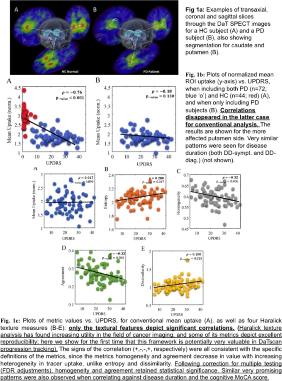
Results For conventional mean uptake analysis in the putamen, we showed significant correlations with clinical measures only when both HC and PD were included (Pearson correlation ρ=-0.76, p-value<0.001). However, this became insignificant when applied to PD subjects only (ρ=-0.18, p-value=0.13), and no such correlations were observed in the caudate. By contrast, for the PD subjects, significant correlations were observed in the caudate when including texture metrics, with (i) UPDRS (p-values<0.01), (ii) DD-sympt (p-values<0.01), (ii) DD-diag. (p-values<0.001), and (iv) MoCA (p-values<0.01), while no correlations were observed for conventional analysis (p-values=0.89, 0.14, 0.52 and 0.85, respectively).

Conclusions Our results demonstrated the ability to capture valuable information using advanced texture metrics from striatal DAT SPECT, enabling significant correlations of striatal DAT binding with clinical, motor and cognitive outcomes, and suggesting that textural features hold potential as biomarkers of PD symptomatology and progression. $$graphic_87EF1F68-967B-47A4-AB44-823D22387A9B$$

Figure
  • Download figure
  • Open in new tab
  • Download powerpoint
Previous
Back to top

In this issue

Journal of Nuclear Medicine
Vol. 57, Issue supplement 2
May 1, 2016
  • Table of Contents
  • Index by author
Article Alerts
Sign In to Email Alerts with your Email Address
Email Article

Thank you for your interest in spreading the word on Journal of Nuclear Medicine.

NOTE: We only request your email address so that the person you are recommending the page to knows that you wanted them to see it, and that it is not junk mail. We do not capture any email address.

Enter multiple addresses on separate lines or separate them with commas.
Application of texture analysis to DaTscan images for enhanced assessment of progression in Parkinson’s disease
(Your Name) has sent you a message from Journal of Nuclear Medicine
(Your Name) thought you would like to see the Journal of Nuclear Medicine web site.
Citation Tools
Application of texture analysis to DaTscan images for enhanced assessment of progression in Parkinson’s disease
Arman Rahmim, Yousef Salimpour, Saurabh Jain, Stephan Blinder, Ivan Klyuzhin, Gwenn Smith, Zoltan Mari, Vesna Sossi
Journal of Nuclear Medicine May 2016, 57 (supplement 2) 73;

Citation Manager Formats

  • BibTeX
  • Bookends
  • EasyBib
  • EndNote (tagged)
  • EndNote 8 (xml)
  • Medlars
  • Mendeley
  • Papers
  • RefWorks Tagged
  • Ref Manager
  • RIS
  • Zotero
Share
Application of texture analysis to DaTscan images for enhanced assessment of progression in Parkinson’s disease
Arman Rahmim, Yousef Salimpour, Saurabh Jain, Stephan Blinder, Ivan Klyuzhin, Gwenn Smith, Zoltan Mari, Vesna Sossi
Journal of Nuclear Medicine May 2016, 57 (supplement 2) 73;
Twitter logo Facebook logo LinkedIn logo Mendeley logo
  • Tweet Widget
  • Facebook Like
  • Google Plus One
Bookmark this article

Jump to section

  • Article
  • Figures & Data
  • Info & Metrics

Related Articles

  • No related articles found.
  • Google Scholar

Cited By...

  • No citing articles found.
  • Google Scholar

More in this TOC Section

Neurosciences Track

  • Impact of cognitive reserve in frontotemporal dementia illustrated by FDG-PET.
  • Quantification of brain cholinergic denervation in dementia with Lewy bodies using PET imaging with 18F-FEOBV
  • Kinetic evaluation of [18F]MOZAT PET imaging in humans.
Show more Neurosciences Track

Neurosciences/Neurology. Motor neuron diseases

  • Diagnostic performance of the visual reading of 123I-ioflupane DaT SPECT images when assessed with or without quantification in patients with movement disorders or dementia
  • Subregional patterns of preferential striatal DAT loss differ in PD, MSA and PSP
  • Longitudinal Decline of Striatal Subregional F-18 FP-CIT Uptake in Parkinson’s Disease
Show more Neurosciences/Neurology. Motor neuron diseases

Similar Articles

SNMMI

© 2025 SNMMI

Powered by HighWire