
Main menu

  • Home
  • Content
    • Current
    • Ahead of print
    • Past Issues
    • JNM Supplement
    • SNMMI Annual Meeting Abstracts
    • Continuing Education
    • JNM Podcasts
  • Subscriptions
    • Subscribers
    • Institutional and Non-member
    • Rates
    • Journal Claims
    • Corporate & Special Sales
  • Authors
    • Submit to JNM
    • Information for Authors
    • Assignment of Copyright
    • AQARA requirements
  • Info
    • Reviewers
    • Permissions
    • Advertisers
  • About
    • About Us
    • Editorial Board
    • Contact Information
  • More
    • Alerts
    • Feedback
    • Help
    • SNMMI Journals
  • SNMMI
    • JNM
    • JNMT
    • SNMMI Journals
    • SNMMI

User menu

  • Subscribe
  • My alerts
  • Log in
  • My Cart

Search

  • Advanced search
Journal of Nuclear Medicine
  • SNMMI
    • JNM
    • JNMT
    • SNMMI Journals
    • SNMMI
  • Subscribe
  • My alerts
  • Log in
  • My Cart
Journal of Nuclear Medicine

Advanced Search

  • Home
  • Content
    • Current
    • Ahead of print
    • Past Issues
    • JNM Supplement
    • SNMMI Annual Meeting Abstracts
    • Continuing Education
    • JNM Podcasts
  • Subscriptions
    • Subscribers
    • Institutional and Non-member
    • Rates
    • Journal Claims
    • Corporate & Special Sales
  • Authors
    • Submit to JNM
    • Information for Authors
    • Assignment of Copyright
    • AQARA requirements
  • Info
    • Reviewers
    • Permissions
    • Advertisers
  • About
    • About Us
    • Editorial Board
    • Contact Information
  • More
    • Alerts
    • Feedback
    • Help
    • SNMMI Journals
  • View or Listen to JNM Podcast
  • Visit JNM on Facebook
  • Join JNM on LinkedIn
  • Follow JNM on Twitter
  • Subscribe to our RSS feeds
Meeting ReportInstrumentation & Data Analysis Track

PSF Overestimation Improves PET Image SUV Quantification

Saeed Ashrafinia, Hassan Mohy-ud-Din, NICOLAS KARAKATSANIS, Michael Casey, Martin Lodge and Arman Rahmim
Journal of Nuclear Medicine May 2016, 57 (supplement 2) 1965;
Saeed Ashrafinia
2Radiology and Radiological Sciences Johns Hopkins University School of Medicine Baltimore MD United States
  • Find this author on Google Scholar
  • Find this author on PubMed
  • Search for this author on this site
Hassan Mohy-ud-Din
5Yale School of Medicine Yale University New Haven CT United States
  • Find this author on Google Scholar
  • Find this author on PubMed
  • Search for this author on this site
NICOLAS KARAKATSANIS
4Translational and Molecular Imaging Institute New York NY United States
  • Find this author on Google Scholar
  • Find this author on PubMed
  • Search for this author on this site
Michael Casey
3Siemens Medical Solutions Knoxville TN United States
  • Find this author on Google Scholar
  • Find this author on PubMed
  • Search for this author on this site
Martin Lodge
1Johns Hopkins University Baltimore MD United States
  • Find this author on Google Scholar
  • Find this author on PubMed
  • Search for this author on this site
Arman Rahmim
1Johns Hopkins University Baltimore MD United States
  • Find this author on Google Scholar
  • Find this author on PubMed
  • Search for this author on this site
  • Article
  • Figures & Data
  • Info & Metrics
Loading

Abstract

1965

Objectives Point spread function (PSF) modeled PET image reconstruction (also known as resolution modeling) improves resolution and contrast, yet may degrade reproducibility and depict edge artifacts (reminiscent of Gibbs ringing). This study investigates the effect of incorporating varying PSF kernels, enabling under- and overestimation of the true PSF, for the potential of enhanced quantitative SUV estimation.

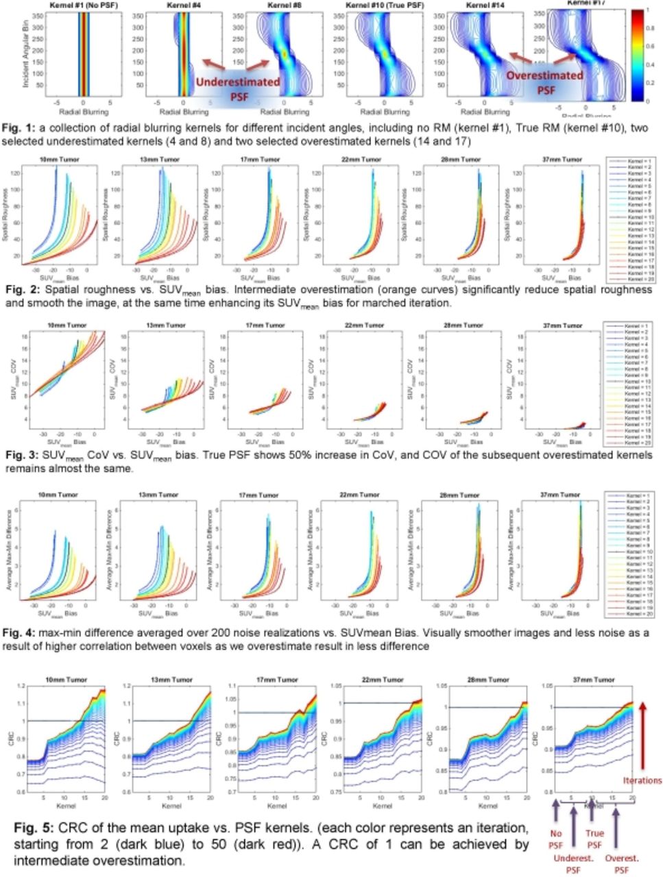
Methods In the context of oncologic FDG PET, we simulated 60min post-injection SUV images of XCAT anthropomorphic phantom (3min/bed) separately including six liver tumors of varying diameters (10, 13, 17, 22, 28 and 37mm). This was based on clinically estimated kinetic parameters from which time-activity curves were generated, followed by temporal integration. Normalization, attenuation, decay correction, and resolution degrading phenomena (including photon non-collinearity, inter-crystal penetration and scattering), as well as clinically realistic noise levels (100 realizations) were incorporated. We introduced controllable scaling factors to generate 20 analytically modeled blurring kernels (incorporating abovementioned physical phenomena with additional scaling in extent), including no/full PSF modeling, as well as 8 underestimated and 10 overestimated kernels. For performance comparison, we performed threshold-based tumor segmentation, and subsequently assessed contrast recovery coefficient (CRC), SUVmean bias, noise (spatial roughness, SUVmean and SUVmax coefficient-of-variability (CoV)), and averaged difference of max and min uptakes in every tumor.

Results For overestimated kernels, CRC was significantly improved, and noise (SUVmean CoV, SUVmax CoV and spatial roughness) vs. bias curves were also typically improved. The latter is because overestimated PSF kernels yield increased inter-voxel correlations, resulting in visually smoothed images and significantly lower spatial roughness (25% less for PSF vs. No PSF, and 43% less for overestimated PSF vs. no PSF, for clinically realistic number of iterations in each method used throughout this work). Furthermore, PSF modeling edge overshoots actually reduced SUVmean bias for the three smallest tumor (10% for true PSF and 15-20% for mid overestimated PSF), and resulted in comparable SUVmean performance in larger tumors. SUVmean CoV (as measure of reproducibility) for smallest tumors had a limited (5-10%) increase compared to true PSF, which was offset by more significant reductions in bias when overestimating. Averaged max-min difference as well as spatial roughness, used as substitutes for visual uniformity assessment (impacted by both presence of noise and edge artifacts), were actually reduced by 35% and 50% for true PSF and overestimated PSF in all six regions, due to overriding reduction in noise relative to enhancements in edge overshoots.

Conclusions Overestimated PSF yields significantly higher contrast for a range of tumors. For reasonable (not excessive) number of iterations, edge enhancement counterintuitively lowers SUVmean bias in small tumors, while inter-voxel correlations suppressing image roughness, and enhance uniformity, in all tumors, only slightly degrading SUVmean reproducibility in the smallest tumors. These results provide perspective on performance of vendor PET images, as some measure the PSF utilizing Ge-68 point sources (that have higher positron range than F18 as commonly used in the clinic), thus implicitly implementing overestimated PSF kernels. This, we suggest, may be enabling enhanced SUV quantitation, while we are exploring impact on measures of heterogeneity. Overall, we propose that overestimated PSF kernels can be pursued as powerful and viable choices in quantitative task-based optimization including prognostication and treatment response assessment.

Figure
  • Download figure
  • Open in new tab
  • Download powerpoint
Previous
Back to top

In this issue

Journal of Nuclear Medicine
Vol. 57, Issue supplement 2
May 1, 2016
  • Table of Contents
  • Index by author
Article Alerts
Sign In to Email Alerts with your Email Address
Email Article

Thank you for your interest in spreading the word on Journal of Nuclear Medicine.

NOTE: We only request your email address so that the person you are recommending the page to knows that you wanted them to see it, and that it is not junk mail. We do not capture any email address.

Enter multiple addresses on separate lines or separate them with commas.
PSF Overestimation Improves PET Image SUV Quantification
(Your Name) has sent you a message from Journal of Nuclear Medicine
(Your Name) thought you would like to see the Journal of Nuclear Medicine web site.
Citation Tools
PSF Overestimation Improves PET Image SUV Quantification
Saeed Ashrafinia, Hassan Mohy-ud-Din, NICOLAS KARAKATSANIS, Michael Casey, Martin Lodge, Arman Rahmim
Journal of Nuclear Medicine May 2016, 57 (supplement 2) 1965;

Citation Manager Formats

  • BibTeX
  • Bookends
  • EasyBib
  • EndNote (tagged)
  • EndNote 8 (xml)
  • Medlars
  • Mendeley
  • Papers
  • RefWorks Tagged
  • Ref Manager
  • RIS
  • Zotero
Share
PSF Overestimation Improves PET Image SUV Quantification
Saeed Ashrafinia, Hassan Mohy-ud-Din, NICOLAS KARAKATSANIS, Michael Casey, Martin Lodge, Arman Rahmim
Journal of Nuclear Medicine May 2016, 57 (supplement 2) 1965;
Twitter logo Facebook logo LinkedIn logo Mendeley logo
  • Tweet Widget
  • Facebook Like
  • Google Plus One
Bookmark this article

Jump to section

  • Article
  • Figures & Data
  • Info & Metrics

Related Articles

  • No related articles found.
  • Google Scholar

Cited By...

  • No citing articles found.
  • Google Scholar

More in this TOC Section

Instrumentation & Data Analysis Track

  • Deep Learning Based Kidney Segmentation for Glomerular Filtration Rate Measurement Using Quantitative SPECT/CT
  • Comparison of 22 partial volume correction methods for amyloid PET imaging with 11C-PiB
  • The Benefit of Time-of-Flight in Digital Photon Counting PET Imaging: Physics and Clinical Evaluation
Show more Instrumentation & Data Analysis Track

MTA II: Image Generation Posters

  • Clinical visual readings of brain region-specific hypometabolism in cognitive impairment patients is independent of attenuation correction method for integrated PET/MR
  • Respiratory gating and phase-matched attenuation correction improves uniformity of regional myocardial blood flow estimates during hypercapnea-induced stress imaging with dynamic rubidium-82 PET
  • Patient specific optimisation of administered activity and acquisition times for F-18 FDG PET imaging
Show more MTA II: Image Generation Posters

Similar Articles

SNMMI

© 2025 SNMMI

Powered by HighWire