
Main menu

  • Home
  • Content
    • Current
    • Ahead of print
    • Past Issues
    • JNM Supplement
    • SNMMI Annual Meeting Abstracts
    • Continuing Education
    • JNM Podcasts
  • Subscriptions
    • Subscribers
    • Institutional and Non-member
    • Rates
    • Journal Claims
    • Corporate & Special Sales
  • Authors
    • Submit to JNM
    • Information for Authors
    • Assignment of Copyright
    • AQARA requirements
  • Info
    • Reviewers
    • Permissions
    • Advertisers
  • About
    • About Us
    • Editorial Board
    • Contact Information
  • More
    • Alerts
    • Feedback
    • Help
    • SNMMI Journals
  • SNMMI
    • JNM
    • JNMT
    • SNMMI Journals
    • SNMMI

User menu

  • Subscribe
  • My alerts
  • Log in
  • My Cart

Search

  • Advanced search
Journal of Nuclear Medicine
  • SNMMI
    • JNM
    • JNMT
    • SNMMI Journals
    • SNMMI
  • Subscribe
  • My alerts
  • Log in
  • My Cart
Journal of Nuclear Medicine

Advanced Search

  • Home
  • Content
    • Current
    • Ahead of print
    • Past Issues
    • JNM Supplement
    • SNMMI Annual Meeting Abstracts
    • Continuing Education
    • JNM Podcasts
  • Subscriptions
    • Subscribers
    • Institutional and Non-member
    • Rates
    • Journal Claims
    • Corporate & Special Sales
  • Authors
    • Submit to JNM
    • Information for Authors
    • Assignment of Copyright
    • AQARA requirements
  • Info
    • Reviewers
    • Permissions
    • Advertisers
  • About
    • About Us
    • Editorial Board
    • Contact Information
  • More
    • Alerts
    • Feedback
    • Help
    • SNMMI Journals
  • View or Listen to JNM Podcast
  • Visit JNM on Facebook
  • Join JNM on LinkedIn
  • Follow JNM on Twitter
  • Subscribe to our RSS feeds
Meeting ReportCardiovascular

Quantitative myocardial perfusion PET parametric imaging at the voxel-level

Hassan Mohy-ud-Din, Martin Lodge and Arman Rahmim
Journal of Nuclear Medicine May 2015, 56 (supplement 3) 1518;
Hassan Mohy-ud-Din
1Electrical and Computer Engineering, Johns Hopkins University, Baltimore, MD
  • Find this author on Google Scholar
  • Find this author on PubMed
  • Search for this author on this site
Martin Lodge
2Radiology and Radiological Sciences, Johns Hopkins University, Baltimore, MD
  • Find this author on Google Scholar
  • Find this author on PubMed
  • Search for this author on this site
Arman Rahmim
2Radiology and Radiological Sciences, Johns Hopkins University, Baltimore, MD
  • Find this author on Google Scholar
  • Find this author on PubMed
  • Search for this author on this site
  • Article
  • Figures & Data
  • Info & Metrics
Loading

Abstract

1518

Objectives To assess performance of a novel approach to obtain quantitative parametric images in cardiac PET.

Methods We have proposed a parametric PET image formation method utilizing spatial constraints via what we term physiological clustering (PC): this iterative kinetic-driven-clustering method observes that ideal clustering depends on underlying physiology of functional regions, and in turn, physiological processes are quantified by kinetic parameter estimation. 30 noise-realizations of Rb-82 datasets were generated based on realistic kinetic parameters and input function. Transmural/non-transmural perfusion defects were simulated. We also validated our approach on n=5 Rb-82 patient studies. Voxel-wise kinetic parameters were obtained using (i) conventional post-smoothing (PS) on reconstructed images (Butterworth, order 4, cut-off 0.2-1 cycles/cm), and (ii) PC, where regression was performed on non-smoothed images via a quadratic penalty that penalizes deviations from mean kinetics as computed using PC of dynamic images.

Results Enhanced noise-bias performance was obtained for both transmural (T) and non-transmural (NT) defects. Specifically, for T, at optimal performance (minimum RMSE), PS reduced noise by 27% with 3.9% increase in bias (38% RMSE reduction). PC reduced noise by 33% with 1.2% increase in bias (46% RMSE reduction). For NT, PS reduced noise by 28% with 4.7% increase in bias (37% RMSE reduction). PC reduced noise by 33.4% with 3.0% increase in bias (47% RMSE reduction). PC, compared to PS, resulted in higher CNR (13.7 as opposed to 7.2 for T; and 11.3 as opposed to 5.7 for NT) at optimal performance. In patient studies, PC resulted in significantly higher CNR at matched K1 value (x 2 improvement at converged values).

Conclusions Parametric imaging based on PC clearly outperformed conventional estimation method in transmural and non-transmural perfusion defects.

Research Support 2014 Bradley-Alavi Fellowship, SNMMI

Figure
  • Download figure
  • Open in new tab
  • Download powerpoint

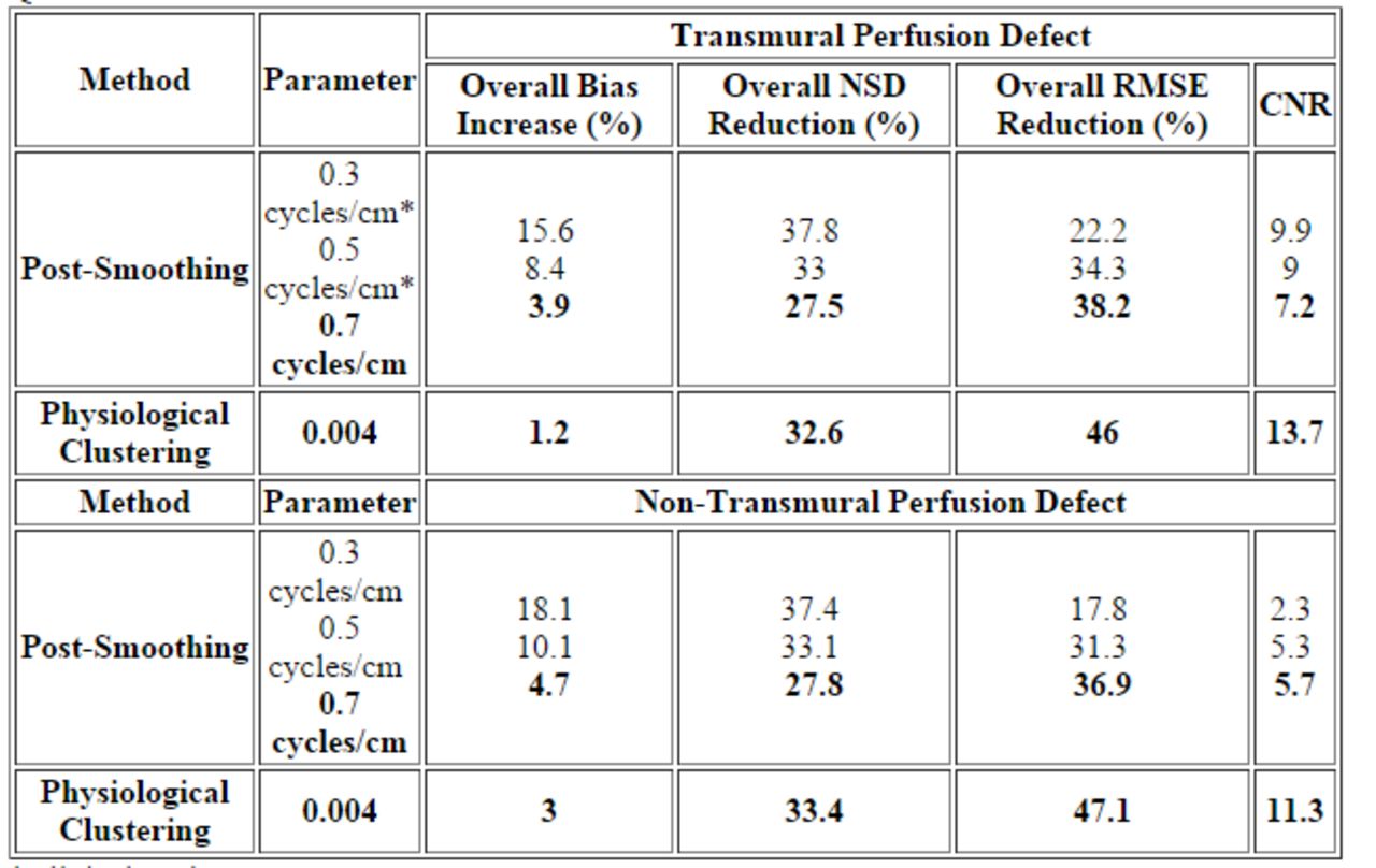
Quantitative Performance

* clinical setting

Previous
Back to top

In this issue

Journal of Nuclear Medicine
Vol. 56, Issue supplement 3
May 1, 2015
  • Table of Contents
  • Index by author
Article Alerts
Sign In to Email Alerts with your Email Address
Email Article

Thank you for your interest in spreading the word on Journal of Nuclear Medicine.

NOTE: We only request your email address so that the person you are recommending the page to knows that you wanted them to see it, and that it is not junk mail. We do not capture any email address.

Enter multiple addresses on separate lines or separate them with commas.
Quantitative myocardial perfusion PET parametric imaging at the voxel-level
(Your Name) has sent you a message from Journal of Nuclear Medicine
(Your Name) thought you would like to see the Journal of Nuclear Medicine web site.
Citation Tools
Quantitative myocardial perfusion PET parametric imaging at the voxel-level
Hassan Mohy-ud-Din, Martin Lodge, Arman Rahmim
Journal of Nuclear Medicine May 2015, 56 (supplement 3) 1518;

Citation Manager Formats

  • BibTeX
  • Bookends
  • EasyBib
  • EndNote (tagged)
  • EndNote 8 (xml)
  • Medlars
  • Mendeley
  • Papers
  • RefWorks Tagged
  • Ref Manager
  • RIS
  • Zotero
Share
Quantitative myocardial perfusion PET parametric imaging at the voxel-level
Hassan Mohy-ud-Din, Martin Lodge, Arman Rahmim
Journal of Nuclear Medicine May 2015, 56 (supplement 3) 1518;
Twitter logo Facebook logo LinkedIn logo Mendeley logo
  • Tweet Widget
  • Facebook Like
  • Google Plus One
Bookmark this article

Jump to section

  • Article
  • Figures & Data
  • Info & Metrics

Related Articles

  • No related articles found.
  • Google Scholar

Cited By...

  • No citing articles found.
  • Google Scholar

More in this TOC Section

Cardiovascular

  • Cardiac β-Adrenergic Receptor Downregulation, Evaluated by Cardiac PET, in Chronotropic Incompetence
  • Diagnostic Performance of PET Versus SPECT Myocardial Perfusion Imaging in Patients with Smaller Left Ventricles: A Substudy of the 18F-Flurpiridaz Phase III Clinical Trial
  • Quantification of Macrophage-Driven Inflammation During Myocardial Infarction with 18F-LW223, a Novel TSPO Radiotracer with Binding Independent of the rs6971 Human Polymorphism
Show more Cardiovascular

MTA II: Cardiovascular Clinical Science Posters

  • Value of FDG-PET/CT and cardiac MR for the diagnosis and therapy monitoring in cardiac sarcoidosis.
  • Atherosclerosis progression in well controlled HIV infected patients who are addicted to cocaine
  • Diagnostic Accuracy of low dose thallium-201 myocardial perfusion imaging with cadmium-zinc-telluride camera compared to coronary angiography and fractional flow reserve
Show more MTA II: Cardiovascular Clinical Science Posters

Similar Articles

SNMMI

© 2025 SNMMI

Powered by HighWire