


Abstract
Targeted α-particle radiation using the radioisotope 225Ac is a promising form of therapy for various types of cancer. Historic obstacles to the use of 225Ac have been the difficulty in finding suitable chelators to stably attach it to targeting vehicles such as peptides and monoclonal antibodies, the low specific activities of the products, and the lack of cost-effective radiolabeling procedures. We initially solved the first problem with a procedure involving 2 chemical steps that has been used as a standard in preclinical and clinical studies. However, this procedure involves the loss of 90% of the input 225Ac. A more efficient, economical process is needed to facilitate the more widespread use of 225Ac. Methods: We conjugated representative antibodies with 2 forms of DOTA as well as other chelators as controls. We developed conditions to radiolabel these constructs in 1 chemical step and characterized their stability, immunoreactivity, biodistribution, and therapeutic efficacy in healthy and tumor-bearing mice. Results: DOTA–antibody constructs were labeled to a wide range of specific activities in 1 chemical step at 37°C. Radiochemical yields were approximately 10-fold higher, and specific activities were up to 30-fold higher than with the previous approach. The products retained immunoreactivity and were stable to serum challenge in vitro and in mice. Labeling kinetics of DOTA–antibody constructs linked through a benzyl isothiocyanate linkage were more favorable than those linked through an N-hydroxysuccinimide linkage. Tissue distribution was similar but not identical between the constructs. The constructs produced specific therapeutic responses in a mouse model of acute myeloid leukemia. Conclusion: We have characterized an efficient, 1-step radiolabeling method that produces stable, therapeutically active conjugates of antibodies with 225Ac at high specific activity. We propose that this technology greatly expands the possible clinical applications of 225Ac monoclonal antibodies.
Radionuclides that emit α particles are promising agents for anticancer therapy, as evidenced by the recent approval by Food and Drug Administration of 223Ra (Xofigo; Bayer) for castration-resistant prostate cancer with bone metastases (1). Because of the high energy (5–8 MeV) and short path length (50–80 μm) of α particles, they have the potential to effectively and selectively target single cells, residual disease, and micrometastatic lesions. Our laboratory has focused on the α-particle generator 225Ac because of its 10-d half-life—which is well suited to the time needed for radiolabeling, injection, and tumor targeting—and the release of 4 net α particles per atom of 225Ac—which delivers massive toxicity to target cells (2).
Early work with 225Ac was limited by difficulty attaching it to targeting vehicles such as peptides and monoclonal antibodies, the low specific activity achievable by the products, and the lack of a cost-effective labeling strategy. Various chelators were investigated, with many failing to chelate the metal at all and others appearing to radiolabel but then releasing 225Ac when subjected to serum challenge (3,4). After testing various additional chelating strategies, our laboratory achieved stable labeling with the chelator DOTA using a procedure in 2 chemical steps that was designed to minimize radiolysis and maximize kinetic stability of the products (5,6). This procedure has since been used as a standard in several successful preclinical studies (7–9) and is currently in human clinical trials in the form of 225Ac-HuM195 to treat advanced myeloid leukemias (10). A major drawback to our 2-step labeling approach is that approximately 90% of the input actinium is conjugated to nonreactive forms of DOTA in the first step of the procedure and is consequently discarded. Because 225Ac is a rare and expensive isotope, a more efficient procedure for preparing actinium–antibody constructs is necessary to promote the more widespread use of these agents. Additionally, the low specific activity currently available limits the type of cellular targets that can be attacked.
The direct 1-step labeling of preformed antibody–DOTA constructs is a potential solution to the above problems but was previously thought to be infeasible at temperatures low enough to be compatible with monoclonal antibodies (5,6). 1-step labelings of peptide–DOTA constructs with 225Ac have been reported (11,12), but they were performed at temperatures of 70°C or higher. In this work, we present a new labeling method in 1 step at 37°C that achieves up to 10-fold-higher radiochemical yield and 30-fold-higher specific activity; demonstrate that the products are stable in vitro and in vivo; and evaluate biodistribution and therapeutic potential of the constructs in healthy and tumor-bearing mice.
MATERIALS AND METHODS
Radionuclides, Reagents, and Monoclonal Antibodies
225Ac was received from Oak Ridge National Laboratory as a nitrate residue, which was dissolved in 0.2 M Optima grade hydrochloric acid (HCl, Fisher Scientific) before use. We measured 225Ac activity using a CRC-15R radioisotope calibrator (Capintec, Inc.) set at 775 and multiplied the displayed activity value by 5. The parent 225Ac was measured when it was in secular equilibrium with its daughters, at least 6 h and typically the next day after sample collection.
The chelating agent DOTA and the bifunctional ligands 2-(4-isothiocyanatobenzyl)-1,4,7,10-tetraazacyclododecane-1,4,7,10-tetraacetic acid (p-SCN-Bn-DOTA) and 2-(4-isothiocyanatobenzyl)-diethylenetriaminepentaacetic acid (p-SCN-Bn-DTPA) were obtained from Macrocyclics. The structures of the DOTA chelating agents and controls are shown in Figure 1, and abbreviated names for the constructs are explained in Table 1 and Figure 2.
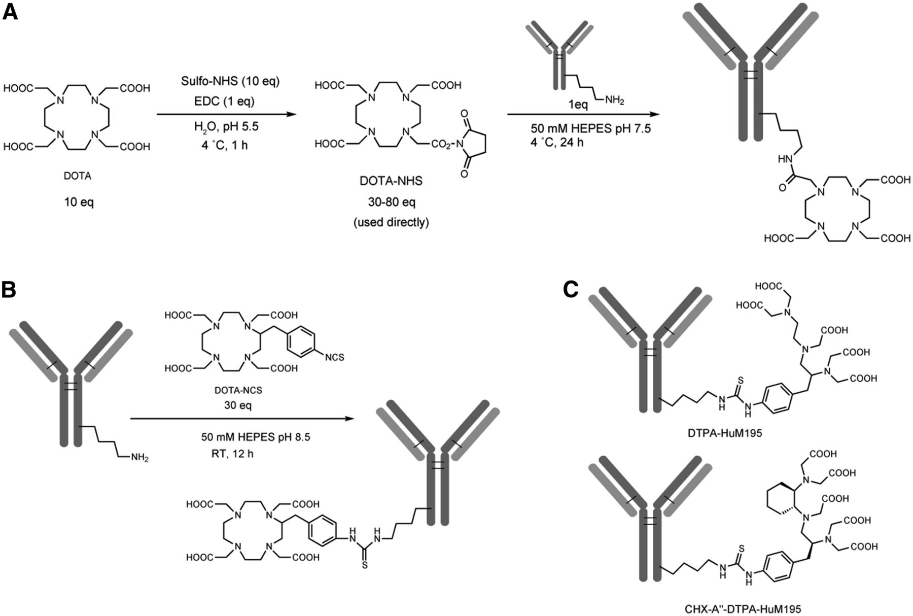
Antibody–chelate constructs for 1-step labeling. (A) Synthesis of 3-arm antibody constructs. (B) Synthesis of 4-arm antibody constructs. (C) Structures of control constructs. EDC = 1-ethyl-3-(3-dimethylaminopropyl) carbodiimide HCl; RT = room temperature.
Statistics on Conjugation of Antibody Constructs
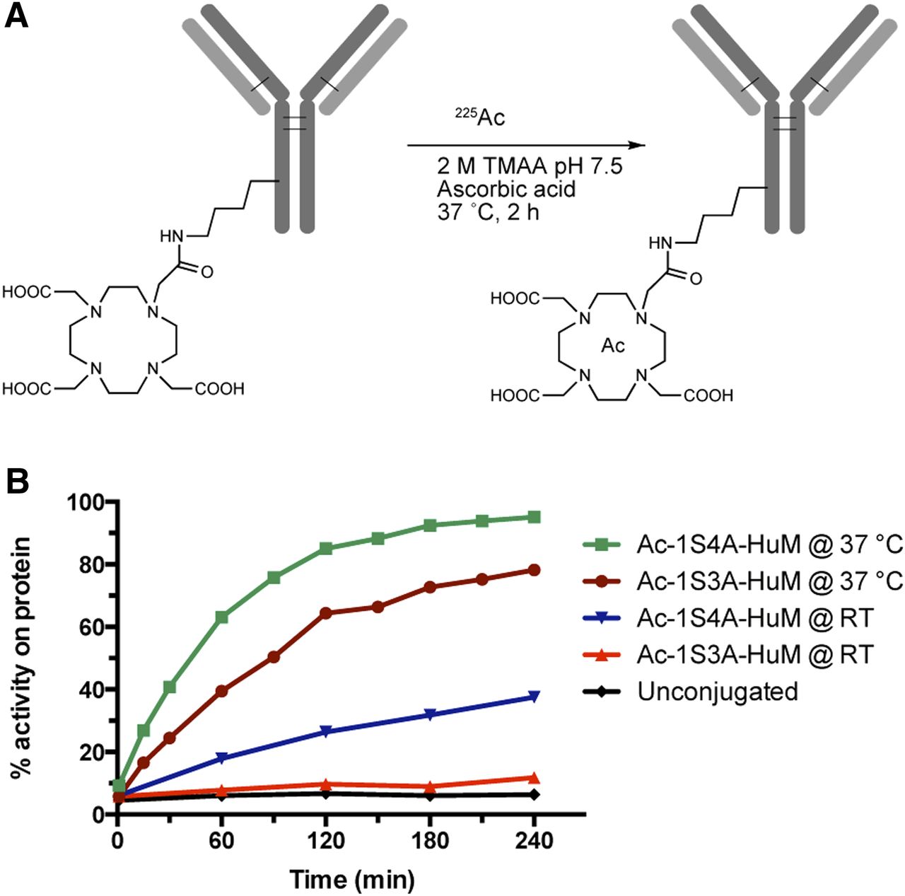
3-arm and 4-arm constructs can be radiolabeled in 1 step at 37°C. (A) Radiolabeling conditions. (B) Time course of labeling at different temperatures. Ac-1S4A-HuM = 225Ac 1-step labeled 4-arm HuM195 construct; RT = room temperature; TMAA = tetramethyl ammonium acetate.
Chemicals used in the conjugation, radiolabeling, and purification steps were American Chemical Society reagent grade or better. Water and buffers were rendered metal-free by passing them through a column of Chelex-100 resin, 200–400 mesh (Bio-Rad Laboratories, Inc.), and were sterile-filtered through a 0.22- or 0.45-μM filter device.
The monoclonal antibodies used were HuM195/Lintuzumab/anti-CD33 (Protein Design Labs) and Rituximab/anti-CD20 (Genentech). The preformed CHX-A″-DTPA–HuM195 construct was obtained from TSI Washington for our previous studies with 213Bi (13).
Synthesis, Purification, and Quality Control of Antibody–Chelate Constructs
The conjugation and radiolabeling procedures were performed using sterile and pyrogen-free plastic ware (Corning, Inc., and Fisher Scientific) and metal-free pipette tips (BioRad Laboratories, Inc.).
Monoclonal antibodies (5–10 mg in 0.5–2 mL of phosphate-buffered saline) were transferred to 15-mL Vivaspin Centrifugal Concentrators with a 10,000-kD molecular weight cutoff (Sartorius Corp.). To render the antibodies metal-free, the Vivaspins were filled to 15 mL with 20 mM (1%) DTPA and allowed to sit at 4°C overnight. The antibody buffer was then exchanged to 50 mM 4-(2-hydroxyethyl)-1-piperazineethanesulfonic acid (HEPES), pH 7.5, by 3 complete rounds of concentration and subsequent dilution. The products were transferred to 1.8-mL Nunc cryovials (Fisher Scientific) at a concentration of greater than 1 mg/mL and subjected to reaction conditions as detailed below.
To form the 3-arm DOTA constructs, we first generated the DOTA N-hydroxysuccinimidyl (NHS) ester in situ using a standard procedure (14,15); full details are in the Supplemental Methods (supplemental materials are available at http://jnm.snmjournals.org). An appropriate amount of activated NHS ester (30–80 equivalents [eq]) was added to the antibody (1 eq) in HEPES buffer at 4°C, and the pH was readjusted to 7.5 by adding sodium hydroxide (NaOH). The reaction was allowed to proceed for 24 h at 4°C. The resulting product was purified via buffer exchange to 20 mM sodium acetate (NaOAc), 150 mM sodium chloride (NaCl), through multiple passes through a Vivaspin 15 centrifugal concentrator as detailed before.
For formation of the 4-arm DOTA constructs and DTPA construct, p-SCN-Bn-DOTA or p-SCN-Bn-DTPA (20–30 eq) dissolved in water (40 mg/mL solution) was added to metal-free antibody (1 eq) in HEPES buffer, prepared via centrifugal concentration as described above. The pH was adjusted to 8.5 by adding NaOH. The reaction was allowed to proceed at room temperature for 12 h, and the product was purified via buffer exchange to 20 mM NaOAc, 150 mM NaCl, through multiple passes through a Vivaspin 15 centrifugal concentrator. Procedures for the quality control of antibody constructs are detailed in the Supplemental Methods.
Radiolabeling Procedures
Our 2-step procedure was performed as previously reported (6).
In a typical 1-step procedure, 225Ac-nitrate (3.7 MBq) dissolved in 0.2 M HCl was added to a 1.0-mL Nunc vial, and the activity was determined exactly using a dose calibrator. To this were added 2 M tetramethyl ammonium acetate buffer (25 μL), l-ascorbic acid (150 g/L; 10 μL), and the appropriate antibody construct (100 μg). The pH of the reaction was determined by spotting 1 μL of the reaction mixture onto Hydrion pH paper (range, 5.0–9.0) (Sigma-Aldrich); pH of a typical reaction was 5.8. The reaction vessel was transferred to a water bath displaying 37.0°C, and the reaction was allowed to proceed for 2 h unless specifically noted. After this, a small aliquot was spotted on a strip of instant thin-layer chromatography (ITLC) silica gel paper to determine the extent of incorporation of actinium onto protein (details are provided in the Supplemental Methods). The reaction was then quenched with 50 mM DTPA (20 μL) and purified using an Econo-Pac 10DG desalting column (Bio-Rad) that had been equilibrated previously with 1% human serum albumin. The product was eluted in approximately 2 mL of 1% human serum albumin and analyzed by ITLC to determine the radiochemical purity.
For the control constructs, the CHX-A″-DTPA construct was radiolabeled and purified with the 1-step procedure. For both the DTPA construct and the unmodified antibody, reactions were not quenched or purified because this would remove a large portion of the free 225Ac. Rather, they were diluted to an approximate final volume of 2 mL with 1% human serum albumin.
Additional procedures for the quality control and stability in vitro of radioimmunoconjugates are listed in the Supplemental Methods.
Animal Studies
A detailed description of the animal studies is provided in the Supplemental Methods. All animal studies were approved by the Institutional Animal Care and Use Committee of Memorial Sloan-Kettering Cancer Center under protocol 96-11-044.
Statistical Analysis
Data were graphed using GraphPad Prism (GraphPad Software Inc.). Unless specifically noted, values reported represent mean ± SD. Statistical comparisons between the experimental groups were performed either via the Student t test with Welch correction (2-group comparison) or via 1-way ANOVA with Bonferroni multiple comparison post hoc test (multiple-group comparison). P values were calculated using GraphPad Prism, with a P value of less than 0.05 considered significant.
RESULTS
Formation of Antibody–Chelate Constructs
We generated constructs of antibodies attached to several different chelating moieties using 2 attachment chemistries. These included 3-arm DOTA constructs, in which 1 of the 4 carboxylic acids of DOTA is used to attach to antibody lysines via N-hydroxysuccinimide chemistry (Fig. 1A), and 4-arm DOTA constructs, in which a benzyl isothiocyanate group attaches to antibody lysines, leaving all 4 carboxylic acids free (Fig. 1B). As controls, we generated antibody constructs with DTPA, which previous reports indicated would not chelate 225Ac (6), and CHX-A″-DTPA, which was reported to chelate 225Ac weakly during the labeling but release the metal on serum challenge (Fig. 1C) (3). Antibodies were conjugated to 2 or more different substitution ratios, and we used constructs with about 10 DOTAs per antibody for future assays. Table 1 lists data on the conjugation of 2 representative antibodies as well as abbreviated names that will be used throughout the rest of the text.
Radiolabeling, Quality Control, and Stability In Vitro
3-arm and 4-arm constructs were radiolabeled to specific activities of approximately 5–7 GBq of protein per gram using conditions shown in Figure 2A. The kinetics of labeling were determined through periodic ITLC of aliquots of the reactions (Fig. 2B). Surprisingly, the 4-arm construct appeared to radiolabel more quickly than the 3-arm construct, with approximately 95% of the activity incorporated onto protein after 4 h as compared with only 78% for the 3-arm construct. Both constructs labeled more slowly at room temperature than at 37°C. For convenience, we decided to radiolabel for only 2 h for future studies.
In a separate experiment, constructs were radiolabeled to a range of specific activities using a 2-h procedure (Table 2). The radiochemical purity of the products was good to excellent, except for the high-specific-activity 3A-HuM labeling, which had too much free 225Ac left over to remove with the 10DG column. The limit of specific activity that could be achieved with the 2-h procedure was about 29.6 GBq/g for the 3-arm construct and about 129 GBq/g for the 4-arm construct. Immunoreactivity for both constructs toward CD33-positive Set2-Luc cells decreased slightly as the amount of 225Ac in the reaction increased, whereas the immunoreactivity toward CD33-negative Ramos cells was negligible in all cases. The sham-labeled construct showed a small amount of background accumulation (∼7%) on both positive and negative cells.
Data from Representative 2-Hour Radiolabelings
Radiolabeled 3-arm and 4-arm constructs and controls were exposed to 90% human serum at 37°C in vitro, challenged with excess DTPA to remove any weakly bound 225Ac, and assayed for actinium remaining on protein by ITLC (Fig. 3A). From 95% to 97% of the 225Ac remained on the constructs after 25 d. By contrast, 225Ac from the unpurified reactions of DTPA construct and unmodified HuM195 did not appear to bind to protein strongly enough to overcome DTPA challenge at any time point. As expected, the CHX-A″-DTPA construct initially bound 225Ac but then released it over time.

Both 3-arm and 4-arm constructs labeled with 1 step are stable to serum challenge at 37°C. (A) Assay in vitro with ITLC to determine percentage actinium on protein. (B) Assay of protein G binding of serum harvested from female BALB/c mice at specified time points. T = 0 is uninjected material. All data are ± SD, n = 3 per point.
Biodistribution and Stability In Vivo
We next injected the radiolabeled 3-arm and 4-arm constructs (11.1 kBq) into healthy BALB/c mice to determine the constructs’ serum stability in vivo and their tissue distribution as compared with the 4-arm 2-step labeled construct. At various time points, we euthanized animals and collected blood and organs for γ counting and assays of stability ex vivo. Constructs harvested from serum at time points of up to 13 d showed nearly undiminished binding to Protein G Agarose (Thermo Scientific) beads as compared with uninjected material, whereas a mixture of 225Ac and unmodified HuM195 showed little binding to the beads (Fig. 3B). At day 13, 225Ac in the serum of animals treated with the 3-arm construct was 80% ± 2% immunoreactive toward Set-2 Luc cells, whereas the corresponding number for the 4-arm construct was 81% ± 2%.
The biodistribution of the constructs indicated that the serum half-life of both 1-step constructs was significantly longer than that of the 2-step construct (Fig. 4A). Radioactivity in many organs correlated with the blood values. When normalized to the blood, the 3 constructs showed similar accumulations in all organs except bone (including marrow), in which the 4-arm constructs labeled with both 1 and 2 steps had significantly higher accumulations than the 3-arm construct (Fig. 4B). All 3 constructs produced a small and stable accumulation of radioactivity in the liver (Fig. 4C). All 3 constructs also had substantial increases in percentage injected dose per gram in the spleen over time, because of transient decreases in spleen weight due to the relatively high dose of 225Ac used, rather than a continued accumulation of activity. Complete graphs of the biodistribution of each construct are given as Supplemental Figures 1–3.
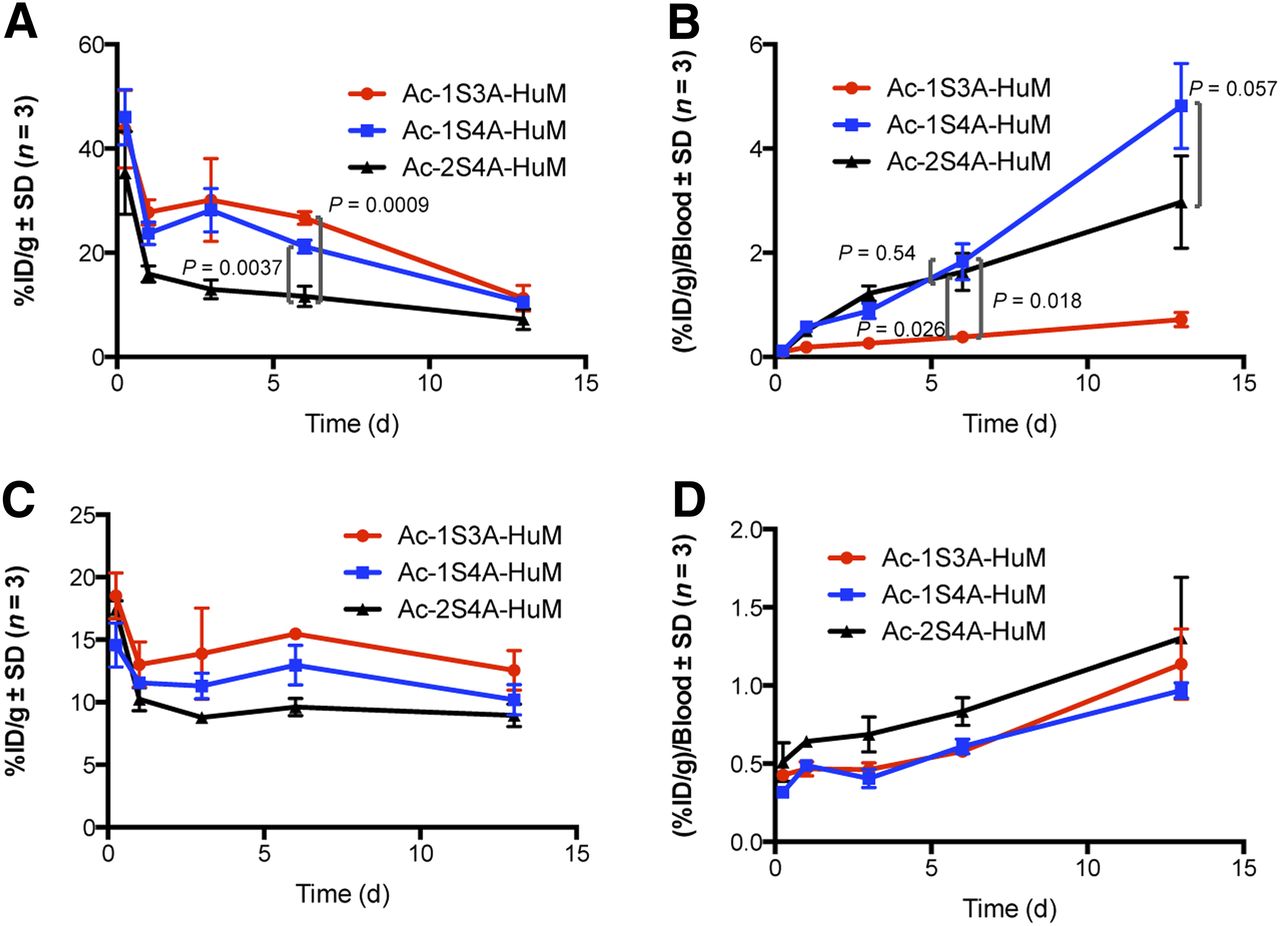
Tissue distribution of 1-step labeled constructs as compared with 4-arm 2-step construct in blood (A), bone plus marrow, normalized to blood (B), and liver without (C) and with (D) normalization to blood. %ID/g = percentage injected dose per gram of tissue.
Therapy of Set-2 AML
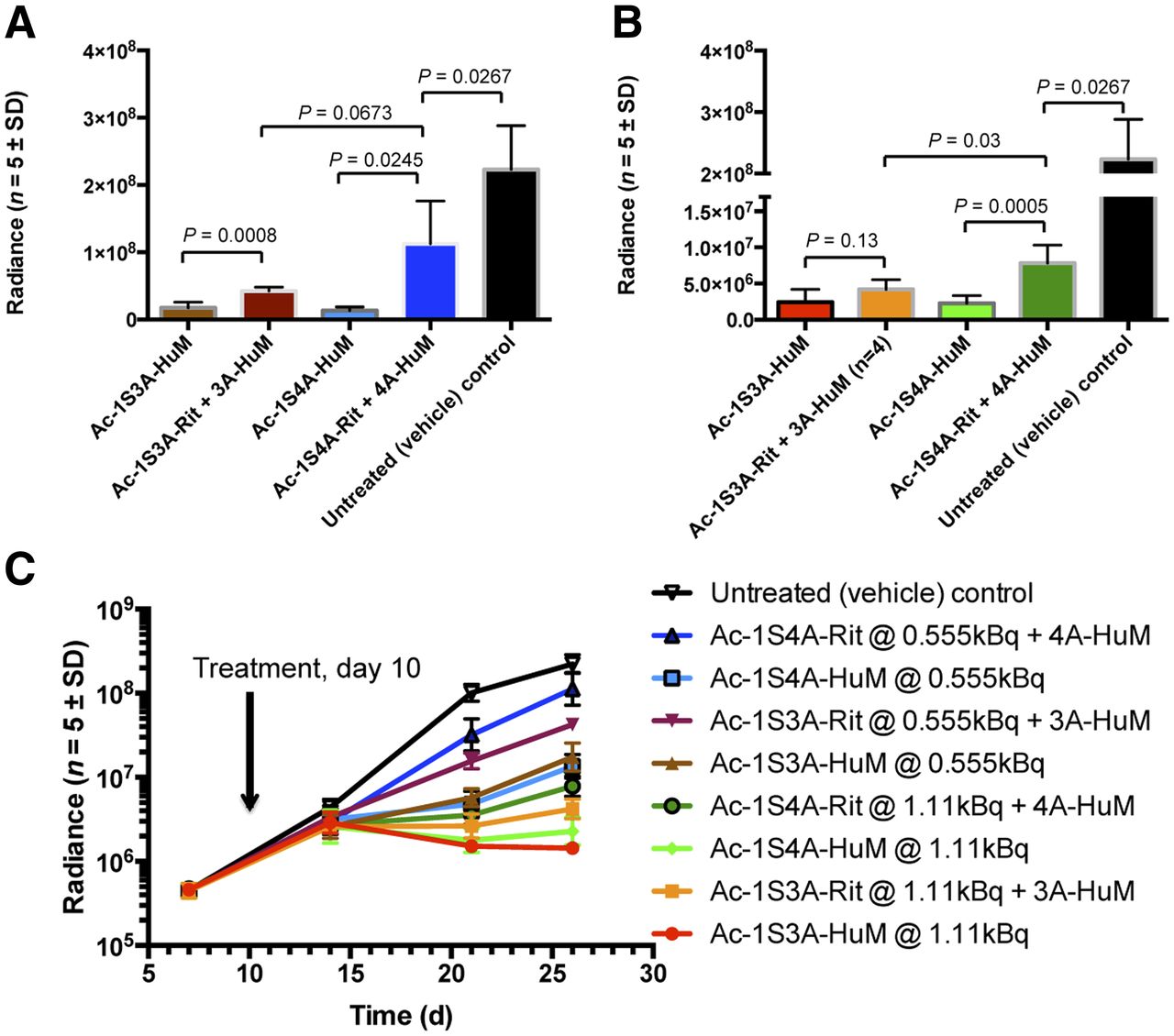
The megakaryoblastic leukemia line Set-2 stably expressing luciferase (Set2-Luc) was determined to bind HuM195 but not rituximab by flow cytometry (Supplemental Fig. 4). Male Nod.Cg-Prkdcscid-Il2rgtm1Wjl/SzJ (Nod scid γ or NSG) mice (n = 5/group) bearing disseminated disease with Set2-Luc cells were treated on day 10 after tumor implantation with a single administration of 225Ac-labeled 3-arm and 4-arm constructs (0.225 μg) labeled to either 0.555 or 1.11 kBq (Fig. 5). One animal in the 3-arm 1.11-kBq dual-control group died on day 17 (day 7 after treatment), possibly from actinium-related toxicity. Tumor burden was monitored by serial bioluminescent imaging. For all radiolabeled constructs, the 1.11-kBq dose produced approximately a 10-fold increased response over the 0.555-kBq dose of corresponding construct (Fig. 5A). The radiolabeled nonspecific antibody plus unlabeled specific construct produced significant responses over vehicle, but in every case the specific construct was substantially more effective than the nonspecific control. This result was statistically significant in every case except the higher dose of 3-arm construct. The 1.11-kBq doses of both specific constructs caused reduction of tumor burden between days 14 and 26 (Fig. 5B). The experiment was terminated after imaging on day 26 before overt morbidity from tumors was observed.

225Ac antibody therapy in mouse model of AML, as determined by bioluminescent intensity. (A) 0.555-kBq treatment groups, day 26 after tumor injection. (B) 1.11-kBq treatment groups, day 26 after tumor injection. (C) Tumor growth curves plotted on log scale.
DISCUSSION
In this work, we designed and characterized a new radiolabeling method of DOTA–antibody constructs in 1 step at 37°C. The 10-fold increased yield (and consequent 10-fold decrease in cost) and up to 30-fold increase in specific activity will have important implications for the preclinical and clinical use of 225Ac on antibodies.
We focused on DOTA because it was successfully used in our 2-step labeling procedure and because it has 2 commonly used chemical forms that might exhibit different radiolabeling or biologic properties. We had initially hypothesized that the 3-arm construct might radiolabel at lower temperatures than the 4-arm construct because of increased kinetic lability afforded by one fewer carboxylic acid. We were therefore surprised to see that the 4-arm DOTA–antibody construct also labeled with 225Ac at 37°C, and that both the kinetics of labeling and the binding capacity for 225Ac appeared to be greater for the 4-arm construct than for the 3-arm one.
The 1-step procedure allows radiochemical yields of up to 80% to be achieved, which is much higher than our former isolated yields of 6%–12% (6). The previous low yields arose from the fact that the labile benzyl isothiocyanate attachment moiety on DOTA was exposed to aqueous solution at 60°C in the first step, causing most of the reactive group to be hydrolyzed before it could react with antibody lysines in the second step. However, the DOTA still chelated 225Ac, causing most of the metal to be discarded in unreactive forms. By contrast, the amount of actinium that can be attached in the 1-step procedure is limited only by the capacity of the antibody construct and the loss of protein in our column purification. Because current high costs and restricted availability of 225Ac limit its use to a small number of laboratories, the 10-fold-higher radiochemical yields should help facilitate the more widespread use of the isotope.
The new procedure is far more attractive from a pharmaceutical and regulatory standpoint. Antibody–chelate constructs can be prepared in a central location, qualified, and stored indefinitely. The end user is only responsible for adding 225Ac and purifying the product, and the specific activity can be adjusted relatively simply by adjusting the amount of 225Ac added to the construct.
Another key advance is that the 1-step procedure afforded products with up to 30-fold-higher specific activities than we have typically achieved with our 2-step procedure. In particular, the 4-arm construct was labeled to 129 GBq/g, versus the 3.7–14.8 GBq/g typically achieved with the 2-step procedure. We posit that this is because the 1-step procedure facilitates the independent control of substitution ratio of DOTA per antibody and amount of 225Ac added, which is more difficult with the 2-step procedure. The highest specific activity we achieved corresponds to approximately 1 molecule of actinium every 25 antibodies. Because it has been estimated that as little as 1 α-particle track through the nucleus can kill a cell (16,17), and 1 in 3 decays at the cell surface will pass through the cell (17), this higher specific activity may facilitate therapy of targets with extremely low cell surface expression or tumor cells that are pharmacologically difficult to access in vivo. This may open the door to a vast array of new cancer or microbial targets.
Tissue distribution of the 3 constructs evaluated was generally similar. Importantly, the absence of time-dependent accumulation of all constructs in the liver was an important indicator of stability because free actinium has been observed to rapidly clear the blood and accumulate in the liver (3). Short-term toxicity was mild in the BALB/c mice used in our biodistribution experiments, despite the relatively high dose of 11.1 kBq needed to obtain sufficient counts. The only obvious toxicity was the reduction in spleen sizes by day 13, but we have observed in other studies that the spleens eventually regrow (not shown). Both 4-arm constructs showed time-dependent accumulation in the bone or marrow, which we attributed to the additional negative charge per DOTA over the 3-arm construct for a net difference of approximately 10 charges. The practical significance of this is unclear, because the two 4-arm constructs behave similarly when corrected for blood half-life, and the 2S4A construct has been used successfully in a variety of preclinical models. Both 1-step constructs had significantly longer serum half-lives than the 2-step construct, which may be due to the effects of the different conjugation and labeling conditions on antibody folding. The pattern of similar initial clearance, delayed clearance of the 1-step constructs over 1–6 d, and then a faster terminal clearance was repeated in an additional biodistribution experiment (Supplemental Fig. 5).
In this study, we have described 3 strategies to stably chelate 225Ac. In some situations, a choice between the 3 chemistries will be obvious, for example, if a preformed DOTA–antibody construct already exists for use with another isotope. If the construct is being prepared de novo, the higher yields and better kinetics of labeling might make the 4-arm 1-step construct the preferred method. Users switching from the 4-arm 2-step strategy may consider lowering the therapeutic dose, given the increased serum half-life of the 1-step constructs.
This study represents the first time, to our knowledge, that 225Ac-HuM195 has been used in a mouse model of leukemia. This was impossible during the initial development of the drug because we lacked a suitable mouse model. The subsequently popularized NSG mice support the growth of AML cell lines in anatomically correct locations, such as the bone marrow for Set2-Luc cells injected intravenously. NSG mice are still not an ideal host for these experiments because of their unusual sensitivity to α-particle irradiation. Although the maximally tolerated dose in some mouse strains is 18.5 kBq or higher (5) and a dose of 11.1 kBq was well tolerated in the BALB/c mice used in the biodistribution study reported here, NSG mice experienced dose-limiting toxicity at as low as 2.22 kBq. We speculate that this is due either to their immunocompromised state, which cannot tolerate even a slight further insult from systemic radiation, or to the lack of circulating antibody, which leads to increased uptake of radiolabeled antibody by nontarget cells with Fc receptors. The target Set2-Luc cells are also highly radiosensitive such that even the control antibody significantly slowed tumor growth. Despite these caveats, we observed dramatic reductions in tumor growth rate and a significant difference between specific and control antibodies. It is likely that in immunocompetent hosts with circulating endogenous IgG, higher or repeated doses can be given and consequently a greater absolute therapeutic effect can be achieved.
CONCLUSION
We have designed an efficient, 1-step radiolabeling method that produces stable, therapeutically active conjugates of antibodies with 225Ac. Because of the large improvements in radiochemical yield, specific activity, and convenience, we propose that this technology can greatly expand preclinical and clinical uses of 225Ac antibodies.
DISCLOSURE
The costs of publication of this article were defrayed in part by the payment of page charges. Therefore, and solely to indicate this fact, this article is hereby marked “advertisement” in accordance with 18 USC section 1734. This study is supported by NIH R01 CA55349 and P01CA23766 and the Metastasis Research Center of MSKCC, NIH RO1 CA166078, and a Medical Scientist Training Program grant from the National Institute of General Medical Sciences of the National Institutes of Health under award number T32GM007739 to the Weill Cornell/Rockefeller/Sloan- Kettering Tri-Institutional MD-PhD Program. The content is solely the responsibility of the authors and does not necessarily represent the official views of the National Institutes of Health. Memorial Sloan Kettering Cancer Center has filed for intellectual property protection for inventions related to this work for David A. Scheinberg, William F. Maguire, Michael R. McDevitt, and Peter M. Smith-Jones. In addition we thank the MSKCC Experimental Therapeutics Center. No other potential conflict of interest relevant to this article was reported.
Acknowledgments
We thank Pharmactinium, Inc., for providing 225Ac.
Footnotes
Published online Jun. 30, 2014.
- © 2014 by the Society of Nuclear Medicine and Molecular Imaging, Inc.
REFERENCES
- Received for publication January 31, 2014.
- Accepted for publication May 30, 2014.


Jump to section
Related Articles
Cited By...
- Cure of Disseminated Human Lymphoma with [225Ac]Ac-Ofatumumab in a Preclinical Model
- Treatment of prostate cancer with CD46 targeted 225Ac alpha particle radioimmunotherapy
- Harnessing {alpha}-Emitting Radionuclides for Therapy: Radiolabeling Method Review
- Genetic signature of prostate cancer mouse models resistant to optimized hK2 targeted {alpha}-particle therapy
- Genetic signature of prostate cancer resistant to optimized hK2 targeted alpha-particle therapy
- {alpha}-Emitters for Radiotherapy: From Basic Radiochemistry to Clinical Studies--Part 1
- Preclinical Development of CD38-Targeted [89Zr]Zr-DFO-Daratumumab for Imaging Multiple Myeloma
- Remodeling the Vascular Microenvironment of Glioblastoma with {alpha}-Particles
- Vascular Targeted Radioimmunotherapy for the Treatment of Glioblastoma