


Abstract
1236
Objectives Clinical whole-body PET/CT imaging currently involves static acquisitions and employs SUV, a semi-quantitative metric with documented limitations. Our objective is to implement quantitative whole body parametric imaging to enhance detectability in regions extending over multiple beds for less FDG-avid tumors and/or with high uptake surrounding tissues.
Methods A clinical whole-body dynamic acquisition protocol as optimized using Monte Carlo (MC) simulations was employed, involving initial 6min scan over the heart (24 frames) followed by 6 whole-body scans. Next, a series of MC simulations were performed, utilizing time-activity curves (TACs) as produced from kinetic parameters, obtained from the literature, and assigned to regions of interest (ROIs), including liver and lung tumors of 5, 10 and 15mm diameter. 25 noise realizations were generated and, for each, 30 dynamic frames were reconstructed (OSEM: 21 subsets, 15 iterations). Subsequently, parametric images were derived, using the Patlak method. Meanwhile, a 3min SUV frame was simulated. A noise vs. bias analysis for each ROI and with increasing iterations was performed for parametric vs. SUV images, and the tumor-to-background ratio (TBR) was calculated. In addition, a series of oncology patient studies (n=5) was conducted.
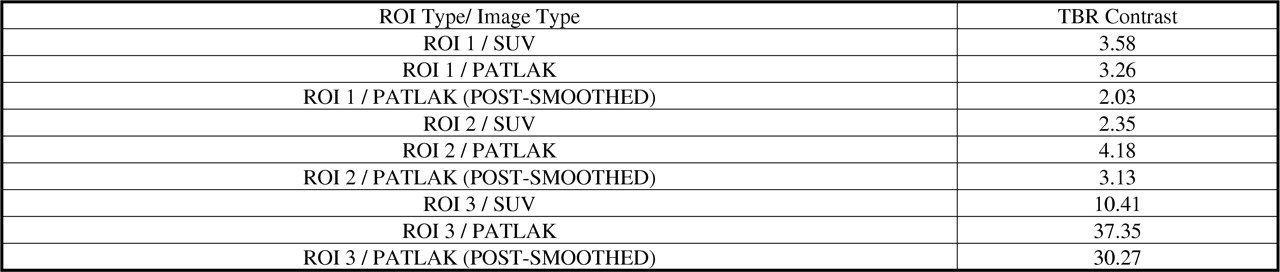
Results Simulated Patlak images exhibited higher TBR values in liver tumors vs. SUV (4.8+/-2.88 vs. 1.8+/-0.54) and in lung tumors (3.3+/- 1.82 vs. 2.7+/-0.8) because of the higher uptake of normal liver. Moreover, clinical cases show improvement of the parametric TBR against SUV as high as 300%.
Conclusions Patlak parametric imaging is sensitive to high levels of noise in short 45sec dynamic PET frames and pre-smoothing before Patlak can reduce noise and enhance parametric TBR. Both simulated and patient data demonstrate enhanced tumor detectability

TBR contrast in clinical data
In this issue


Jump to section
Related Articles
Cited By...
- No citing articles found.