Main menu

  • Home
  • Content
    • Current
    • Ahead of print
    • Past Issues
    • JNM Supplement
    • SNMMI Annual Meeting Abstracts
    • Continuing Education
    • JNM Podcasts
  • Subscriptions
    • Subscribers
    • Institutional and Non-member
    • Rates
    • Journal Claims
    • Corporate & Special Sales
  • Authors
    • Submit to JNM
    • Information for Authors
    • Assignment of Copyright
    • AQARA requirements
  • Info
    • Reviewers
    • Permissions
    • Advertisers
  • About
    • About Us
    • Editorial Board
    • Contact Information
  • More
    • Alerts
    • Feedback
    • Help
    • SNMMI Journals
  • SNMMI
    • JNM
    • JNMT
    • SNMMI Journals
    • SNMMI

User menu

  • Subscribe
  • My alerts
  • Log in
  • Log out
  • My Cart

Search

  • Advanced search
Journal of Nuclear Medicine
  • SNMMI
    • JNM
    • JNMT
    • SNMMI Journals
    • SNMMI
  • Subscribe
  • My alerts
  • Log in
  • Log out
  • My Cart
Journal of Nuclear Medicine

Advanced Search

  • Home
  • Content
    • Current
    • Ahead of print
    • Past Issues
    • JNM Supplement
    • SNMMI Annual Meeting Abstracts
    • Continuing Education
    • JNM Podcasts
  • Subscriptions
    • Subscribers
    • Institutional and Non-member
    • Rates
    • Journal Claims
    • Corporate & Special Sales
  • Authors
    • Submit to JNM
    • Information for Authors
    • Assignment of Copyright
    • AQARA requirements
  • Info
    • Reviewers
    • Permissions
    • Advertisers
  • About
    • About Us
    • Editorial Board
    • Contact Information
  • More
    • Alerts
    • Feedback
    • Help
    • SNMMI Journals
  • View or Listen to JNM Podcast
  • Visit JNM on Facebook
  • Join JNM on LinkedIn
  • Follow JNM on Twitter
  • Subscribe to our RSS feeds
Research ArticleThe Illustrated Post

[68Ga]Ga-PSMA-11 PET/CT–Positive Hepatic Inflammatory Pseudotumor: Possible PSMA-Avid Pitfall in Nuclear Imaging

Fabio Monastero, Luigia Vetrone, Lina Cardisciani, Matteo Renzulli, Enrico Prosperi, Matteo Cescon, Matteo Ravaioli, Stefano Fanti, Andrea Farolfi and Francesco Vasuri
Journal of Nuclear Medicine October 2024, 65 (10) 1658-1659; DOI: https://doi.org/10.2967/jnumed.124.267518
Fabio Monastero
1Nuclear Medicine, Alma Mater Studiorum, University of Bologna, Bologna, Italy;
  • Find this author on Google Scholar
  • Find this author on PubMed
  • Search for this author on this site
Luigia Vetrone
2Nuclear Medicine, IRCCS Azienda Ospedaliero-Universitaria di Bologna, Bologna, Italy;
  • Find this author on Google Scholar
  • Find this author on PubMed
  • Search for this author on this site
Lina Cardisciani
3School of Anatomic Pathology, Department of Biomedical and Neuromotor Sciences, University of Bologna, Bologna, Italy;
  • Find this author on Google Scholar
  • Find this author on PubMed
  • Search for this author on this site
Matteo Renzulli
4Department of Radiology, IRCCS Azienda Ospedaliero-Universitaria di Bologna, Bologna, Italy;
  • Find this author on Google Scholar
  • Find this author on PubMed
  • Search for this author on this site
Enrico Prosperi
5Department of Hepatobiliary Surgery and Liver Transplant, IRCCS Azienda Ospedaliero-Universitaria di Bologna, Bologna, Italy; and
  • Find this author on Google Scholar
  • Find this author on PubMed
  • Search for this author on this site
Matteo Cescon
5Department of Hepatobiliary Surgery and Liver Transplant, IRCCS Azienda Ospedaliero-Universitaria di Bologna, Bologna, Italy; and
  • Find this author on Google Scholar
  • Find this author on PubMed
  • Search for this author on this site
Matteo Ravaioli
5Department of Hepatobiliary Surgery and Liver Transplant, IRCCS Azienda Ospedaliero-Universitaria di Bologna, Bologna, Italy; and
  • Find this author on Google Scholar
  • Find this author on PubMed
  • Search for this author on this site
Stefano Fanti
2Nuclear Medicine, IRCCS Azienda Ospedaliero-Universitaria di Bologna, Bologna, Italy;
  • Find this author on Google Scholar
  • Find this author on PubMed
  • Search for this author on this site
Andrea Farolfi
2Nuclear Medicine, IRCCS Azienda Ospedaliero-Universitaria di Bologna, Bologna, Italy;
  • Find this author on Google Scholar
  • Find this author on PubMed
  • Search for this author on this site
Francesco Vasuri
6Pathology Unit, IRCCS Azienda Ospedaliero-Universitaria di Bologna, Bologna, Italy
  • Find this author on Google Scholar
  • Find this author on PubMed
  • Search for this author on this site
  • Article
  • Figures & Data
  • Info & Metrics
  • PDF
Loading

Prostate-specific membrane antigen (PSMA) is a transmembrane glycoprotein highly overexpressed in many cancers and benign conditions, and [68Ga]Ga-PSMA-11 PET/CT represents a successful diagnostic imaging method in nuclear medicine for prostate cancer.

This is a case of a 64-y-old woman enrolled in the Monocentric Observational Experience with PSMA study in Bologna, Italy, a prospective study examining in vivo and in vitro PSMA expression in patients with nonprostatic neoplasms, with histopathology as the reference standard. The study protocol was approved by the local ethics committee (code 538/2021/Oss/AOUBo), and subjects provided written informed consent. The patient’s contrast-enhanced MRI revealed a lesion in segment 5 of the liver, with late enhancement in wash-in and wash-out phases, consistent with hepatocellular carcinoma. [68Ga]Ga-PSMA-11 PET/CT was performed 17 d later (injected activity, 146 MBq; uptake time, 85 min), showing increased PSMA uptake at segment 5 (SUVmax, 11.3; SUVmax liver background, 7.9) and matching the MRI lesion. A wedge resection of segment 5 of the liver was executed 8 days later.

A yellowish well-defined 2-cm nodule made of an inflammatory component (mainly comprising histiocytic xanthogranulomatous cells and multinucleated giant cell macrophages containing biliary pigment, with stromal fibrotic and fibroblastic proliferation) was observed within noncirrhotic liver parenchyma. Eosinophilic nodular deposits surrounded by plasma cells, foamy macrophages or neutrophils, and activated myofibroblasts were also observed. Immunohistochemistry showed no cytokeratin-positive epithelial cells within the lesion, whereas the histiocytic component was CD68PGM1-positive/s100-negative with an IgG4-to-IgG ratio of less than 40%. The final diagnosis was a hepatic inflammatory pseudotumor (fibrohistiocytic type) (1).

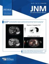
Both PSMA immunohistochemistry and immunofluorescence were performed. The results are shown in Figure 1.

FIGURE 1.
  • Download figure
  • Open in new tab
  • Download powerpoint
FIGURE 1.

(A) Maximum-intensity projection image showing increased [68Ga]Ga-PSMA-11 uptake (arrowhead) in liver (SUVmax, 11.3). (B–E) Gadolinium ethoxybenzyl diethylenetriamine pentaacetic acid MRI showing nodule in segment 5 (arrow) hyperintense in T2-weighted image (B), highly hyperintense in diffusion-weighted image (C), highly hyperintense in arterial phase (D), and with targetoid sign in transitional phase (E). (F) Axial-fused [68Ga]Ga-PSMA-11 PET/CT showing increased PSMA uptake (SUVmax, 11.3) at segment 5. (G) PSMA immunohistochemistry image (×20) with sparce positive endothelial cells in intralesional microvascular component (circles) and negative inflammatory cells. (H) Negative PSMA immunofluorescence image (×10) in normal liver parenchyma (asterisk) with adjacent inflammatory infiltrate and sparce but strongly positive endothelial cells in intralesional microvascular component (circles).

Interestingly, only endothelial cells within the pseudotumor expressed PSMA, whereas inflammatory cells and normal liver endothelial cells were negative for PSMA. Hypothetically, endothelial cells within the lesion are stimulated by the tumor microenvironment to express PSMA, even if the mechanism beyond is still not known.

Hepatic inflammatory pseudotumor is a rare disease with less than 300 cases described in the literature. Differential diagnosis between a pseudotumor and hepatocellular carcinoma with CT and MRI is challenging. One multicentric study showed that 50% of patients with a final diagnosis of pseudotumor received a previous hepatocellular carcinoma misdiagnosis at CT or MRI (2). However, the prognosis and the treatment of pseudotumors are different and often resolvable with more conservative approaches, avoiding unnecessary invasive procedures.

The hereby described clinical case highlights that characterization of liver lesions is challenging with PSMA ligands because of the presence of several PSMA-avid diseases (e.g., hepatocellular carcinoma, prostate cancer metastases, and hemangioma) along with the physiologic PSMA background uptake in the liver parenchyma. Moreover, unlike prostate cancer, in which PSMA is overexpressed directly by cancer cells, PSMA in an inflammatory pseudotumor is expressed by endothelial cells. Therefore, PSMA images necessitate careful interpretation when evaluating the liver, as different mechanisms of PSMA overexpression can mimic neoplastic lesions.

DISCLOSURE

No potential conflict of interest relevant to this article was reported.

Footnotes

  • Published online Jun. 13, 2024.

  • © 2024 by the Society of Nuclear Medicine and Molecular Imaging.

REFERENCES

  1. 1.↵
    1. Zen Y,
    2. Fujii T,
    3. Sato Y,
    4. Masuda S,
    5. Nakanuma Y
    . Pathological classification of hepatic inflammatory pseudotumor with respect to IgG4-related disease. Mod Pathol. 2007;20:884–894.
    OpenUrlCrossRefPubMed
  2. 2.↵
    1. Arora KS,
    2. Anderson MA,
    3. Neyaz A,
    4. et al
    . Fibrohistiocytic variant of hepatic pseudotumor: an antibiotic responsive tumefactive lesion. Am J Surg Pathol. 2021;45:1314–1323.
    OpenUrl
  • Received for publication February 5, 2024.
  • Accepted for publication May 13, 2024.
PreviousNext
Back to top

In this issue

Journal of Nuclear Medicine: 65 (10)
Journal of Nuclear Medicine
Vol. 65, Issue 10
October 1, 2024
  • Table of Contents
  • Table of Contents (PDF)
  • About the Cover
  • Index by author
  • Complete Issue (PDF)
Print
Download PDF
Article Alerts
Sign In to Email Alerts with your Email Address
Email Article

Thank you for your interest in spreading the word on Journal of Nuclear Medicine.

NOTE: We only request your email address so that the person you are recommending the page to knows that you wanted them to see it, and that it is not junk mail. We do not capture any email address.

Enter multiple addresses on separate lines or separate them with commas.
[68Ga]Ga-PSMA-11 PET/CT–Positive Hepatic Inflammatory Pseudotumor: Possible PSMA-Avid Pitfall in Nuclear Imaging
(Your Name) has sent you a message from Journal of Nuclear Medicine
(Your Name) thought you would like to see the Journal of Nuclear Medicine web site.
Citation Tools
[68Ga]Ga-PSMA-11 PET/CT–Positive Hepatic Inflammatory Pseudotumor: Possible PSMA-Avid Pitfall in Nuclear Imaging
Fabio Monastero, Luigia Vetrone, Lina Cardisciani, Matteo Renzulli, Enrico Prosperi, Matteo Cescon, Matteo Ravaioli, Stefano Fanti, Andrea Farolfi, Francesco Vasuri
Journal of Nuclear Medicine Oct 2024, 65 (10) 1658-1659; DOI: 10.2967/jnumed.124.267518

Citation Manager Formats

  • BibTeX
  • Bookends
  • EasyBib
  • EndNote (tagged)
  • EndNote 8 (xml)
  • Medlars
  • Mendeley
  • Papers
  • RefWorks Tagged
  • Ref Manager
  • RIS
  • Zotero
Share
[68Ga]Ga-PSMA-11 PET/CT–Positive Hepatic Inflammatory Pseudotumor: Possible PSMA-Avid Pitfall in Nuclear Imaging
Fabio Monastero, Luigia Vetrone, Lina Cardisciani, Matteo Renzulli, Enrico Prosperi, Matteo Cescon, Matteo Ravaioli, Stefano Fanti, Andrea Farolfi, Francesco Vasuri
Journal of Nuclear Medicine Oct 2024, 65 (10) 1658-1659; DOI: 10.2967/jnumed.124.267518
Twitter logo Facebook logo LinkedIn logo Mendeley logo
  • Tweet Widget
  • Facebook Like
  • Google Plus One
Bookmark this article

Jump to section

  • Article
    • DISCLOSURE
    • Footnotes
    • REFERENCES
  • Figures & Data
  • Info & Metrics
  • PDF

Related Articles

  • No related articles found.
  • PubMed
  • Google Scholar

Cited By...

  • No citing articles found.
  • Google Scholar

More in this TOC Section

  • Uncommon Anomalous Biodistribution of 18F-DCFPyL Prostate-Specific Membrane Antigen: A Case Series
  • [68Ga]Ga-FAPI PET/CT Monitors Response to Receptor Activator of Nuclear Factor-κB Ligand Inhibitor in a Giant Cell Tumor of Bone: Correlation with Histopathology
  • Evaluating Bone Healing with [18F]NaF PET/CT During Bone Segment Transport in Femoral Fracture Treatment
Show more The Illustrated Post

Similar Articles

SNMMI

© 2025 SNMMI

Powered by HighWire