


Visual Abstract

Abstract
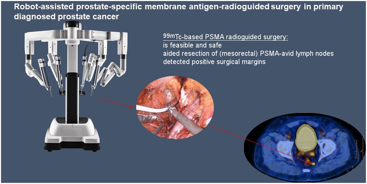
The objective of this study was to evaluate the safety and feasibility of 99mTc-based prostate-specific membrane antigen (PSMA) robot-assisted–radioguided surgery to aid or improve the intraoperative detection of lymph node metastases during primary robot-assisted radical prostatectomy (RARP) for prostate cancer (PCa). Methods: Men with primary high-risk PCa (≥ cT3a, International Society of Urological Pathology (ISUP) grade group ≥ 3 or prostate-specific antigen of ≥ 15 ng/mL) with potential lymph node metastasis (Briganti nomogram risk > 10% or on preoperative imaging) were enrolled in the study. Patients underwent staging 68Ga-PSMA PET/CT scanning. Preoperatively, a 99mTc-labeled PSMA ligand (99mTc PSMA I&S; 500 MBq) was administered followed by SPECT/CT. A RARP including extended pelvic lymph node dissection was performed, with intraoperative tracing of PSMA-avid tissues using a prototype DROP-IN γ-probe. Resected specimens were also measured ex vivo. Histopathologic concordance with probe findings was evaluated. A radiotracer count of ≥ 1.5 times the background reference (in vivo), and ≥ 10 (absolute count) in the ex vivo setting, was considered positive. Results: Twelve patients were included (median age, 68 y, and prostate-specific antigen, 9.15 ng/mL). Most of the patients harbored ISUP 5 PCa (75%) and had avid lymph nodes on preoperative PSMA PET (64%). The DROP-IN probe aided resection of PSMA-avid (out-of-template) lymph nodes and residual disease at the prostate bed. Eleven metastatic lymph nodes were identified by the probe that were not observed on preoperative 68Ga-PSMA PET/CT. Of the 74 extraprostatic tissue specimens that were resected, 22 (29.7%) contained PCa. The sensitivity, specificity, positive predictive value, and negative predictive value of inpatient use of the γ-probe were 76% (95% CI, 53%–92%), 69% (95% CI, 55%–81%), 50%, and 88%, respectively. Ex vivo, the diagnostic accuracy was superior: 76% (95% CI, 53%–92%), 96% (95% CI, 87%–99%), 89%, and 91%, respectively, for sensitivity, specificity, positive predictive value, and negative predictive value. Of the missed lymph nodes in vivo (n = 5) and ex vivo (n = 5), 90% were micrometastasis (≤3 mm). No complications greater than Clavien–Dindo Grade I occurred. Conclusion: Robot-assisted 99mTc-based PSMA-radioguided surgery is feasible and safe in the primary setting, optimizing the detection of nodal metastases at the time of RARP and extended pelvic lymph node dissection. Further improvement of the detector technology may optimize the capabilities of robot-assisted 99mTc-based PSMA-radioguided surgery.
- image-guided surgery
- prostate-specific membrane antigen
- prostate cancer
- robot-assisted surgery
- extended pelvic lymph node dissection
The ability to accurately determine the location and extent of lymph node involvement in prostate cancer (PCa) has significant implications on decision-making regarding treatment modality and planning. In men with higher risk PCa, bilateral extended pelvic lymph node dissection (ePLND) during robot-assisted radical prostatectomy (RARP) remains an important staging procedure (1). Additionally, ePLND has been observed to provide a chance of cure in patients with limited nodal metastases (2,3). Despite these valuable considerations, ePLND has several shortcomings. Importantly, ePLND is associated with increased patient morbidity (4). The extent of dissection has an impact on the detection and removal of any nodal disease (5). Even with an extended template dissection, 35% of lymph nodes (potentially containing PCa) will not be removed at the time of surgery, either being out of the surgical template or being missed within it (6).
The emergence of prostate-specific membrane antigen PET/CT (PSMA PET) has greatly improved the detection of small lymph node metastases when compared with conventional imaging. Although PSMA PET imaging has demonstrated excellence for the detection of lymph node metastases in the setting of both primary staging and biochemical recurrence (BCR), it is intrinsically limited by its spatial resolution of 2–4 mm given radioisotope decay and detector distance from the tumor (7,8). Several studies correlating preoperative PSMA PET findings to histopathologic findings from a subsequent ePLND have demonstrated poor sensitivity in the detection of nodal metastases < 4 mm (9–11). Other studies showed that patients with nodal metastatic disease on PSMA PET/CT have an increased risk of developing BCR after RARP (12). It may be the subset of patients with limited nodal micrometastases (1–2 nodes) who are most likely to benefit from an (optimized) ePLND (3).
PSMA-targeted radioguided surgery has been suggested as an auxiliary technique to improve the intraoperative detection and clearance of lymph nodes harboring (limited) metastatic disease. Recently, Maurer et al. described the use of 99mTc-based PSMA-radioguided surgery in the setting of an open salvage ePLND following BCR after surgery with promising results, detecting metastatic deposits as small as 3 mm (13). It has been possible to translate radioguided surgery to the robotic setting using a new DROP-IN γ-detector (Eurorad S.A., Eckbolsheim, France) technology (14,15), raising the question whether 99mTc-based PSMA-radioguided surgery in the primary treatment setting could improve treatment outcomes by optimizing metastatic lymph node detection and clearance. Furthermore, no data exist on the ability to perioperatively detect significant positive surgical margins.
To date, there are no results of prospective trials evaluating the radioguided identification of lymph node metastases at the time of RARP and ePLND in the primary treatment setting. The DETECT trial is a single-site, prospective pilot study that aimed to describe the technique and feasibility of robot-assisted radioguided surgery using the molecular targeting of PSMA to aid intraoperative localization of nodal metastases at the time of RARP and ePLND in a primary treatment setting.
MATERIALS AND METHODS
Study Population
Twelve men with histologically proven PCa and potential lymph node metastasis were included in this prospective clinical trial between July 2019 and May 2021. Men 18 y or older with high-risk PCa (≥ cT3a or International Society of Urological Pathology [ISUP] grade group ≥ 3 or preoperative prostate-specific antigen [PSA] ≥ 15 ng/mL), with a Briganti nomogram risk of ≥10%, were included in the trial. Exclusion criteria were prior PCa treatment and evidence of distant (lymph node) metastatic disease. The institutional review board approved this study (HREC/16/SVH/157), and written informed consent was obtained from all men. The trial is registered at ANZCTR (ACTRN12621001728820), and funding was obtained from the Prostate Cancer Foundation of Australia.
Preoperative Radiotracer Administration and Imaging
Patients underwent 68Ga-PSMA PET/CT scanning as part of preoperative staging (<3 mo before surgery). These scans were centrally reviewed by 2 experienced nuclear physicians who were masked to pathology. In the case of conflicting results, a second consensus read was performed. Patients underwent intravenous administration of 500 MBq of an 99mTc-labeled PSMA-targeted ligand (99mTc PSMA I&S) approximately 18 h before surgery (16). On the morning of their surgery, SPECT/CT imaging was performed to cross-validate the findings of the preoperative 68Ga-PSMA PET/CT scan and to serve as a quality control for radiotracer injection and distribution. The results of the 99mTc-PSMA SPECT/CT were not used to guide surgery. In patients suspected of harboring metastatic disease in out-of-template mesorectal lymph node regions, a percutaneous CT-guided hookwire was inserted at the time of 99mTc-PSMA SPECT/CT to assist localization at time of dissection.
Surgical Procedure
Patients underwent a RARP and ePLND via a standard 6-port, transperitoneal approach with a 4-arm da Vinci XI system (Intuitive Surgical Inc., USA). A prototype DROP-IN γ-probe connected to the Europrobe 3.2 console (Eurorad, S.A.) (Fig. 1A) was inserted through a 12-mm assistant port via the Alexis laparoscopic system (Applied Medical Corp., USA) placed above the left iliac crest (Fig. 1B) (15,17). Once inserted, the DROP-IN γ-probe (Figs. 1C and 1D) was articulated with da Vinci ProGrasp forceps to take measurements. Real-time feedback was provided both acoustically and numerically via a read-out console placed in the surgical theater. Before resection, γ-probe measurements were systemically taken at the anterior abdominal wall muscle as a background reference measurement, at each template lymph node site (external iliac, internal iliac, and obturator pelvic lymph nodes) bilaterally, the prostate, the prostate bed after resection, and other out-of-template sites clinically suspected of harboring disease (based on preoperative imaging). To exclude the influence of in vivo background interference, ex vivo γ-probe measurements were taken of each resected tissue specimen using the same DROP-IN probe.

(A) Europrobe 3.2 console (Eurorad, S.A.) providing visual and acoustic count feedback. (B) Insertion of DROP-IN γ-probe and 12-mm assistant laparoscopic port through the Alexis system. (C and D) Intraoperative use of the DROP-IN γ-probe to obtain counts from left obturator lymph node template region (C) and prostate bed (D).
Histopathologic Correlation with 99mTc-PSMA γ-Probe Measurements
Histopathologic evaluation was performed by a specialist uropathologist masked to preoperative imaging and γ-probe findings. Regions with γ-probe measurements were correlated with final tissue histopathologic findings on a per-region basis. Several radiotracer cutoffs were evaluated on their diagnostic performance as no literature existed on the radiotracer activity count in the primary treatment setting. Probe findings were rated positive when tissue specimens had a radiotracer activity count of ≥1.5 times the background reference value in the in vivo setting and ≥10 (absolute count) in the ex vivo setting.
Follow-up
Patients were first followed-up at 6 wk postoperatively. Subsequent interval PSA measurements or adjuvant therapy was determined by the treating urologist on the basis of the patient’s biochemical response and final pathology. A BCR after surgery was defined as PSA > 0.1 ng/mL after initial biochemical control. In the case of persistent PSA after surgery (PSA > 0.1 ng/mL) or BCR, adjuvant pelvic radiotherapy with androgen deprivation therapy (ADT) was discussed. Postoperative complications were categorized by Clavien–Dindo classification. Any subsequent PSA measurements or adjuvant therapy was monitored during the course of this study.
Statistical Analysis
Preoperative imaging and DROP-IN γ-probe results were compared with corresponding final pathology. Sensitivity, specificity, positive predictive value (PPV), and negative predictive value (NPV) were derived from 2 × 2 contingency tables. Quantitative data are reported as the median value and interquartile ranges (IQRs). Categoric data are reported as absolute and relative frequencies. Differences in radiotracer counts between malignant and benign tissue were compared with a χ2 test. All statistical analyses were conducted with the SPSS statistical software package (version 26.0; IBM).
RESULTS
Preoperative Patient Characteristics
Preoperative patient characteristics are summarized in Table 1. The median age was 68 y (IQR, 57–69 y), and preoperative PSA was 9.15 ng/mL (IQR, 6.0–21.2 ng/mL). Nine patients had ISUP 5, and 3 patients had ISUP 4 PCa. Seven patients demonstrated evidence of pelvic nodal disease on 68Ga-PSMA PET/CT across 11 anatomic regions. Four of those lesions were within the standard ePLND template whereas the other 7 lesions were outside the standard surgical template (mesorectal, presacral, below the aortic bifurcation). Figure 2 shows an example of a mesorectal node. Because of the known limitations (sensitivity and resolution) of 99mTc-PSMA SPECT/CT, only 4 lesions were observed on 99mTc-PSMA SPECT/CT (13). These corresponded with large lymph nodes (>9 mm) on 68Ga-PSMA PET/CT and were all histologically confirmed as cancer.
Preoperative Patient Characteristics

Abdominal CT scan (top), PSMA PET/CT scan (middle), and SPECT/CT scan (bottom). Suspicion for left mesorectal lymph node involvement on preoperative 68Ga-PSMA PET/CT in patient 12 (SUVmax, 49). Probe successfully identified node (in vivo node count of 183) and final pathology showed 8-mm malignant lymph node. Red circles indicate a lymph node metastasis suspected on preoperative imaging, that was subsequently correctly identified by the probe and histologically confirmed to be malignant.
Safety Outcomes
No postoperative complications greater than Clavien–Dindo grade 1 were observed in the follow-up period. Only 1 patient suffered from a surgical postoperative complication (lymphoedema) that was conservatively managed. No complications relating to the administration of 99mTc-PSMA radiotracer or the use of the prototype DROP-IN γ-probe were seen.
99mTc-PSMA γ-Probe Guidance During Surgery and Concordance with Preoperative Imaging
A total of 11 preoperative lesions were visible on 68Ga-PSMA PET/CT, and 8 demonstrated metastatic disease on final histopathology. Of the remaining 3 lesions, 2 lesions (both in patient 11) demonstrated no disease (however, the patient had persistent PSA after surgery, therefore the lesions may not have been surgically removed), and the other lesion (patient 10, left mesorectal node) was irresectable. In contrast, 11 metastatic lymph nodes were identified by in vivo and ex vivo use of the probe that were not observed on preoperative 68Ga-PSMA PET/CT, with 1 lymph node harboring 15 mm of PCa.
The DROP-IN γ-probe was utilized in vivo in patient 2 to correctly identify 6-mm residual PCa at the right apical margin of the prostate bed that would have otherwise remained undiscovered (Fig. 1D). For 2 patients, the probe was successfully used in conjunction with percutaneously placed hookwires to localize metastatic out-of-template mesorectal lymph nodes.
99mTc-PSMA γ-Probe Concordance with ePLND Histopathology
Detailed γ-probe findings with histopathologic correlation in the in vivo and ex vivo setting are included in Supplemental Tables 1 and 2, respectively (supplemental materials are available at http://jnm.snmjournals.org). A total of 74 extraprostatic (nodal packages and periprostatic tissue) specimens were resected (median, 6 per patient; IQR, 5–7), which included 213 lymph nodes (median, 17 per patient; IQR, 12–22).
Twenty-two of the 74 resected specimens (29.7%) demonstrated evidence of PCa on histopathology. Histopathology-positive specimens had a median in vivo and ex vivo count of 35 (IQR, 20–63) and 19 (IQR, 8–29), whereas negative specimens showed a median in vivo and ex vivo count of 17 (IQR, 10–34) and 4 (IQR, 1–6), respectively. The count difference between malignant and benign lymph nodes was not statistically significantly different in vivo (P = 0.32) while being significantly different ex vivo (P = 0.001). The signal-to-background ratio (SBR) was 2.1 during surgery (in vivo) and 4.8 ex vivo.
In vivo, the DROP-IN probe readings were rated positive in 32 locations. Of these positive locations, 16 contained PCa whereas 16 contained no cancer. The DROP-IN probe readings were rated negative in 41 locations, of which 36 contained no cancer and 5 harbored PCa, resulting in an in vivo sensitivity, specificity, PPV, and NPV of 76% (95% CI, 53%–92%), 69% (95% CI, 55%–81%), 50%, and 88%, respectively. The locations of false-positive results varied between patients (Supplemental Table 1).
Ex vivo, with no background signal from the body, a superior accuracy was observed using the same probe. DROP-IN probe readings were rated positive in 18 resected specimens. Of these positive specimens, 16 contained PCa whereas only 2 contained no PCa. The DROP-IN probe readings were rated negative in 55 specimens, of which 50 contained no PCa and 5 harbored PCa, resulting in an ex vivo sensitivity, specificity, PPV, and NPV of 76% (95% CI, 53%–92%), 96% (95% CI, 87%–99%), 89%, and 91%, respectively.
The median size of metastatic foci that were correctly identified by the DROP-IN probe was 9 mm (IQR, 6.5–11.2 mm) for both in vivo and ex vivo use. The smallest nodes detected in vivo and ex vivo were 0.4 and 0.1 mm, respectively. In the 5 specimens with metastatic disease that the probe did not detect in vivo, none was avid on either 68Ga-PSMA PET/CT or 99mTc-PSMA SPECT/CT, indicating low tracer uptake. Although the tracer uptake in 4 of these instances could be related to size (patients 1, 2, and 8: <1 mm; patient 2: 3 mm), the other 6-mm metastatic lesion could be due to low PSMA expression (patient 8). In the 5 specimens with metastatic disease that were unsuccessfully detected by the probe ex vivo, none was avid on either 68Ga-PSMA PET/CT or 99mTc-PSMA SPECT/CT, probably due to size (<3 mm).
Follow-up and Oncologic Outcomes
Median follow-up was 13 mo (4–21) after surgery. At 6 wk after surgery, 5 of the 12 patients had an undetectable PSA (<0.03 ng/mL), 2 patients had an equivocal PSA persistence (<0.1 and ≥0.03 ng/mL), and 5 patients had a persistent PSA (>0.1 ng/mL). All 5 patients who had a complete metabolic response had no BCR during follow-up and required no adjuvant therapy. Final pathology in those 5 patients showed limited lymph node metastasis in 80% (patients 1, 6, 8, 9, and 12, Supplemental Tables 1 and 2).
The 2 patients with equivocal PSA persistence after surgery both experienced a BCR. Of the 7 patients with detectable PSA (BCR, n = 2; persistent PSA, n = 5), 6 patients received ADT in combination with adjuvant radiotherapy as part of multimodal therapy whereas the remaining patient was ineligible to receive adjuvant radiotherapy. Of these 7 patients, 3 had a complete metabolic response (after withdrawal from hormonal treatment > 12 mo), and at the time of this article the remaining 4 patients were still on ADT as part of their adjuvant therapy and therefore cannot be biochemically assessed. At present none of the included patients have metastatic disease; PCa-specific and overall mortality is 0%.
DISCUSSION
To our knowledge, this is the first prospective series on robot-assisted PSMA-targeted surgery and ePLND in the primary treatment setting. Prior PSMA-targeted surgery studies were performed in the salvage pelvic lymph node and local recurrence dissection setting (13,18,19). The technical setup used in our study is a combination of Meershoek et al. (robotic use of DROP-IN γ-probe) and Maurer et al. (PSMA-radioguided surgery during open surgery) (13,17). A small γ-probe head is “dropped in” to the abdominopelvic cavity through an Alexis working port and manipulated using the da Vinci ProGrasp forceps. Using this technique, we were able to identify the far majority of the PSMA PET–avid foci (>3 mm), indicating that the DROP-IN technology can bridge pre- and intraoperative findings. Interestingly, the probe helped identify 11 foci that were not seen on PSMA PET/CT or SPECT. Nevertheless, not all foci could be identified but particularly micrometastasis (foci < 3 mm) were missed.
The propensity for the PSMA-targeted approach to miss small PSMA PET–negative foci suggests this approach cannot substitute final histopathologic analysis. Consequently, PSMA-radioguided surgery may not safely omit probe-negative template regions from standard ePLND. From our results to date, we still advise that the complete ePLND template should be surgically removed and be indicated by preoperative nomograms (e.g., Briganti). However, the aim of the DROP-IN probe development was to aid resection of preoperative identified lesions on PSMA PET/CT and potentially improve the sensitivity to detect pelvic lymph node metastasis during surgery.
A significantly poorer specificity of the probe was observed in vivo when compared with the very high specificity seen ex vivo. Only 1 false-positive probe measurement was observed ex vivo. Consistent with prior literature findings, the false-positive result is most likely due to the pharmacokinetics of 99mTc-PSMA I&T, which results in in vivo background radiotracer activity (1). Probe measurements were extremely sensitive to any angulation toward secondary sources of radiotracer activity, particularly, the prostate (basal PSMA expression; median count, 95), bladder, and ureters (99mTc-PSMA I&S is renally excreted). Prostate background could be neutralized by first performing a RARP, but this then comes with urine contaminations. Altogether, this suggests that in vivo performance can be improved, by either refining the detector or using a tracer with less background uptake.
Compared with radioguided sentinel node identification studies, including those using the same prototype DROP-IN γ-probe, this study indicates not only that the effect of background signals is more explicit for PSMA-directed resections, but also that the absolute count rates are up to 10-fold lower (15). The combination of these features complicated identification of foci. From Supplemental Tables 1 and 2, it can be concluded that no single radiotracer cutoff would include all true-positive and -negative results. The radiotracer activity count cutoff at SBR > 1.5 as applied in this study was aimed to identify most regions containing lymph node metastasis. As the tracer pharmacokinetics are defined, and may be subject to individual variation, further refinement of the detector technology could help resolve these issues. Another consideration is the use of alternative PSMA-targeting tracers that do not suffer from deeper lying background signals (20). In a recent series, potential mitigation of such interference was explored (ex vivo) using 68Ga-PSMA-11 as a radiotracer, with a novel DROP-IN β-probe using the limited tissue penetration exhibited by β-particle emission (21). These findings demonstrated high β-radiation attenuation when more than 1.5 mm of normal tissue separated the probe from the tumor. Further work is required to evaluate whether this remains feasible in aiding the in vivo detection of PCa. Future development of 18F-based PSMA-targeting radiotracers detectable by such a β-probe could serve to abate background urinary activity (21). A downside of this approach is that the surgical staff is exposed to 511-keV radiation doses. As fluorescence is also attenuated by tissue, tracers that contain a 99mTc and fluorescent signature may help improve the accuracy in the future (22).
With consideration of the high ex vivo specificity, we conclude that one of the most promising uses of this technology is ex vivo probe confirmation of removal of suspicious metastatic nodes identified on preoperative 68Ga-PSMA PET at the time of ePLND. Negative ex vivo probe measurements should prompt further in vivo assessment and consideration of regional reresection. Furthermore, the in vivo utility of the probe in detecting out-of-template lymph nodes in surgically challenging regions was valuable. In 2 patients, the probe was crucial in the detection of deep mesorectal lymph nodes avid on preoperative 68Ga-PSMA PET/CT and deemed unlikely to be achievable by the preinserted hookwire alone. With the emergence of PSMA PET/CT imaging, PCa metastases to mesorectal lymph nodes have been demonstrated to be more prevalent than previously thought and may occasionally be included in lymph node dissections (23).
Last, we found that examination of the prostate bed after prostatectomy was a useful maneuver to evaluate surgical margins. A small focus of tissue at the right apical prostate demonstrated high counts on inspection with the DROP-IN probe and was found to harbor residual cancerous tissue in patient 2. Caution needs to be taken given potential urinary contamination of the 99mTc-PSMA radiotracer.
The limitations of our study include limited cohort size and short-term follow-up. This especially affects the oncologic results. Additionally, given the novelty of this study, many technical aspects remain relatively experimental, including the time interval from radiotracer administration to surgery, positive threshold count values, and learning curve associated with intraoperative probe manipulation. Despite these limitations, the use of this technique has been observed to be safe and feasible throughout the study period, with promising short-term oncologic results. Whether an improved oncologic resection by PSMA-guided radiosurgery results in superior long-term oncologic outcomes, especially in the era of PSMA PET--directed whole-pelvis radiotherapy, can be derived only from a prospective randomized trial. This need for a prospective randomized trial also comprises the query that potential (mesorectal) dose reduction in adjuvant radiotherapy results in less patient morbidity.
On the basis of the preliminary results, we have decided to redesign the study toward patients suspected of lymph node metastasis on preoperative imaging or those with locally advanced disease in which the probe may detect residual PCa after removal of the prostate. Further technologic advancement of the DROP-IN probe and radiotracers convinced us to prematurely close this study and evaluate these developments in the aforementioned patient population.
CONCLUSION
99mTc-based PSMA radioguided surgery is both feasible and safe in aiding the detection of nodal metastases at the time of primary RARP and ePLND for higher risk PCa. Preliminary experience of this technology in the primary setting has found it valuable for intraoperative detection of suspected (out-of-template) lymph nodes and particularly in ex vivo confirmation of successful resection of malignant nodes. Furthermore, it was also able to detect residual cancerous tissue in the prostate bed after prostatectomy. PSMA-radioguided surgery holds promise for improving the intraoperative identification and removal of PCa tissue.
DISCLOSURE
This trial was funded by the Cancer Institute NSW Grant, Ramsay Foundation, St Vincent’s Prostate Cancer Research Centre, and Prostate Cancer Foundation of Australia. No other potential conflict of interest relevant to this article was reported.
KEY POINTS
QUESTION: Is it safe and feasible to aid or improve the intraoperative detection of lymph node metastases during primary RARP with 99mTc-based PSMA-radioguided surgery?
PERTINENT FINDINGS: 99mTc-based PSMA robot-assisted radioguided surgery was safe and aided resection of PSMA-avid (out-of-template) lymph nodes and residual disease at the prostate bed.
IMPLICATIONS FOR PATIENT CARE: Additional metastatic lymph nodes were identified by the probe that were not observed on preoperative 68Ga-PSMA PET/CT, whereas predominantly micrometastasis were missed.
ACKNOWLEDGMENTS
We acknowledge the IT Applications Group and CANSTO Database at Garvan Institute.
Footnotes
Published online Mar. 3, 2022.
- © 2022 by the Society of Nuclear Medicine and Molecular Imaging.
REFERENCES
- Received for publication January 10, 2022.
- Revision received February 17, 2022.
In this issue


Jump to section
Related Articles
Cited By...
- Evaluation of Surgical Margins with Intraoperative PSMA PET/CT and Their Prognostic Value in Radical Prostatectomy
- Strong Correlation Between SUVmax on PSMA PET/CT and Numeric Drop-In {gamma}-Probe Signal for Intraoperative Identification of Prostate Cancer Lesions
- Prostate-Specific Membrane Antigen-Targeted Radioguided Pelvic Lymph Node Dissection in Newly Diagnosed Prostate Cancer Patients with a Suspicion of Locoregional Lymph Node Metastases: The DETECT Trial