
Main menu

  • Home
  • Content
    • Current
    • Ahead of print
    • Past Issues
    • JNM Supplement
    • SNMMI Annual Meeting Abstracts
    • Continuing Education
    • JNM Podcasts
  • Subscriptions
    • Subscribers
    • Institutional and Non-member
    • Rates
    • Journal Claims
    • Corporate & Special Sales
  • Authors
    • Submit to JNM
    • Information for Authors
    • Assignment of Copyright
    • AQARA requirements
  • Info
    • Reviewers
    • Permissions
    • Advertisers
  • About
    • About Us
    • Editorial Board
    • Contact Information
  • More
    • Alerts
    • Feedback
    • Help
    • SNMMI Journals
  • SNMMI
    • JNM
    • JNMT
    • SNMMI Journals
    • SNMMI

User menu

  • Subscribe
  • My alerts
  • Log in
  • My Cart

Search

  • Advanced search
Journal of Nuclear Medicine
  • SNMMI
    • JNM
    • JNMT
    • SNMMI Journals
    • SNMMI
  • Subscribe
  • My alerts
  • Log in
  • My Cart
Journal of Nuclear Medicine

Advanced Search

  • Home
  • Content
    • Current
    • Ahead of print
    • Past Issues
    • JNM Supplement
    • SNMMI Annual Meeting Abstracts
    • Continuing Education
    • JNM Podcasts
  • Subscriptions
    • Subscribers
    • Institutional and Non-member
    • Rates
    • Journal Claims
    • Corporate & Special Sales
  • Authors
    • Submit to JNM
    • Information for Authors
    • Assignment of Copyright
    • AQARA requirements
  • Info
    • Reviewers
    • Permissions
    • Advertisers
  • About
    • About Us
    • Editorial Board
    • Contact Information
  • More
    • Alerts
    • Feedback
    • Help
    • SNMMI Journals
  • View or Listen to JNM Podcast
  • Visit JNM on Facebook
  • Join JNM on LinkedIn
  • Follow JNM on Twitter
  • Subscribe to our RSS feeds
Research ArticleBrief Communication

68Ga-Pentixafor PET/CT for Detection of Chemokine Receptor CXCR4 Expression in Myeloproliferative Neoplasms

Sabrina Kraus, Alexander Dierks, Leo Rasche, Olivia Kertels, Malte Kircher, Andreas Schirbel, Josip Zovko, Torsten Steinbrunn, Raoul Tibes, Hans-Jürgen Wester, Andreas K. Buck, Hermann Einsele, K. Martin Kortüm, Andreas Rosenwald and Constantin Lapa
Journal of Nuclear Medicine January 2022, 63 (1) 96-99; DOI: https://doi.org/10.2967/jnumed.121.262206
Sabrina Kraus
1Department of Internal Medicine II, University Hospital of Würzburg, Würzburg, Germany;
  • Find this author on Google Scholar
  • Find this author on PubMed
  • Search for this author on this site
Alexander Dierks
2Department of Nuclear Medicine, University Hospital of Würzburg, Würzburg, Germany;
3Nuclear Medicine, Medical Faculty, University of Augsburg, Augsburg, Germany;
  • Find this author on Google Scholar
  • Find this author on PubMed
  • Search for this author on this site
Leo Rasche
1Department of Internal Medicine II, University Hospital of Würzburg, Würzburg, Germany;
  • Find this author on Google Scholar
  • Find this author on PubMed
  • Search for this author on this site
Olivia Kertels
4Department of Diagnostic Radiology, University Hospital of Würzburg, Würzburg, Germany;
  • Find this author on Google Scholar
  • Find this author on PubMed
  • Search for this author on this site
Malte Kircher
2Department of Nuclear Medicine, University Hospital of Würzburg, Würzburg, Germany;
3Nuclear Medicine, Medical Faculty, University of Augsburg, Augsburg, Germany;
  • Find this author on Google Scholar
  • Find this author on PubMed
  • Search for this author on this site
Andreas Schirbel
2Department of Nuclear Medicine, University Hospital of Würzburg, Würzburg, Germany;
  • Find this author on Google Scholar
  • Find this author on PubMed
  • Search for this author on this site
Josip Zovko
1Department of Internal Medicine II, University Hospital of Würzburg, Würzburg, Germany;
  • Find this author on Google Scholar
  • Find this author on PubMed
  • Search for this author on this site
Torsten Steinbrunn
1Department of Internal Medicine II, University Hospital of Würzburg, Würzburg, Germany;
  • Find this author on Google Scholar
  • Find this author on PubMed
  • Search for this author on this site
Raoul Tibes
1Department of Internal Medicine II, University Hospital of Würzburg, Würzburg, Germany;
5Division of Hematology and Medical Oncology, New York University School of Medicine, New York, New York;
  • Find this author on Google Scholar
  • Find this author on PubMed
  • Search for this author on this site
Hans-Jürgen Wester
6Pharmaceutical Radiochemistry, Technical University of Munich, Munich, Germany; and
  • Find this author on Google Scholar
  • Find this author on PubMed
  • Search for this author on this site
Andreas K. Buck
2Department of Nuclear Medicine, University Hospital of Würzburg, Würzburg, Germany;
  • Find this author on Google Scholar
  • Find this author on PubMed
  • Search for this author on this site
Hermann Einsele
1Department of Internal Medicine II, University Hospital of Würzburg, Würzburg, Germany;
  • Find this author on Google Scholar
  • Find this author on PubMed
  • Search for this author on this site
K. Martin Kortüm
1Department of Internal Medicine II, University Hospital of Würzburg, Würzburg, Germany;
  • Find this author on Google Scholar
  • Find this author on PubMed
  • Search for this author on this site
Andreas Rosenwald
7Institute of Pathology, University of Würzburg, Würzburg, Germany
  • Find this author on Google Scholar
  • Find this author on PubMed
  • Search for this author on this site
Constantin Lapa
2Department of Nuclear Medicine, University Hospital of Würzburg, Würzburg, Germany;
3Nuclear Medicine, Medical Faculty, University of Augsburg, Augsburg, Germany;
  • Find this author on Google Scholar
  • Find this author on PubMed
  • Search for this author on this site
  • Article
  • Figures & Data
  • Supplemental
  • Info & Metrics
  • PDF
Loading

Abstract

C-X-C motif chemokine receptor 4 (CXCR4) is an attractive target for cancer diagnosis and treatment, as it is overexpressed in many solid and hematologic malignancies. This study investigated the feasibility of CXCR4-directed imaging with PET/CT using 68Ga-pentixafor to visualize and quantify disease involvement in myeloproliferative neoplasms (MPNs). Methods: Twelve patients with MPNs (4 with primary myelofibrosis, 6 with essential thrombocythemia, and 2 with polycythemia vera) and 5 controls underwent 68Ga-pentixafor PET/CT. Imaging findings were compared with immunohistochemical stainings, laboratory data, and splenic volume. Results: 68Ga-pentixafor PET/CT was visually positive in 12 of 12 patients, and CXCR4 target specificity could be confirmed by immunohistochemical staining. A significantly higher tracer uptake could be detected in the bone marrow of MPN patients (SUVmean, 6.45 ± 2.34 vs. 4.44 ± 1.24). Dynamic changes in CXCR4 expression determined by 68Ga-pentixafor PET/CT corresponded with treatment response. Conclusion: 68Ga-pentixafor PET/CT represents a novel diagnostic tool to noninvasively detect and quantify the extent of disease involvement in MPNs.

  • myeloproliferative neoplasms
  • CXCR4
  • molecular imaging
  • PET
  • theranostics

Myeloproliferative neoplasms (MPNs) are a heterogeneous group of rare, potentially life-threatening hematopoietic stem cell disorders characterized by aberrant proliferation of one or more myeloid lineages (1). Because of similarities in pathogenesis and symptoms, diagnosis is often challenging. However, no imaging technique is presently established for the assessment of bone marrow (BM) involvement in MPNs

C-X-C motif chemokine receptor 4 (CXCR4) is a widely studied transmembrane chemokine receptor involved in tumor growth, metastasis, and hematopoietic stem cell or progenitor homing and retention in hematopoietic sites (2). In addition, previous studies have shown CXCR4 overexpression in more than 30 different tumor entities (3–5), including multiple myeloma (6), diffuse large B-cell lymphoma (7), and small cell lung cancer (8). Recently, the radiolabeled CXCR4-targeted ligand 68Ga-pentixafor has been developed for PET imaging and has been shown to noninvasively visualize CXCR4 expression in multiple hematologic malignancies as well as inflammatory disease conditions in vivo (3,5,7,9,10). However, there is limited knowledge regarding imaging features of MPNs.

The aim of this proof-of-principle study was to assess the feasibility of noninvasive CXCR4-directed imaging with PET/CT in patients with MPNs.

MATERIALS AND METHODS

Subjects and Study Design

Between April 2015 and May 2017, 12 patients (6 men and 6 women; age range, 37–73 y; mean age, 58.2 ± 9.1 y) with MPNs underwent molecular imaging with 68Ga-pentixafor PET/CT. All patients had a clinically, molecularly, or histologically confirmed myeloproliferative disorder (primary myelofibrosis, n = 4; polycythemia vera, n = 2; essential thrombocythemia, n = 6). Detailed characteristics of the patient cohort are shown in Table 1. Five nononcologic patients (3 men and 2 women; mean age, 59 ± 8 y) were included as a control group (detailed in the supplemental methods; supplemental materials are available at http://jnm.snmjournals.org).

View this table:
  • View inline
  • View popup
TABLE 1

Patient Characteristics

View this table:
  • View inline
  • View popup
TABLE 2

BM CXCR4 and Splenic Uptake in MPNs

PET/CT Imaging

68Ga-pentixafor was prepared as previously described (11) (detailed in the supplemental methods). After injection of 68Ga-pentixafor (median, 130 MBq; range, 74–190 MBq), all PET/CT scans were performed on a dedicated PET/CT scanner (Biograph mCT 64; Siemens Medical Solutions) using standard acquisition and reconstruction protocols (detailed in the supplemental methods).

Image Analysis

PET/CT scans were visually assessed by 2 board-certified nuclear medicine physicians. First, a visual inspection of scans for elevated intramedullary tracer uptake (higher than mediastinal blood pool) was performed. For semiquantitative analysis, SUVmean was determined as follows: in an initial step, transaxial slices in the middle of the L2–L4 vertebral bodies were selected. Next, tracer uptake in each vertebral body was determined by placing a region of interest of 10-mm diameter in the center of each vertebra. The individual overall SUVmean (L2–L4) was calculated as the mean of the respective SUVmean of these 3 regions of interest. For assessment of the spleen, a region of interest with a diameter of 4 cm was used. Background activity was measured by placing a 15-mm region of interest in the center of the right atrium (blood pool SUV). Mean tumor-to-blood ratios were calculated by dividing the L2–L4 SUVmean by the blood pool SUV. Splenic volumes were assessed by means of CT.

Immunohistochemistry Stainings of Patient Biopsy Material

Immunohistochemistry stainings were performed on 10% formalin-fixed, paraffin-embedded BM biopsy samples from 9 of 12 MPN patients (detailed in the supplemental methods).

Statistical Analysis

Statistical analyses were performed using Prism software (version 6.0; GraphPad Software, Inc.). Results are shown as mean ± SD or median and range as indicated. All statistical tests were performed 2-sided, and a P value of less than 0.05 was considered to indicate statistical significance.

RESULTS

CXCR4-directed PET imaging with 68Ga-pentixafor was visually positive in all patients. 68Ga-pentixafor PET/CT depicted significantly increased tracer uptake both in the BM and in extramedullary hematopoietic sites as compared with controls, with an SUVmean of 4.44 ± 1.3 in the BM (controls, 2.67 ± 0.41; P = 0.01) and an SUVmean of 6.45 ± 2.34 in the spleen (controls, 4.44 ± 1.24; P = 0.09) (example given in Fig. 1). Individual imaging results can be found in Supplemental Tables 1 and 2, respectively.

FIGURE 1.
  • Download figure
  • Open in new tab
  • Download powerpoint
FIGURE 1.

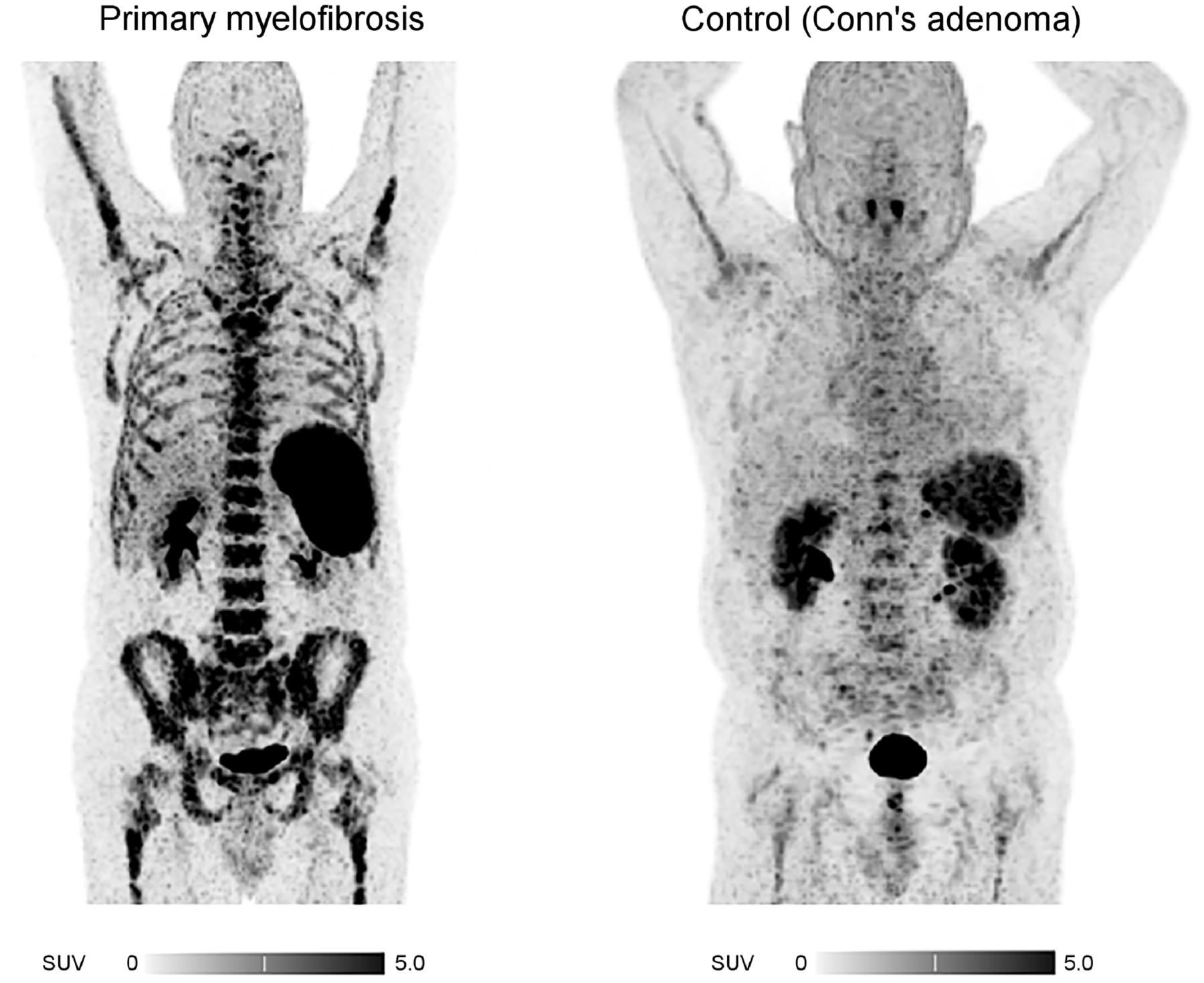
Display of patient (patient 6) with primary myelofibrosis. 68Ga-pentixafor PET/CT (maximum-intensity projections) depicts significantly increased tracer uptake in BM as well as spleen compared with control group.

No significant differences were observed between the SUVmean of the different subtypes of MPN (Table 2). Patients with JAK2V617F-positive or CALR-positive mutations did not show higher BM uptake than JAK2V617F-negative patients (r = 0.19, P = not statistically significant). Additional BM biopsies were available in 9 of 12 patients and confirmed moderate to strong CXCR4 expression in dysplastic cells of the megakaryocytic lineage in all samples (Supplemental Fig. 1).

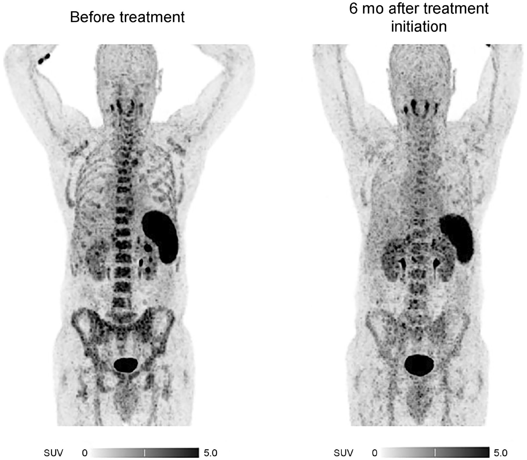
Three patients additionally underwent follow-up CXCR4-directed PET/CT after a median of 6 mo (range, 4–7 mo) after treatment initiation with ruxolitinib (patients 1 and 6) and hydroxyurea (patient 5). In these patients, initially high tracer accumulation in the spleen and BM (with an SUVmean of 6.12 ± 2.15, 3.60 ± 1.14, and 4.10 ± 0.32, respectively) declined (SUVmean: BM, 3.97 vs. 2.86; spleen, 7.72 vs. 4.89) in response to treatment (Fig. 2). Furthermore, the decrease in SUVmean after treatment corresponded with a spleen volume reduction and normalization of hemoglobin, peripheral leukocyte count, thrombocyte count, and lactate dehydrogenase level (Supplemental Table 2).

FIGURE 2.
  • Download figure
  • Open in new tab
  • Download powerpoint
FIGURE 2.

Response assessment with CXCR4-directed PET/CT imaging (maximum-intensity projections). Shown is example of therapy-induced CXCR4 downregulation in patient with essential thrombocythemia (patient 5).

DISCUSSION

Although PET/CT imaging is widely used for the diagnosis, staging, and response assessment of various types of hematologic malignancies (12), it is not routinely used in patients with MPNs. Currently, with the diagnosis being based solely on the assessment of clinical, hematologic, histopathologic, and genetic parameters (1), no imaging technique is established for the assessment of BM involvement. Here, we present the first (to our knowledge) proof-of-principle study investigating the in vivo application of 68Ga-pentixafor in patients with MPNs. We demonstrate the feasibility of 68Ga-pentixafor PET/CT to noninvasively detect and quantify the extent of BM involvement, with MPN patients demonstrating significantly higher tracer uptake in the BM and extramedullary hematopoietic sites than was seen in nonmalignant controls. With SUVmean ranging between 2.9 and 7.4, the intensity of 68Ga-pentixafor uptake in the BM of MPN patients was similar to that previously reported for BM involvement in patients with chronic lymphatic leukemia (13) and acute myeloid leukemia (14).

Interestingly, in our small cohort, the highest tracer accumulation was detected in the BM of a patient with essential thrombocythemia at the time of initial diagnosis. However, further research to establish and validate cutoffs for both detection of BM involvement and differentiation between various disease types is still needed.

Our findings are consistent with the concept of MPNs as chronic inflammatory diseases (15) in conjunction with an impaired microenvironment that favors malignant over normal hematopoiesis through profound changes in the BM stromal compartment and increased cytokine levels. Interestingly, megakaryocytes have been shown to contribute to MPN pathology and to be a major driver of BM fibrosis (16). In line with that observation, our immunohistochemistry data show CXCR4 expression predominantly on the surface of dysplastic cells of the megakaryocytic lineage. In this context, the extent of CXCR4 uptake may, therefore, also serve as a prognostic factor to further stratify MPN patients.

We monitored 3 newly diagnosed MPN patients (3 of our total of 12 patients) over a median of 6 mo after treatment initiation, showing that CXCR4 uptake determined by 68Ga-pentixafor PET/CT might correlate with treatment response. The extent of both BM and splenic uptake (SUVmean: BM, 3.97 vs. 2.86; spleen, 7.72 vs. 4.89) decreased with treatment initiation. Interestingly, tracer uptake in BM and spleen corresponded with hematologic parameters and splenic volume. The reduction in SUVmean after treatment correlated with normalization of hemoglobin, peripheral leukocyte count, thrombocyte count, and lactate dehydrogenase. Whereas recent data suggest additional utility for CXCR4-directed PET/CT imaging for response assessment in hematologic malignancies such as extranodal marginal zone lymphoma (17) or central nervous system B-cell lymphoma (18), the extent to which 68Ga-pentixafor PET/CT can be used for response assessment in MPNs needs to be investigated in future prospective studies.

Our pilot observation suffers from several limitations. The radiation exposure associated with PET/CT prevented the use of a control group of healthy individuals. Instead, we included Conn adenoma patients as a control group who received CXCR4 PET/CT imaging as part of their endocrinologic investigation (19), and 68Ga-pentixafor uptake in other controls might be even lower than in our endocrinologic control group. However, when compared with previously reported upper limits of physiologic BM uptake measurements in cancer patients without BM involvement, values agree broadly with our study (SUVmean: BM for pancreatic adenocarcinoma, 1.7; BM for mucosa-associated lymphoid tissue lymphoma, 2.3) (13). Furthermore, although immunohistochemical analysis could demonstrate CXCR4 expression in all BM biopsies, CXCR4 expression was relatively low and did not clearly correspond with the intensity of the PET signal. However, surface expression of CXCR4 is a dynamic process and can be influenced by therapeutic interventions (20).

CONCLUSION

To our knowledge, our data are the first to demonstrate that CXCR4-directed imaging with 68Ga-pentixafor PET/CT is feasible to visualize and quantify disease involvement in MPN patients. Further evaluation in larger, prospective studies is warranted to determine the clinical impact in primary staging and response assessment and to evaluate the potential for a theranostic approach.

DISCLOSURE

Hans-Jürgen Wester is the founder and shareholder of Scintomics. No other potential conflict of interest relevant to this article was reported.

KEY POINTS

QUESTION: Is CXCR4-directed PET imaging with 68Ga-pentixafor feasible to visualize and quantify disease involvement in MPNs?

PERTINENT FINDINGS: This retrospective analysis revealed that CXCR4-directed imaging is positive in all investigated patients and showed a significantly higher tracer uptake in the BM of patients with MPNs.

IMPLICATIONS FOR PATIENT CARE: CXCR4-directed PET imaging with 68Ga-pentixafor is feasible to noninvasively detect and quantify the extent of BM involvement in patients with MPNs.

Footnotes

  • Published online May 28, 2021.

  • © 2022 by the Society of Nuclear Medicine and Molecular Imaging.

REFERENCES

  1. 1.↵
    1. Spivak JL
    . Myeloproliferative neoplasms. N Engl J Med. 2017;377:895–896.
    OpenUrl
  2. 2.↵
    1. Jacobson O,
    2. Weiss ID
    . CXCR4 chemokine receptor overview: biology, pathology and applications in imaging and therapy. Theranostics. 2013;3:1–2.
    OpenUrlPubMed
  3. 3.↵
    1. Zhao H,
    2. Guo L,
    3. Zhao H,
    4. Zhao J,
    5. Weng H,
    6. Zhao B
    . CXCR4 over-expression and survival in cancer: a system review and meta-analysis. Oncotarget. 2015;6:5022–5040.
    OpenUrlCrossRefPubMed
  4. 4.
    1. Domanska UM,
    2. Kruizinga RC,
    3. Nagengast WB,
    4. et al
    .A review on CXCR4/CXCL12 axis in oncology: no place to hide. Eur J Cancer. 2013;49:219–230.
    OpenUrlCrossRefPubMed
  5. 5.↵
    1. Kircher M,
    2. Herhaus P,
    3. Schottelius M,
    4. et al
    .CXCR4-directed theranostics in oncology and inflammation. Ann Nucl Med. 2018;32:503–511.
    OpenUrl
  6. 6.↵
    1. Lapa C,
    2. Schreder M,
    3. Schirbel A,
    4. et al
    .[68Ga]pentixafor-PET/CT for imaging of chemokine receptor CXCR4 expression in multiple myeloma: comparison to [18F]FDG and laboratory values. Theranostics. 2017;7:205–212.
    OpenUrl
  7. 7.↵
    1. Lapa C,
    2. Hanscheid H,
    3. Kircher M,
    4. et al
    .Feasibility of CXCR4-directed radioligand therapy in advanced diffuse large B-cell lymphoma. J Nucl Med. 2019;60:60–64.
    OpenUrlAbstract/FREE Full Text
  8. 8.↵
    1. Lapa C,
    2. Luckerath K,
    3. Rudelius M,
    4. et al
    .[68Ga]pentixafor-PET/CT for imaging of chemokine receptor 4 expression in small cell lung cancer: initial experience. Oncotarget. 2016;7:9288–9295.
    OpenUrl
  9. 9.↵
    1. Derlin T,
    2. Hueper K
    . CXCR4-targeted therapy in breast cancer. Lancet Oncol. 2018;19:e370.
    OpenUrl
  10. 10.↵
    1. Reiter T,
    2. Kircher M,
    3. Schirbel A,
    4. et al
    .Imaging of C-X-C motif chemokine receptor CXCR4 expression after myocardial infarction with [68Ga]pentixafor-PET/CT in correlation with cardiac MRI. JACC Cardiovasc Imaging. 2018;11:1541–1543.
    OpenUrlFREE Full Text
  11. 11.↵
    1. Martin R,
    2. Juttler S,
    3. Muller M,
    4. Wester HJ
    . Cationic eluate pretreatment for automated synthesis of [68Ga]CPCR4.2. Nucl Med Biol. 2014;41:84–89.
    OpenUrlCrossRefPubMed
  12. 12.↵
    1. Valls L,
    2. Badve C,
    3. Avril S,
    4. et al
    .FDG-PET imaging in hematological malignancies. Blood Rev. 2016;30:317–331.
    OpenUrl
  13. 13.↵
    1. Mayerhoefer ME,
    2. Jaeger U,
    3. Staber P,
    4. et al
    .[68Ga]Ga-pentixafor PET/MRI for CXCR4 imaging of chronic lymphocytic leukemia: preliminary results. Invest Radiol. 2018;53:403–408.
    OpenUrl
  14. 14.↵
    1. Herhaus P,
    2. Habringer S,
    3. Philipp-Abbrederis K,
    4. et al
    .Targeted positron emission tomography imaging of CXCR4 expression in patients with acute myeloid leukemia. Haematologica. 2016;101:932–940.
    OpenUrlAbstract/FREE Full Text
  15. 15.↵
    1. Longhitano L,
    2. Li Volti G,
    3. Giallongo C,
    4. et al
    .The role of inflammation and inflammasome in myeloproliferative disease. J Clin Med. 2020;9:2334.
    OpenUrl
  16. 16.↵
    1. Ciurea SO,
    2. Merchant D,
    3. Mahmud N,
    4. et al
    .Pivotal contributions of megakaryocytes to the biology of idiopathic myelofibrosis. Blood. 2007;110:986–993.
    OpenUrlAbstract/FREE Full Text
  17. 17.↵
    1. Herhaus P,
    2. Habringer S,
    3. Vag T,
    4. et al
    .Response assessment with the CXCR4-directed positron emission tomography tracer [68Ga]pentixafor in a patient with extranodal marginal zone lymphoma of the orbital cavities. EJNMMI Res. 2017;7:51.
    OpenUrl
  18. 18.↵
    1. Herhaus P,
    2. Lipkova J,
    3. Lammer F,
    4. et al
    .CXCR4-targeted PET imaging of central nervous system B-cell lymphoma. J Nucl Med. 2020;61:1765–1771.
    OpenUrlAbstract/FREE Full Text
  19. 19.↵
    1. Rieckmann M,
    2. Delgobo M,
    3. Gaal C,
    4. et al
    .Myocardial infarction triggers cardioprotective antigen-specific T helper cell responses. J Clin Invest. 2019;129:4922–4936.
    OpenUrl
  20. 20.↵
    1. Lapa C,
    2. Luckerath K,
    3. Kircher S,
    4. et al
    .Potential influence of concomitant chemotherapy on CXCR4 expression in receptor directed endoradiotherapy. Br J Haematol. 2019;184:440–443.
    OpenUrl
  • Received for publication February 27, 2021.
  • Revision received April 1, 2021.
PreviousNext
Back to top

In this issue

Journal of Nuclear Medicine: 63 (1)
Journal of Nuclear Medicine
Vol. 63, Issue 1
January 1, 2022
  • Table of Contents
  • Table of Contents (PDF)
  • About the Cover
  • Index by author
  • Complete Issue (PDF)
Print
Download PDF
Article Alerts
Sign In to Email Alerts with your Email Address
Email Article

Thank you for your interest in spreading the word on Journal of Nuclear Medicine.

NOTE: We only request your email address so that the person you are recommending the page to knows that you wanted them to see it, and that it is not junk mail. We do not capture any email address.

Enter multiple addresses on separate lines or separate them with commas.
68Ga-Pentixafor PET/CT for Detection of Chemokine Receptor CXCR4 Expression in Myeloproliferative Neoplasms
(Your Name) has sent you a message from Journal of Nuclear Medicine
(Your Name) thought you would like to see the Journal of Nuclear Medicine web site.
Citation Tools
68Ga-Pentixafor PET/CT for Detection of Chemokine Receptor CXCR4 Expression in Myeloproliferative Neoplasms
Sabrina Kraus, Alexander Dierks, Leo Rasche, Olivia Kertels, Malte Kircher, Andreas Schirbel, Josip Zovko, Torsten Steinbrunn, Raoul Tibes, Hans-Jürgen Wester, Andreas K. Buck, Hermann Einsele, K. Martin Kortüm, Andreas Rosenwald, Constantin Lapa
Journal of Nuclear Medicine Jan 2022, 63 (1) 96-99; DOI: 10.2967/jnumed.121.262206

Citation Manager Formats

  • BibTeX
  • Bookends
  • EasyBib
  • EndNote (tagged)
  • EndNote 8 (xml)
  • Medlars
  • Mendeley
  • Papers
  • RefWorks Tagged
  • Ref Manager
  • RIS
  • Zotero
Share
68Ga-Pentixafor PET/CT for Detection of Chemokine Receptor CXCR4 Expression in Myeloproliferative Neoplasms
Sabrina Kraus, Alexander Dierks, Leo Rasche, Olivia Kertels, Malte Kircher, Andreas Schirbel, Josip Zovko, Torsten Steinbrunn, Raoul Tibes, Hans-Jürgen Wester, Andreas K. Buck, Hermann Einsele, K. Martin Kortüm, Andreas Rosenwald, Constantin Lapa
Journal of Nuclear Medicine Jan 2022, 63 (1) 96-99; DOI: 10.2967/jnumed.121.262206
Twitter logo Facebook logo LinkedIn logo Mendeley logo
  • Tweet Widget
  • Facebook Like
  • Google Plus One
Bookmark this article

Jump to section

  • Article
    • Abstract
    • MATERIALS AND METHODS
    • RESULTS
    • DISCUSSION
    • CONCLUSION
    • DISCLOSURE
    • Footnotes
    • REFERENCES
  • Figures & Data
  • Supplemental
  • Info & Metrics
  • PDF

Related Articles

  • This Month in JNM
  • PubMed
  • Google Scholar

Cited By...

  • Imaging of C-X-C Motif Chemokine Receptor 4 Expression in 690 Patients with Solid or Hematologic Neoplasms Using 68Ga-Pentixafor PET
  • Google Scholar

More in this TOC Section

  • Feasibility of an Ultra-Low-Dose PET Scan Protocol with CT-Based and LSO-TX–Based Attenuation Correction Using a Long–Axial-Field-of-View PET/CT Scanner
  • Prostate Cancer Recurrence Due to Isolated Testicular Metastases Detected by PSMA PET/CT
  • First-in-Human Serum Stability Studies of [177Lu]Lu-AMTG: A Step Toward Improved GRPR-Targeted Radiopharmaceutical Therapy
Show more BRIEF COMMUNICATION

Similar Articles

Keywords

  • myeloproliferative neoplasms
  • CXCR4
  • molecular imaging
  • PET
  • theranostics
SNMMI

© 2025 SNMMI

Powered by HighWire