
Main menu

  • Home
  • Content
    • Current
    • Ahead of print
    • Past Issues
    • JNM Supplement
    • SNMMI Annual Meeting Abstracts
    • Continuing Education
    • JNM Podcasts
  • Subscriptions
    • Subscribers
    • Institutional and Non-member
    • Rates
    • Journal Claims
    • Corporate & Special Sales
  • Authors
    • Submit to JNM
    • Information for Authors
    • Assignment of Copyright
    • AQARA requirements
  • Info
    • Reviewers
    • Permissions
    • Advertisers
  • About
    • About Us
    • Editorial Board
    • Contact Information
  • More
    • Alerts
    • Feedback
    • Help
    • SNMMI Journals
  • SNMMI
    • JNM
    • JNMT
    • SNMMI Journals
    • SNMMI

User menu

  • Subscribe
  • My alerts
  • Log in
  • Log out
  • My Cart

Search

  • Advanced search
Journal of Nuclear Medicine
  • SNMMI
    • JNM
    • JNMT
    • SNMMI Journals
    • SNMMI
  • Subscribe
  • My alerts
  • Log in
  • Log out
  • My Cart
Journal of Nuclear Medicine

Advanced Search

  • Home
  • Content
    • Current
    • Ahead of print
    • Past Issues
    • JNM Supplement
    • SNMMI Annual Meeting Abstracts
    • Continuing Education
    • JNM Podcasts
  • Subscriptions
    • Subscribers
    • Institutional and Non-member
    • Rates
    • Journal Claims
    • Corporate & Special Sales
  • Authors
    • Submit to JNM
    • Information for Authors
    • Assignment of Copyright
    • AQARA requirements
  • Info
    • Reviewers
    • Permissions
    • Advertisers
  • About
    • About Us
    • Editorial Board
    • Contact Information
  • More
    • Alerts
    • Feedback
    • Help
    • SNMMI Journals
  • View or Listen to JNM Podcast
  • Visit JNM on Facebook
  • Join JNM on LinkedIn
  • Follow JNM on Twitter
  • Subscribe to our RSS feeds
Research ArticleHot Topics

Is Tau Imaging More Than Just Upside-Down 18F-FDG Imaging?

Thilo van Eimeren, Gérard N. Bischof and Alexander Drzezga
Journal of Nuclear Medicine September 2017, 58 (9) 1357-1359; DOI: https://doi.org/10.2967/jnumed.117.190082
Thilo van Eimeren
1Multimodal Neuroimaging Group, Department of Nuclear Medicine, University of Cologne, Cologne, Germany; and
2German Research Center for Neurodegenerative Diseases (DZNE), Bonn/Cologne, Germany
  • Find this author on Google Scholar
  • Find this author on PubMed
  • Search for this author on this site
Gérard N. Bischof
1Multimodal Neuroimaging Group, Department of Nuclear Medicine, University of Cologne, Cologne, Germany; and
  • Find this author on Google Scholar
  • Find this author on PubMed
  • Search for this author on this site
Alexander Drzezga
1Multimodal Neuroimaging Group, Department of Nuclear Medicine, University of Cologne, Cologne, Germany; and
2German Research Center for Neurodegenerative Diseases (DZNE), Bonn/Cologne, Germany
  • Find this author on Google Scholar
  • Find this author on PubMed
  • Search for this author on this site
  • Article
  • Figures & Data
  • Info & Metrics
  • PDF
Loading

Several neurodegenerative disorders exhibit pathologic aggregation of the tau protein in the brain and are therefore summarized under the term tauopathies (1). Alzheimer disease (AD) is considered a tauopathy because, in addition to extraneuronal β-amyloid plaques, it is characterized by pathologic tau aggregation in the form of intraneuronal neurofibrillary tangles (2). In other tauopathies—including progressive supranuclear palsy, corticobasal degeneration, and some other disorders of the spectrum of frontotemporal lobar degenerative disorders—tau pathology may occur as the leading form of protein aggregation abnormality (3). The disappointing results of antiamyloid therapy approaches in AD (4,5) and the onset of novel anti-tau therapeutic concepts (6,7) have increased interest in the in vivo detection of tau pathology, which has been limited to postmortem examinations so far.

Several PET tracers for cerebral tau deposits have been developed, and some have already been applied in vivo to patients with neurodegenerative disorders. The most widely used tracers are 18F-AV-1451, 18F-THK5351, and 11C-PBB3 (8,9). More recently, tracers such as 18F-MK-6240, 18F-RO6958948, 18F-PI-2620, and 18F-JNJ64349311 have been introduced, and further compounds are under evaluation (10–12). Although some of these compounds are already being tested in clinical trials (up to phase 3), all of the available tracers can currently still be considered in an exploratory stage and the available literature is still limited.

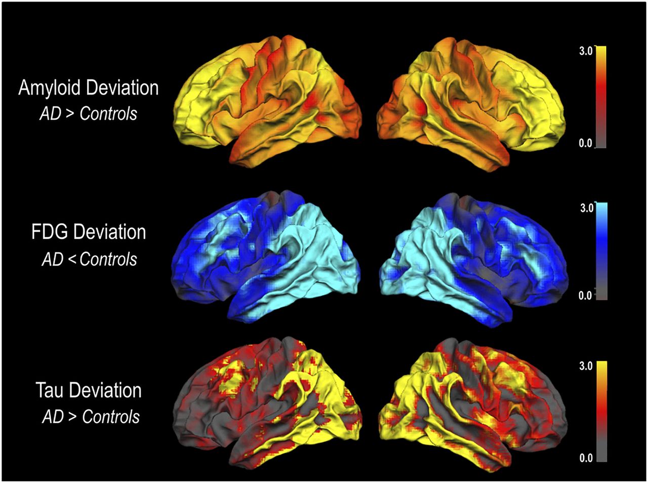
In several studies, a strong similarity between the distribution of tau tracer retention in the brain and the extent of hypometabolic abnormalities (as measured with 18F-FDG PET) has been observed (Fig. 1) (13–16). Furthermore, these studies statistically demonstrated an inverse quantitative relationship between tau deposition and metabolism. While underscoring the hypothesis that tau pathology may be causally involved in the development of neuronal dysfunction, this upside-down similarity between the two methods raises the question of whether tau PET has additional clinical value over established 18F-FDG PET.

FIGURE 1.
  • Download figure
  • Open in new tab
  • Download powerpoint
FIGURE 1.

Spatial correspondence of mean 18F-FDG deviation and mean tau deviation but not amyloid deviation in patients with AD. Deviation images were projected on lateral surface of left and right hemispheres using FreeSurfer. (Reprinted with permission of (13).)

STATUS OF TRACER VALIDATION

18F-FDG PET imaging has proven to have high diagnostic value at relatively low costs in the assessment of neurodegeneration (17–21). The tracer is broadly available, and automated analytic approaches and international guidelines are established (20,21).

In contrast, validation and standardization of tau tracers is far less advanced. This may stem not only from the novelty of the tracers but also from the complexity of tau as a target itself. Tau deposition can occur in different splicing isoforms (3R vs. 4R), different primary forms of fibrous assembly (paired helical vs. straight filaments), and different aggregation states (neurofibrils, pretangles, mature tangles, and coiled bodies, among others). Moreover, tau can be found in different cellular locations (intraneuronal, intraglial) and macroscopic anatomic locations (basal ganglia, cortex) (22,23). This variance in the appearance of tau pathology hampers the development and evaluation of tau imaging tracers.

Recently developed tau tracers have been promising insofar as in vitro analyses suggested distinct binding to tau pathology in the absence of relevant binding to amyloid (8). Furthermore, in vivo tracer retention patterns showed good agreement with the expected distribution of tau pathology (2,24). However, considerable between-tracer differences in regional capture of tau pathology may exist in AD (25–27) and in non-AD tauopathies such as corticobasal degeneration or progressive supranuclear palsy. Additionally, some evidence questions the specificity of currently available tau tracers. This refers to non-tau pathology such as TDP-43 and to off-target binding, which has been observed in vivo for several of the tracers in various regions (basal ganglia, brain stem, choroid plexus, and extracerebral structures) (24,28,29).

Elevated binding to melanin and monoaminooxydase may explain some of the off-target binding (30–32). Particularly worrisome are recent findings demonstrating a potential contribution of monoaminooxydase-B binding to tau PET imaging not only in subcortical but also in cortical brain regions (33). Such a contribution by unspecific binding could hamper quantitative assessment, particularly in longitudinal tau PET studies.

In summary, comparability between existing tau tracers is currently not warranted. To gain critical insight into the differential sensitivity and specificity of the currently available tracers, further evaluation is required, particularly with regard to systematic in vivo or postmortem cross validation. Corresponding studies are currently under way.

POTENTIAL FOR EARLY DIAGNOSIS

The value of 18F-FDG PET for early diagnosis of AD, particularly regarding short-term prediction of conversion to AD-type dementia in patients with mild cognitive impairment, has been repeatedly demonstrated (20,34,35). Similar data are not yet available for tau PET imaging. Supporting current hypothetical disease models, some studies suggest that tau pathology may be detected subsequent to elevated amyloid pathology by means of amyloid PET (36). Others suggest that mesial temporal tau aggregation may exist in some healthy elderly subjects in the absence of amyloid pathology, but the pathologic relevance of these findings is yet unclear (24). Regarding the obvious similarity between 18F-FDG PET and tau PET findings documented so far, similar sensitivities with regard to early detection may be expected. However, preliminary data from our and other groups (directly comparing 18F-FDG and tau PET in patients with mild cognitive impairment and AD) suggest that tau abnormalities may be somewhat more pronounced than hypometabolic changes and possibly can be detected earlier (Fig. 2) (37). Also, hot lesions in tau PET may be more salient than cold lesions in 18F-FDG PET.

FIGURE 2.
  • Download figure
  • Open in new tab
  • Download powerpoint
FIGURE 2.

Clearly abnormal tau PET findings but barely abnormal 18F-FDG PET findings in 66-y-old man with mild cognitive impairment and cerebrospinal fluid values indicative of AD pathology. Axial slices of SUV ratio images are shown for 18F-AV-1451 and 18F-FDG PET. Global maximum intensities were set to 100 for scaling.

POTENTIAL FOR DIFFERENTIAL DIAGNOSIS

The valuable role of 18F-FDG PET in differential diagnosis of neurodegenerative disorders has repeatedly been demonstrated, including various forms of dementias, movement disorders, and atypical Parkinsonian syndromes (38,39). In a similar vein, tau PET imaging shows specific patterns of tau tracer retention in typical and atypical variants of AD and other tauopathies (14,15,40–43). However, unlike 18F-FDG, tau PET at the same time provides insights on underlying neuropathology, which may allow tauopathies (e.g., corticobasal degeneration or progressive supranuclear palsy) to be distinguished from non-tauopathies (e.g., multiple-system atrophy).

POTENTIAL AS A BIOMARKER IN CLINICAL TRIALS

For 18F-FDG PET, a reliable association between hypometabolism and the type and severity of cognitive deficits has been demonstrated (44,45). This association qualifies 18F-FDG PET as a valuable tool to track clinical progression. However, 18F-FDG PET results may be affected by functional processes such as diaschisis or compensatory effects. In this respect, tau imaging may more directly mirror the extent and progression of neuropathology. Moreover, as a major advantage over amyloid PET, tau tracer uptake may better track cognitive decline than does amyloid deposition, as expected from neuropathologic studies. Most importantly, tau PET may be used for the selection of patients in future therapeutic trials directed toward the removal of tau pathology (7). Results from tau PET may also serve as a surrogate endpoint in clinical trials, since tau PET may reflect more proximal processes in the cascade of neurodegeneration than does hypometabolism. Tau PET could therefore surpass 18F-FDG PET as a “reasonably likely surrogate endpoint” in clinical trials (46).

CONCLUSION

Tau imaging represents an exciting, novel approach aiming to assess a basic pathology of neurodegeneration in vivo. Although similarities between patterns of hypometabolism and tau tracer deposition have been documented, tau imaging certainly offers more than just upside-down 18F-FDG PET information. An added clinical value of tau PET over the well-established 18F-FDG PET can be expected but remains to be proven. Preliminary data indicate that tau imaging could be slightly more sensitive than 18F-FDG PET in detecting the onset of neuronal injury in early stages of AD. Tau PET may also have advantages with regard to differential diagnosis. In addition to measuring the extent and localization of pathology, tau PET may allow differentiation between tauopathies and non-tauopathies. These questions may gain relevance with the advent of anti-tau therapy trials. Specific monitoring of anti-tau treatment may also represent a most important future application for tau PET. Although tau PET imaging certainly holds great potential in various diseases and in different domains spanning from early and differential diagnosis to disease monitoring, many questions, particularly with regard to validation of the tracers, still remain open. Much more needs to be learned about the variability of the signal due to unspecific contributions, across different tracers, and in different disease populations.

DISCLOSURE

Thilo van Eimeren received speaker and consulting honoraria and/or research support from AVID Radiopharmaceuticals and Eli Lilly. Alexander Drzezga received speaker and consulting honoraria and/or research support from AVID Radiopharmaceuticals, Eli Lilly, GE Healthcare, Siemens Healthcare, and Piramal. No other potential conflict of interest relevant to this article was reported.

Footnotes

  • Published online May 10, 2017.

  • © 2017 by the Society of Nuclear Medicine and Molecular Imaging.

REFERENCES

  1. 1.↵
    1. Lee VM,
    2. Goedert M,
    3. Trojanowski JQ
    . Neurodegenerative tauopathies. Annu Rev Neurosci. 2001;24:1121–1159.
    OpenUrlCrossRefPubMed
  2. 2.↵
    1. Braak H,
    2. Braak E
    . Staging of Alzheimer’s disease-related neurofibrillary changes. Neurobiol Aging. 1995;16:271–284.
    OpenUrlCrossRefPubMed
  3. 3.↵
    1. Neary D,
    2. Snowden J,
    3. Mann D
    . Frontotemporal dementia. Lancet Neurol. 2005;4:771–780.
    OpenUrlCrossRefPubMed
  4. 4.↵
    1. Hawkes N
    . Merck ends trial of potential Alzheimer’s drug verubecestat. BMJ. 2017;356:j845.
    OpenUrlFREE Full Text
  5. 5.↵
    1. Salloway S,
    2. Sperling R,
    3. Fox NC,
    4. et al
    . Two phase 3 trials of bapineuzumab in mild-to-moderate Alzheimer’s disease. N Engl J Med. 2014;370:322–333.
    OpenUrlCrossRefPubMed
  6. 6.↵
    1. Yanamandra K,
    2. Kfoury N,
    3. Jiang H,
    4. et al
    . Anti-tau antibodies that block tau aggregate seeding in vitro markedly decrease pathology and improve cognition in vivo. Neuron. 2013;80:402–414.
    OpenUrlCrossRefPubMed
  7. 7.↵
    1. Golde TE,
    2. Lewis J,
    3. McFarland NR
    . Anti-tau antibodies: hitting the target. Neuron. 2013;80:254–256.
    OpenUrl
  8. 8.↵
    1. Saint-Aubert L,
    2. Lemoine L,
    3. Chiotis K,
    4. Leuzy A,
    5. Rodriguez-Vieitez E,
    6. Nordberg A
    . Tau PET imaging: present and future directions. Mol Neurodegener. 2017;12:19.
    OpenUrl
  9. 9.↵
    1. Villemagne VL,
    2. Okamura N
    . In vivo tau imaging: obstacles and progress. Alzheimers Dement. 2014;10(suppl):S254–S264.
    OpenUrlCrossRefPubMed
  10. 10.↵
    1. Hostetler ED,
    2. Walji AM,
    3. Zeng Z,
    4. et al
    . Preclinical characterization of 18F-MK-6240, a promising PET tracer for in vivo quantification of human neurofibrillary tangles. J Nucl Med. 2016;57:1599–1606.
    OpenUrlAbstract/FREE Full Text
  11. 11.
    1. Furumoto S,
    2. Tago T,
    3. Harada R,
    4. Kudo Y,
    5. Okamura N
    . 18F-labeled 2-arylquinoline derivatives for tau imaging: chemical, radiochemical, biological and clinical features. Curr Alzheimer Res. 2017;14:178–185.
    OpenUrl
  12. 12.↵
    1. Declercq L,
    2. Rombouts F,
    3. Koole M,
    4. et al
    . Preclinical evaluation of 18F-JNJ64349311, a novel PET tracer for tau imaging. J Nucl Med. 2017;58:975–981.
    OpenUrlAbstract/FREE Full Text
  13. 13.↵
    1. Bischof GN,
    2. Jessen F,
    3. Fliessbach K,
    4. et al
    . Impact of tau and amyloid burden on glucose metabolism in Alzheimer’s disease. Ann Clin Transl Neurol. 2016;3:934–939.
    OpenUrl
  14. 14.↵
    1. Dronse J,
    2. Fliessbach K,
    3. Bischof GN,
    4. et al
    . In vivo patterns of tau pathology, amyloid-β burden, and neuronal dysfunction in clinical variants of Alzheimer’s disease. J Alzheimers Dis. 2017;55:465–471.
    OpenUrl
  15. 15.↵
    1. Ossenkoppele R,
    2. Schonhaut DR,
    3. Schöll M,
    4. et al
    . Tau PET patterns mirror clinical and neuroanatomical variability in Alzheimer’s disease. Brain. 2016;139:1551–1567.
    OpenUrlCrossRefPubMed
  16. 16.↵
    1. Ossenkoppele R,
    2. Schonhaut DR,
    3. Baker SL,
    4. et al
    . Tau, amyloid, and hypometabolism in a patient with posterior cortical atrophy. Ann Neurol. 2015;77:338–342.
    OpenUrlCrossRefPubMed
  17. 17.↵
    1. Silverman DH,
    2. Small GW,
    3. Chang CY,
    4. et al
    . Positron emission tomography in evaluation of dementia: regional brain metabolism and long-term outcome. JAMA. 2001;286:2120–2127.
    OpenUrlCrossRefPubMed
  18. 18.
    1. Chételat G,
    2. Desgranges B,
    3. de la Sayette V,
    4. Viader F,
    5. Eustache F,
    6. Baron J-C
    . Mild cognitive impairment: can FDG-PET predict who is to rapidly convert to Alzheimer’s disease? Neurology. 2003;60:1374–1377.
    OpenUrlCrossRefPubMed
  19. 19.
    1. Moulin-Romsee G,
    2. Maes A,
    3. Silverman D,
    4. Mortelmans L,
    5. Van Laere K
    . Cost-effectiveness of 18F-fluorodeoxyglucose positron emission tomography in the assessment of early dementia from a Belgian and European perspective. Eur J Neurol. 2005;12:254–263.
    OpenUrlCrossRefPubMed
  20. 20.↵
    1. Minoshima S,
    2. Frey KA,
    3. Koeppe RA,
    4. Foster NL,
    5. Kuhl DE
    . A diagnostic approach in Alzheimer’s disease using three-dimensional stereotactic surface projections of fluorine-18-FDG PET. J Nucl Med. 1995;36:1238–1248.
    OpenUrlAbstract/FREE Full Text
  21. 21.↵
    1. Varrone A,
    2. Asenbaum S,
    3. Vander Borght T,
    4. et al
    . EANM procedure guidelines for PET brain imaging using [18F]FDG, version 2. Eur J Nucl Med Mol Imaging. 2009;36:2103–2110.
    OpenUrlCrossRefPubMed
  22. 22.↵
    1. Majounie E,
    2. Cross W,
    3. Newsway V,
    4. et al
    . Variation in tau isoform expression in different brain regions and disease states. Neurobiol Aging. 2013;34:1922e7–1922e12.
    OpenUrl
  23. 23.↵
    1. Wang Y,
    2. Mandelkow E
    . Tau in physiology and pathology. Nat Rev Neurosci. 2016;17:5–21.
    OpenUrlCrossRefPubMed
  24. 24.↵
    1. Schöll M,
    2. Lockhart SN,
    3. Schonhaut DR,
    4. et al
    . PET imaging of tau deposition in the aging human brain. Neuron. 2016;89:971–982.
    OpenUrlCrossRefPubMed
  25. 25.↵
    1. Villemagne VL,
    2. Doré V,
    3. Bourgeat P,
    4. et al
    . Aβ-amyloid and tau imaging in dementia. Semin Nucl Med. 2017;47:75–88.
    OpenUrl
  26. 26.
    Chiotis K, Stenkrona P, Almkvist O, et al. Head-to-head comparison of tau-specific tracers in Alzheimer’s disease: [11C]THK5351 vs [11C]PBB3 PET imaging. Presented at the 11th Human Amyloid Imaging Conference; Miami, Florida; January 2017.
  27. 27.↵
    Jang YK, Rabinovici G, Kim H, Lee JS, Kim KW, Seo SW. Comparison of tau distribution according to tau tracers in various neurodegenerative diseases: using [18F]AV1451 and [18F]THK-5351. Presented at the 11th Human Amyloid Imaging Conference; Miami, Florida; January 2017.
  28. 28.↵
    1. Hansen AK,
    2. Knudsen K,
    3. Lillethorup TP,
    4. et al
    . In vivo imaging of neuromelanin in Parkinson’s disease using 18F-AV-1451 PET. Brain. 2016;139:2039–2049.
    OpenUrlCrossRefPubMed
  29. 29.↵
    1. Lowe VJ,
    2. Curran G,
    3. Fang P,
    4. et al
    . An autoradiographic evaluation of AV-1451 tau PET in dementia. Acta Neuropathol Commun. 2016;4:58.
    OpenUrl
  30. 30.↵
    1. Marquié M,
    2. Normandin MD,
    3. Vanderburg CR,
    4. et al
    . Validating novel tau positron emission tomography tracer [F-18]-AV-1451 (T807) on postmortem brain tissue. Ann Neurol. 2015;78:787–800.
    OpenUrlCrossRefPubMed
  31. 31.
    1. Ng KP,
    2. Pascoal TA,
    3. Mathotaarachchi S,
    4. et al
    . Monoamine oxidase B inhibitor, selegiline, reduces 18F-THK5351 uptake in the human brain. Alzheimers Res Ther. 2017;9:25.
    OpenUrl
  32. 32.↵
    1. Harada R,
    2. Okamura N,
    3. Furumoto S,
    4. et al
    . [18F]THK-5117 PET for assessing neurofibrillary pathology in Alzheimer’s disease. Eur J Nucl Med Mol Imaging. 2015;42:1052–1061.
    OpenUrlCrossRefPubMed
  33. 33.↵
    Guo Q. Evaluation of the selectivity of tau PET radioligand THK5351 in AD brain in vitro and nonhuman primate brain in vivo. Presented at the 11th Human Amyloid Imaging Conference; Miami, Florida; January 12, 2017.
  34. 34.↵
    1. Drzezga A,
    2. Grimmer T,
    3. Riemenschneider M,
    4. et al
    . Prediction of individual clinical outcome in MCI by means of genetic assessment and 18F-FDG PET. J Nucl Med. 2005;46:1625–1632.
    OpenUrlAbstract/FREE Full Text
  35. 35.↵
    1. Ishii K,
    2. Minoshima S
    . PET is better than perfusion SPECT for early diagnosis of Alzheimer’s disease. Eur J Nucl Med Mol Imaging. 2005;32:1463–1465.
    OpenUrlCrossRefPubMed
  36. 36.↵
    1. Jack CR, Jr,
    2. Knopman DS,
    3. Jagust WJ,
    4. et al
    . Tracking pathophysiological processes in Alzheimer’s disease: an updated hypothetical model of dynamic biomarkers. Lancet Neurol. 2013;12:207–216.
    OpenUrlCrossRefPubMed
  37. 37.↵
    1. Schonhaut DR,
    2. Ossenkoppele R,
    3. Bejanin A,
    4. et al
    . Tau-PET patterns overlap and exceed hypometabolism in Alzheimer’s disease. Alzheimers Dement. 2016;12(suppl):P545–P547.
    OpenUrl
  38. 38.↵
    1. Hellwig S,
    2. Amtage F,
    3. Kreft A,
    4. et al
    . [18F]FDG-PET is superior to [123I]IBZM-SPECT for the differential diagnosis of parkinsonism. Neurology. 2012;79:1314–1322.
    OpenUrlCrossRefPubMed
  39. 39.↵
    1. Foster NL,
    2. Heidebrink JL,
    3. Clark CM,
    4. et al
    . FDG-PET improves accuracy in distinguishing frontotemporal dementia and Alzheimer’s disease. Brain. 2007;130:2616–2635.
    OpenUrlCrossRefPubMed
  40. 40.↵
    1. Hammes J,
    2. Bischof GN,
    3. Giehl K,
    4. et al
    . Elevated in vivo [18F]-AV-1451 uptake in a patient with progressive supranuclear palsy. Mov Disord. 2017;32:170–171.
    OpenUrl
  41. 41.
    1. Kikuchi A,
    2. Okamura N,
    3. Hasegawa T,
    4. et al
    . In vivo visualization of tau deposits in corticobasal syndrome by 18F-THK5351 PET. Neurology. 2016;87:2309–2316.
    OpenUrl
  42. 42.
    1. Passamonti L,
    2. Vázquez Rodríguez P,
    3. Hong YT,
    4. et al
    . 18F-AV-1451 positron emission tomography in Alzheimer’s disease and progressive supranuclear palsy. Brain. 2017;140:781–791.
    OpenUrl
  43. 43.↵
    1. Whitwell JL,
    2. Lowe VJ,
    3. Tosakulwong N,
    4. et al
    . [18F]AV-1451 tau positron emission tomography in progressive supranuclear palsy. Mov Disord. 2017;32:124–133.
    OpenUrl
  44. 44.↵
    1. Chételat G,
    2. Eustache F,
    3. Viader F,
    4. et al
    . FDG-PET measurement is more accurate than neuropsychological assessments to predict global cognitive deterioration in patients with mild cognitive impairment. Neurocase. 2005;11:14–25.
    OpenUrlCrossRefPubMed
  45. 45.↵
    1. Kuczynski B,
    2. Reed B,
    3. Mungas D,
    4. Weiner M,
    5. Chui HC,
    6. Jagust W
    . Cognitive and anatomic contributions of metabolic decline in Alzheimer disease and cerebrovascular disease. Arch Neurol. 2008;65:650–655.
    OpenUrlCrossRefPubMed
  46. 46.↵
    FDA-NIH Biomarker Working Group. BEST (Biomarkers, EndpointS, and other Tools) Resource. Bethesda, Maryland: National Institutes of Health; 2016.
  • Received for publication April 4, 2017.
  • Accepted for publication May 5, 2017.
PreviousNext
Back to top

In this issue

Journal of Nuclear Medicine: 58 (9)
Journal of Nuclear Medicine
Vol. 58, Issue 9
September 1, 2017
  • Table of Contents
  • Table of Contents (PDF)
  • About the Cover
  • Index by author
Print
Download PDF
Article Alerts
Sign In to Email Alerts with your Email Address
Email Article

Thank you for your interest in spreading the word on Journal of Nuclear Medicine.

NOTE: We only request your email address so that the person you are recommending the page to knows that you wanted them to see it, and that it is not junk mail. We do not capture any email address.

Enter multiple addresses on separate lines or separate them with commas.
Is Tau Imaging More Than Just Upside-Down 18F-FDG Imaging?
(Your Name) has sent you a message from Journal of Nuclear Medicine
(Your Name) thought you would like to see the Journal of Nuclear Medicine web site.
Citation Tools
Is Tau Imaging More Than Just Upside-Down 18F-FDG Imaging?
Thilo van Eimeren, Gérard N. Bischof, Alexander Drzezga
Journal of Nuclear Medicine Sep 2017, 58 (9) 1357-1359; DOI: 10.2967/jnumed.117.190082

Citation Manager Formats

  • BibTeX
  • Bookends
  • EasyBib
  • EndNote (tagged)
  • EndNote 8 (xml)
  • Medlars
  • Mendeley
  • Papers
  • RefWorks Tagged
  • Ref Manager
  • RIS
  • Zotero
Share
Is Tau Imaging More Than Just Upside-Down 18F-FDG Imaging?
Thilo van Eimeren, Gérard N. Bischof, Alexander Drzezga
Journal of Nuclear Medicine Sep 2017, 58 (9) 1357-1359; DOI: 10.2967/jnumed.117.190082
Twitter logo Facebook logo LinkedIn logo Mendeley logo
  • Tweet Widget
  • Facebook Like
  • Google Plus One
Bookmark this article

Jump to section

  • Article
    • STATUS OF TRACER VALIDATION
    • POTENTIAL FOR EARLY DIAGNOSIS
    • POTENTIAL FOR DIFFERENTIAL DIAGNOSIS
    • POTENTIAL AS A BIOMARKER IN CLINICAL TRIALS
    • CONCLUSION
    • DISCLOSURE
    • Footnotes
    • REFERENCES
  • Figures & Data
  • Info & Metrics
  • PDF

Related Articles

  • This Month in JNM
  • PubMed
  • Google Scholar

Cited By...

  • Tau Imaging with 18F-MK6240 across the Alzheimers Disease spectrum
  • One-Stop Shop: 18F-Flortaucipir PET Differentiates Amyloid-Positive and -Negative Forms of Neurodegenerative Diseases
  • Google Scholar

More in this TOC Section

  • RECIP 1.0: A Roadmap for Clinical Implementation
  • Diagnostic Radiopharmaceutical Trial Design: Is It Time to Change Nomenclature?
  • From Stabilization to Depletion: Molecular Imaging to Measure Therapeutic Response in ATTR-CA
Show more Hot Topics

Similar Articles

SNMMI

© 2025 SNMMI

Powered by HighWire